Author: Emily Carlson

Headshot of emily Carlson

Posts by Emily Carlson

The Simple Rules Bacteria Follow to Survive

1 comment
Left: Football stadium. Right: Colored contoured lines showing the periodic stops in the growth of a bacterial colony
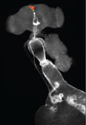
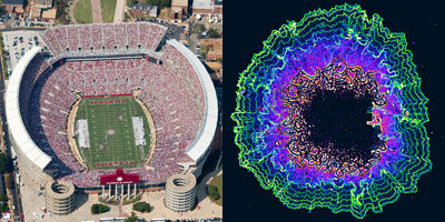
Football image credit: Stock image. The colored contoured lines show the periodic stops in the growth of a bacterial colony. Credit: Süel Lab, UCSD.

What do these images of football fans and bacterial cells have in common? By following simple rules, each individual allows the group to accomplish tasks none of them could do alone—a stadium wave that ripples through the crowd or a cell colony that rebounds after antibiotic treatment.

These collective behaviors are just a few examples of what scientists call emergent phenomena. While the reasons for the emergence of such behavior in groups of birds, fish, ants and other creatures is well understood, they’ve been less clear in bacteria. Two independent research teams have now identified some of the rules bacterial cells follow to enable the colony to persist. Continue reading “The Simple Rules Bacteria Follow to Survive”

Food for Thought: Nutrient-Detecting Brain Sensor in Flies

0 comments

If you participated in a cupcake taste test, do you think you’d be able to distinguish a treat made with natural sugar from one made with artificial sweetener? Scientists have known for decades that animals can tell the difference, but what’s been less clear is how.

Fruit fly neurons in the brain (red) with nerve fibers (white) that extend to the gut.
For fruit flies, nutritive sugars activate a set of neurons in the brain (red) with nerve fibers (white) that extend to the gut. Credit: Jason Lai and Greg Suh, New York University School of Medicine.

Now, researchers at the New York University School of Medicine have identified a collection of specialized nerve cells in fruit flies that acts as a nutrient-detecting sensor, helping them select natural sugar over artificial sweetener to get the energy they need to survive.

“How specific sensory stimuli trigger specific behaviors is a big research question,” says NIGMS’ Mike Sesma. “Food preferences involve more than taste and hunger, and this study, which was done in an organism with many of the same cellular components as humans, gives us a glimpse of the complex interplay among the many factors.”

The study, described in the July 15 issue of Neuron, builds on the researchers’ earlier studies of feeding behavior that showed hungry fruit flies, even ones lacking the ability to taste, selected calorie-packed sugars over zero-calorie alternatives. The scientists, led by Greg Suh and Monica Dus Exit icon, suspected that the flies had a molecular system for choosing energy-replenishing foods, especially during periods of starvation. Continue reading “Food for Thought: Nutrient-Detecting Brain Sensor in Flies”

Mapping Our Skin’s Microbes and Molecules

0 comments

Last month, we shared some facts about the microbes that inhabit us. Here’s another: From head to toe, our skin bacteria coexist with chemicals in hygiene products, fibers from clothes and proteins shed by dead or dying skin cells.

These images highlight the complex composition of our body’s largest organ. They show the association between microbial diversity (top images) and skin chemistry (middle images). The different colors note the abundance of a certain bacterium or molecule—red is high, and blue is low. The skin maps remind NIH Director Francis Collins of a 60’s rock album cover.

Continue reading “Mapping Our Skin’s Microbes and Molecules”

Field Focus: Our Microbial Menagerie

0 comments

Trillions of microorganisms inhabit us—inside and out. Scientists are surveying these microbial metropolises to learn more about their role in health. Microbiologists Darren Sledjeski of NIGMS and Andrew Goodman Exit icon of Yale University share a few details of what researchers have learned so far.

Vitruvian man filled with bacteria.
Researchers are surveying the microbes that inhabit us to learn more about their role in health. Credit: Andrew Goodman, Yale University.
  1. The majority of the microbes that inhabit us are bacteria. The rest of the microbial menagerie is fungi and viruses, including ones that infect the bacteria! Collectively, our resident microorganisms are referred to as the human microbiota, and their genomes are called the human microbiome.
  2. Our bodies harbor more bacterial cells than human ones. Even so, the microbiota accounts for less than 3 percent of a person’s body mass. That’s because our cells are up to 10,000 times bigger in volume than bacterial cells.
  3. Your collection of bacteria has more genes than you do. Scientists estimate that the genomes of gut bacteria contain 100-fold or more genes than our own genomes. For this reason, the human microbiome is sometimes called our second genome.
  4. Most of our microbes are harmless, and some are helpful. For example, harmless microbes on the skin keep infectious microbes from occupying that space. Microbes in the colon break down lactose and other complex carbohydrates that our bodies can’t naturally digest.
  5. Different microbes occupy different parts of the body. Some skin bacteria prefer the oily nooks near the nose, while others like the dry terrain of the forearm. Bacteria don’t all fare well in the same environment and have adapted to live in certain niches. The NIGMS Findings Magazine article Body Bacteria: Exploring the Skin’s Microbial Metropolis shows what types of bacteria colonize where.
Screenshot from the iBiology video.
Are we more microbial than human? Richard Losick, a microbiologist at Harvard University, explores that question in this video lecture produced by iBiology Exit icon.
  1. Each person’s microbiota is unique. The demographics of microbiota differ among individuals. Diet is one reason. Also, while a type of microbe might be part of one person’s normal microbial flora, it might not be part of another’s, and could potentially make that person sick.
  2. Host-microbial interactions are universal. Microbial communities may vary from person to person, but everyone’s got them, including other creatures. For this reason, researchers can use model organisms to tease apart the complexities of host-microbial interactions and develop broad principles for understanding them. The mouse is the most widely used animal model for microbiome studies.
  3. The role of microbiota in our health isn’t entirely clear. While it’s now well accepted that the microbial communities that inhabit us are actively involved in a range of conditions—from asthma to obesity—research studies have not yet pinpointed why or how. In other words, the results may suggest that the presence of a bacterial community is associated with a disease, but they don’t show cause and effect.
  4. Most of our microbes have not been grown in the lab. Microbes require a certain mix of nutrients and other microbes to survive, making it challenging to replicate their natural environments in a petri dish. New culturing techniques are enabling scientists to study previously uncultivated microbes.
  5. The impact of probiotic and prebiotic products isn’t clear. Fundamental knowledge gaps remain regarding how these products may work and what effects they might have on host-microbial interactions. A new NIH effort to stimulate research in this area is under way.
  6. There’s even more we don’t know! Additional areas of research include studying the functions of microbial genes and the effects of gut microbes on medicines. The more we learn from these and other studies, the more we’ll understand how our normal microbiota interacts with us and how to apply that knowledge to promote our health.

5 Reasons Biologists Love Math

4 comments

Biologists use math in a variety of ways, from designing experiments to mapping complex biological systems. Credit: Stock image.

On Saturday (at 9:26:53 to be exact), math lovers and others around the world will celebrate Pi—that really long number that represents the ratio of the circumference of a circle to its diameter. I asked our scientific experts why math is important to biomedical research. Here are a few reasons.

  1. Math allows biologists to describe how molecules move in and out of cells, how bacteria shuttle through blood vessels, how drugs get broken down in the body and many other physiological processes.
  2. Studying the geometry, topology and other physical characteristics of DNA, proteins and cellular structures has shed light on their functions and on approaches for enhancing or disrupting those functions.
  3. Math helps scientists design their experiments, including clinical trials, so they result in meaningful data, a.k.a statistical significance.
  4. Scientists use math to piece together all the different parts of a cell, an organ or an entire organism to better understand how the parts interact and how perturbations in these complex systems may contribute to disease.
  5. Sometimes it’s impossible or too difficult to answer a research question through traditional lab experiments, so biologists rely on math to develop models that represent the system they’re studying, whether it’s a metastasizing cancer cell or an emerging infectious disease. These approaches allow scientists to indicate the likelihood of certain outcomes as well as refine the research questions.

Want more? Here’s a video with 10 reasons biologists should know some math.

Simulating the Potential Spread of Measles

0 comments
Try out FRED Measles:

  1. Go to http://fred.publichealth.
    pitt.edu/measles
    Exit icon
  2. Select “Get Started”
  3. Pick a state and city
  4. Play both simulations

To help the public better understand how measles can spread, a team of infectious disease computer modelers at the University of Pittsburgh has launched a free, mobile-friendly tool that lets users simulate measles outbreaks in cities across the country.

The tool is part of the Pitt team’s Framework for Reconstructing Epidemiological Dynamics, or FRED, that it previously developed to simulate flu epidemics. FRED is based on anonymized U.S. census data that captures demographic and geographic distributions of different communities. It also incorporates details about the simulated disease, such as how contagious it is.

Screenshot of the FRED simulation.

A free, mobile-friendly tool lets users simulate potential measles outbreaks in cities across the country. Credit: University of Pittsburgh Graduate School of Public Health.

Continue reading “Simulating the Potential Spread of Measles”

Unprecedented Views of HIV

0 comments

Visualizations can give scientists unprecedented views of complex biological processes. Here’s a look at two new ones that shed light on how HIV enters host cells.

Animation of HIV’s Entry Into Host Cells

Screen shot of the video
This video animation of HIV’s entry into a human immune cell is the first one released in Janet Iwasa’s current project to visualize the virus’ life cycle. As they’re completed, the animations will be posted at http://scienceofhiv.org Exit icon.

We previously introduced you to Janet Iwasa, a molecular animator who’s visualized complex biological processes such as cells ingesting materials and proteins being transported across a cell membrane. She has now released several animations from her current project of visualizing HIV’s life cycle Exit icon. The one featured here shows the virus’ entry into a human immune cell.

“Janet’s animations add great value by helping us consider how complex interactions between viruses and their host cells actually occur in time and space,” says Wes Sundquist, who directs the Center for the Structural Biology of Cellular Host Elements in Egress, Trafficking, and Assembly of HIV Exit icon at the University of Utah. “By showing us how different steps in viral replication must be linked together, the animations suggest hypotheses that hadn’t yet occurred to us.”

Continue reading “Unprecedented Views of HIV”

Meet Maureen L. Mulvihill

1 comment
Maureen L. Mulvihill, Ph.D.
Credit: Actuated Medical, Inc.
Maureen L. Mulvihill, Ph.D.
Fields: Materials science, logistics
Works at: Actuated Medical, Inc., a small company that develops medical devices
Second job (volunteer): Bellefonte YMCA Swim Team Parent Boost Club Treasurer
Best skill: Listening to people
Last thing she does every night: Reads to her 7- and 10-year-old children until “one of us falls asleep”

If you’re a fan of the reality TV show Shark Tank, you tune in to watch aspiring entrepreneurs present their ideas and try to get one of the investors to help develop and market the products. Afterward, you might start to think about what you could invent.

Maureen L. Mulvihill has never watched the show, but she lives it every day. She is co-founder, president and CEO of Actuated Medical, Inc. (AMI), a Pennsylvania-based company that develops specialized medical devices. The devices include a system for unclogging feeding tubes, motors that assist MRI-related procedures and needles that gently draw blood.

AMI’s products rely on the same motion-control technologies that allow a quartz watch to keep time, a microphone to project sound and even a telescope to focus on a distant object in a sky. In general, the devices are portable, affordable and unobtrusive, making them appealing to doctors and patients.

Mulvihill, who’s trained in an area of engineering called materials science, says, “I’m really focused on how to translate technologies into ways that help people.” Continue reading “Meet Maureen L. Mulvihill”

Untangling a Trending Topic

0 comments

It’s not every day that we log into Facebook and Twitter to see conversations about denaturing proteins and the possibility of reducing biotechnology costs, but that changed last week when a story about “unboiling” eggs became a trending topic.

Since NIGMS partially funded the research advance Exit icon that led to the media scramble, we asked our scientific expert Jean Chin to tell us more about it.

What’s the advance?

Gregory Weiss of the University of California, Irvine, and his collaborators have designed a device that basically unties proteins that have been tangled together. Continue reading “Untangling a Trending Topic”

Forecasting Infectious Disease Spread with Web Data

0 comments

Just as you might turn to Twitter or Facebook for a pulse on what’s happening around you, researchers involved in an infectious disease computational modeling project are turning to anonymized social media and other publicly available Web data to improve their ability to forecast emerging outbreaks and develop tools that can help health officials as they respond.

Mining Wikipedia Data

Screen shot of the Wikipedia site
Incorporating real-time, anonymized data from Wikipedia and other novel sources of information is aiding efforts to forecast and respond to emerging outbreaks. Credit: Stock image.

“When it comes to infectious disease forecasting, getting ahead of the curve is problematic because data from official public health sources is retrospective,” says Irene Eckstrand of the National Institutes of Health, which funds the project, called Models of Infectious Disease Agent Study (MIDAS). “Incorporating real-time, anonymized data from social media and other Web sources into disease modeling tools may be helpful, but it also presents challenges.”

To help evaluate the Web’s potential for improving infectious disease forecasting efforts, MIDAS researcher Sara Del Valle of Los Alamos National Laboratory conducted proof-of-concept experiments involving data that Wikipedia releases hourly to any interested party. Del Valle’s research group built models based on the page view histories of disease-related Wikipedia pages in seven languages. The scientists tested the new models against their other models, which rely on official health data reported from countries using those languages. By comparing the outcomes of the different modeling approaches, the Los Alamos team concluded that the Wikipedia-based modeling results for flu and dengue fever performed better than those for other diseases. Continue reading “Forecasting Infectious Disease Spread with Web Data”