
Last month, we shared some facts about the microbes that inhabit us. Here’s another: From head to toe, our skin bacteria coexist with chemicals in hygiene products, fibers from clothes and proteins shed by dead or dying skin cells.
These images highlight the complex composition of our body’s largest organ. They show the association between microbial diversity (top images) and skin chemistry (middle images). The different colors note the abundance of a certain bacterium or molecule—red is high, and blue is low. The skin maps remind NIH Director Francis Collins of a 60’s rock album cover.
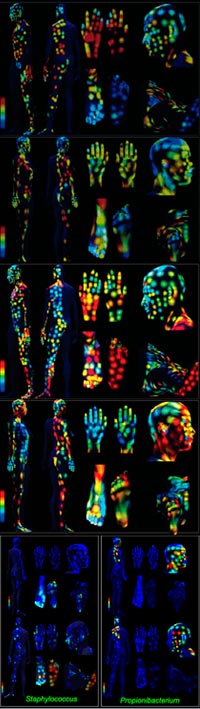
To create the maps, researchers led by Pieter Dorrestein of the University of California, San Diego, started by swabbing the skin of two volunteers who had abstained from bathing, wearing deodorant, moisturizing and applying any other hygiene or beauty product for three days. The research team then ran the swabs through different machines to identify the bacterial species present in the samples as well as the molecular and chemical compositions of them. The scientists used this data to construct the whole body maps.
Earlier work by others mapped the distribution of skin microbes, showing that ones such as Propionibacterium like oily areas of the face while others such as Staphylococcus prefer sweaty spots between the toes. Dorrestein’s maps show a similar distribution (see bottom images).
The new maps go beyond this microbial picture to also show what shares those spaces. “This is the first study of its kind to characterize the surface distribution of skin molecules and pair that data with microbial diversity,” says Dorrestein in a UCSD news release.
By doing so, his team learned, for instance, that Propionibacterium on the head coexists with large amounts of molecules found in skin cell membranes as well as ingredients found in sunscreens and cosmetics. Whether there’s a cause-and-effect relationship still needs to be investigated.
Dorrestein’s skin mapping approach may be a starting point for those investigations. The work offers a proof of concept, says UCSD’s Trey Ideker, who leads the NIGMS-funded systems biology center that supported the project through its seed grant program to further stimulate the development of new approaches for studying complex biomedical problems.
“It demonstrates that we can now take detailed molecular and microbiological readings from all over the body, and that all of these measurements differ depending on where you look,” Ideker explains. “The big question for future research is which of these measurements can be linked to the health of a person or what they can tell us about disease.”