Tag: Systems Biology

Shedding Light on Sepsis

1 comment

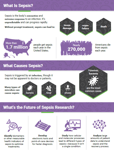
Sepsis is the body’s overactive and extreme response to an infection. It’s unpredictable, can progress rapidly, and affects more than 1.7 million people in the United States each year. Without prompt treatment, it can lead to tissue damage, organ failure, and death. NIGMS supports state-of-the-art sepsis research, including the development of rapid diagnostics and new therapeutics. September is Sepsis Awareness Month, and we’re highlighting a few resources that offer more information about this condition.

Our infographic provides details at a glance on basic statistics and the future of sepsis research. It’s also available in Spanish.

Continue reading “Shedding Light on Sepsis”

Sepsis: Using Big Data to Cut a Killer Down to Size

0 comments
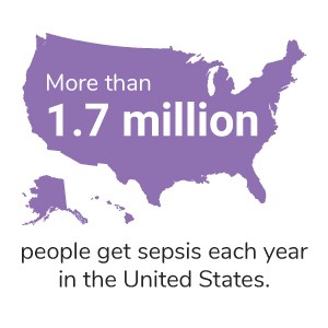
A geographical outline of the U.S. with the text More than 1.7 million people get sepsis each year in the United States. View the full infographic for more facts about sepsis.

Sepsis is a serious medical condition caused by an overwhelming response to infection that damages tissues and organs. It’s unpredictable, progresses quickly, can strike anyone, and is a leading cause of hospital-related deaths. In the U.S. alone, nearly 270,000 people die each year from sepsis. Those who survive sepsis often end up in the hospital again, and some have long-term health complications. Early treatment is key for many patients to survive sepsis, yet doctors can’t easily diagnose it because it’s so complex and each patient is different.

Despite decades of research, sepsis remains a poorly understood condition with limited diagnostic tools and treatment. To tackle these obstacles, scientists Vincent Liu, Christopher Seymour, and Hallie Prescott have started using a “big data” approach, which relies on complex computer programs to sift through huge amounts of information. In this case, the computers analyze data such as demographic information, vital signs, and routine blood tests in the electronic health records of sepsis patients. The goal is to find patterns in the data that might help doctors understand, predict, and treat sepsis more effectively.

Continue reading “Sepsis: Using Big Data to Cut a Killer Down to Size”

PECASE Honoree Elizabeth Nance Highlights the Importance of Collaboration in Nanotechnology

0 comments
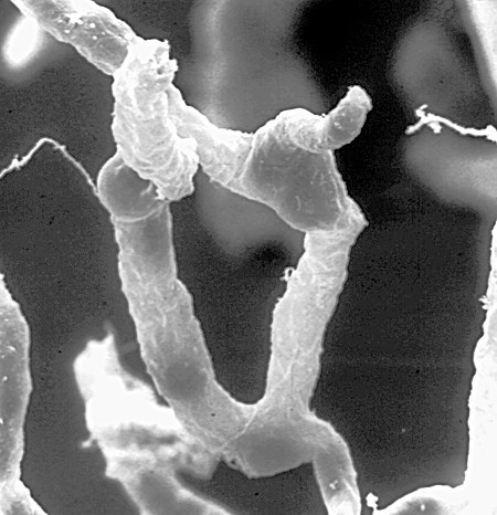
Black and white microscopic image of a capillary supplying blood to brain cells. A network of capillaries supplies brain cells with nutrients. Tight seals in their walls keep blood toxins—and many beneficial drugs—out of the brain. Credit: Dan Ferber, PLOS Biol 2007 Jun; (5)6:E169. CC by 2.5 Link to external web site.

The blood-brain barrier—the ultra-tight seal in the walls of the brain’s capillaries—is an important part of the body’s defense system. It keeps invaders and other toxins from entering the human brain by screening out dangerous molecules. But the intricate workings of this extremely effective barrier also make it challenging to design therapeutics that would help us. And as it turns out, getting a drug across the blood-brain barrier is only half the battle. Once it’s across, the drug needs to effectively target the right cells in the brain tissue. With this in mind, it’s no surprise that challenges this complex are solved through collaboration among scientists from several different specialties.

Elizabeth Nance Link to external web site, an assistant professor of chemical engineering at the University of Washington in Seattle and a recent recipient of the Presidential Early Career Award for Scientists and Engineers (PECASE), focuses her research on understanding the barriers in the brain and other cell- and tissue-based barriers in the body to see how nanoparticles interact with them. Her lab uses nanoparticles to package therapies that will treat newborn brain injury, which can occur when the brain loses oxygen and blood flow, often during or immediately prior to delivery. This damage can lead to cerebral palsy, developmental delays, or sometimes death. Early interventions for newborn brain injury can be valuable, but they need to target specific, injured cells without harming healthy ones.

Continue reading “PECASE Honoree Elizabeth Nance Highlights the Importance of Collaboration in Nanotechnology”

Get Kids Excited About Science: Free STEM Resources

0 comments
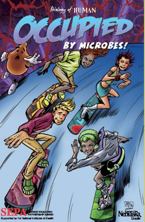
Cover of the graphic novel Occupied by Microbes!, showing four teens racing downhill on skateboards. Credit: University of Nebraska, Lincoln.

We have a new Science Education and Partnership Award (SEPA) webpage, featuring free, easy-to-access, SEPA-funded Link to external web site resources that educators nationwide can use to engage their students in science. The SEPA program supports innovative STEM Link to external web site and informal science education Link to external web site  projects for pre-kindergarten through grade 12. The program includes tools that teachers, scientists, and parents can use to excite kids about science and research, such as:

Continue reading “Get Kids Excited About Science: Free STEM Resources”

Advances in 3D Printing of Replacement Tissue

0 comments
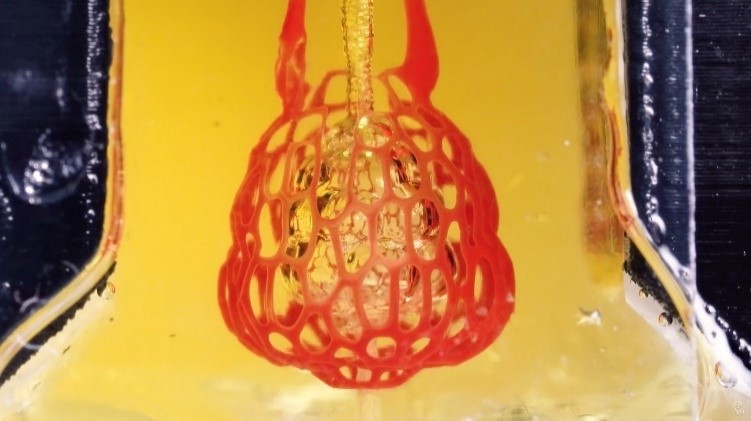
A bioprint of the small air sac in the lungs with red blood cells moving through a vessel network supplying oxygen to living cells. Credit: Rice University. A bioprint of the small air sac in the lungs with red blood cells moving through a vessel network supplying oxygen to living cells. Credit: Rice University.

A team of bioengineers, funded in part by NIGMS, has devised a way to use 3D bioprinting technology to construct the small air sacs in the lungs and intricate blood vessels. When hooked up to a machine, the air sacs can “breathe,” and the blood flowing through the tiny blood vessels can take up oxygen, much like they would in an animal’s body. In the long term, this technology may allow the production of replacement organs for patients who need them. Visit the NIH Director’s Blog to read more and watch a video from Rice University’s Miller Lab.

Mapping Our Skin’s Microbes and Molecules

0 comments

Last month, we shared some facts about the microbes that inhabit us. Here’s another: From head to toe, our skin bacteria coexist with chemicals in hygiene products, fibers from clothes and proteins shed by dead or dying skin cells.

These images highlight the complex composition of our body’s largest organ. They show the association between microbial diversity (top images) and skin chemistry (middle images). The different colors note the abundance of a certain bacterium or molecule—red is high, and blue is low. The skin maps remind NIH Director Francis Collins of a 60’s rock album cover.

Continue reading “Mapping Our Skin’s Microbes and Molecules”

Meet Karen Carlson

0 comments
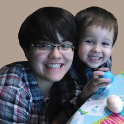
Karen Carlson
Credit: Karen Carlson
Karen Carlson
Fields: Systems biology, bacterial biofilms
Born and raised in: Alaska
Undergraduate student at: The University of Alaska, Anchorage
When not in the lab, she’s: Out and about with her 3-year-old son, friends and family
Secret talent: “I make some really good cookies.”

Karen Carlson got a surprise in her 10th grade biology class. Not only did she find out that she enjoyed science (thanks to an inspiring teacher), but, as she puts it, “I realized that I was really good at it.”

In particular, she says, “I was good at putting all the pieces [of a scientific question] together. And that’s what I had the most fun with—looking at systems: how things fit together and the flow between them.”

These are perfect interests for a budding systems biologist, which is what Carlson is on her way to becoming. She’s a senior in college on track to graduate this year with a bachelor’s degree in biology from the University of Alaska, Anchorage (UAA). Next, she plans to enroll in a master’s degree program at UAA, and eventually to pursue a Ph.D. in a biomedical field. Continue reading “Meet Karen Carlson”

Knowing Networks

1 comment
Artist's rendition of a network diagram. Credit: Allison Kudla, Institute for Systems Biology.
Artist’s rendition of a network diagram. Credit: Allison Kudla, Institute for Systems Biology.

Networks—both real and virtual—are everywhere, from our social media circles to the power grid that delivers electricity. The interactions of genes, proteins and other molecules in a cell are examples of networks, too.

Scientists working in a field called systems biology study and chart living networks to learn how the individual parts work together to make a functioning whole and what happens when these complex, dynamic systems go awry. For example, the network diagram here depicts yeast cells (superimposed circles) and the biochemical “chatter” between them (lines) that tells the cells to gather together in clumps. This clumping helps them survive stressful conditions like a shortage of nutrients.

Network diagrams provide more than just hub-and-spoke pictures. They can yield information that helps us better understand—and potentially influence—complex phenomena that affect our health.

Read more about network analysis and systems biology in this Inside Life Science article.

Meet Ravi Iyengar

0 comments
Ravi Iyengar
Ravi Iyengar
Fields: Systems pharmacology and systems biology
Works at: Mount Sinai School of Medicine, New York, NY
Favorite sports team: Yankees
Favorite subject in high school: Math
Recently read book: The Signal and the Noise by Nate Silver
Credit: Pedro Martinez, Systems Biology Center New York

Ravi Iyengar, a professor at Mount Sinai School of Medicine, stood in an empty lecture hall, primed to tell thousands of students about systems biology, a holistic approach to studying fundamental life processes. To prepare for this moment, he had spent 4 months reading hundreds of scientific papers and distilling the research into understandable nuggets. But that day, his only student was a videographer.

Together, they recorded 15 different lectures about systems biology—many related to Iyengar’s own research—that thousands of people would stream or download as part of a MOOC, or massive open online course.

Trained in biochemistry, Iyengar built his research career around studying molecules and developing a list of all the parts that help nerve, kidney and skin cells to function. As he obtained more information, he realized he needed to know how all the components worked together. To achieve this comprehensive understanding, Iyengar turned to computational techniques and mathematical analyses—cornerstones of systems biology.

For more than a decade, he has been using and developing systems biology approaches to explore a range of biomedical questions, from very basic to translational ones with immediate relevance to human health.

Iyengar’s Findings

In his earlier work, Iyengar used mathematical analyses to show that molecules within cells connect with one another to form switches that produce cellular memory. This may allow, for instance, an immune cell to remember a foreign object and secrete an antibody. In recent work, he and his team developed a mathematical model showing that the shape of a cell influences the flow of information across the membrane, possibly contributing to disease states and offering a way to study and identify them under the microscope. In another study, they analyzed a database of drug side effects to find combinations of medications that produce fewer adverse reactions and then created a cell biology interaction network that explains why a certain drug pair had this beneficial outcome. The approach could point to other combinations of FDA-approved drugs that reduce serious side effects and thereby guide clinical practice.

“Systems biology is a powerful way to explore important biological and medical questions, and it’s relevant to many fields of science,” said Iyengar. But he added that the majority of educational institutions, including liberal arts and community colleges, don’t have systems biology courses. So, Iyengar teamed with colleagues to create a series of MOOCs.

The first course, offered last summer and taught by Iyengar, presented all the facets of systems biology. The syllabus included lessons on genomics and bioinformatics, fields that have contributed to systems biology; gathering and integrating data; and the use of modeling in drug development.

“My goal was for the students to get the general gestalt of systems biology,” explained Iyengar, who directs an NIH-funded center focused on the systems-level study of medicine and therapeutics.

In total, more than 12,000 participants watched at least one video lecture, 3,000 submitted one or more of the weekly quizzes and 1,800 took a mid-term or final exam. The online discussions forum included nearly 400 topics with about 5,000 posts. The students, most enrolled in a graduate program or working full-time, had some training in the biological, biomedical, computer and information sciences.

“The stats tell me that many people are in fields adjacent to systems biology and don’t have access to more traditional systems biology courses,” concluded Iyengar. “Through the MOOC, we can reach them in a substantial way.”

The second course, which covers network analysis, wrapped up in early December, and the third course, which covers dynamical modeling methods, began in January. Iyengar plans to offer the intro course again in late March.

Learn more:
MOOC Systems Biology Courses Exit icon

Meet Jasmine Johnson and Gabe Vela

0 comments
Jasmine Johnson and Gabriel 'Gabe' Vela
Jasmine Johnson and Gabriel “Gabe” Vela
Field: Genetics of sleep and obesity
Worked as researchers at: The Jackson Laboratory, Bar Harbor, Maine
Graduated from high school at: Rockdale Magnet School for Science and Technology in Conyers, Ga.
Now freshman at: Stanford University in Palo Alto, Calif. (Johnson) and Southern Polytechnic State University in Marietta, Ga. (Vela)
Fascinating fact: Johnson presented her research at the 2013 White House Science Fair
Credit: Joe Piergrossi

Jasmine Johnson and Gabe Vela might still be teenagers, but they are also seasoned scientists. It all started 3 years ago, when, as high school juniors, they took the research course Independent Studies in Computational Biology at The Jackson Laboratory in Bar Harbor, Maine. They were hooked. They continued to do research until they graduated, working part-time for 2 academic years and full-time for 2 summers.

They worked with statistical geneticist Gary Churchill, using computational biology to explore the relationship between sleep and obesity. They focused on finding genes that regulate sleep and understanding how sleep affects the body. One goal of the research is to tease out a genetic explanation for why sleep deprivation increases the risk of obesity.

Working in a lab “completely changed what I thought I was going to do with my life,” said Vela. “Now I’m going to focus more on research than anything else.”

For Johnson, the experience provided the opportunity to present her research at the 2013 White House Science Fair, where she hobnobbed with some political hot shots.

“It was an amazing experience,” she said. Having “important White House officials be interested in my project … inspired me.”

Johnson and Vela visited NIH a few months ago and talked with us about their research experiences, their lives and their future goals. Jasmine Johnson & Gabriel Vela on their experience as high school researchers at The Jackson Laboratory in Bar Harbor, Maine.

Learn More
Article Exit icon about Johnson and Vela and other young researchers, from The Jackson Laboratory’s magazine The Search.
Article about the work of Gary Churchill, from NIH’s Findings magazine.