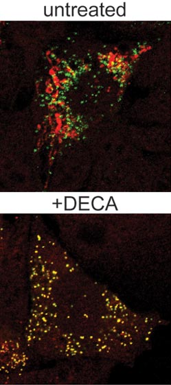
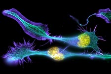
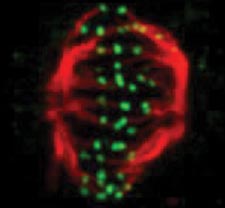
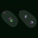
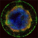
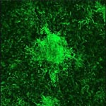
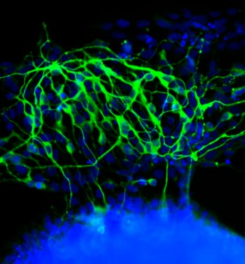
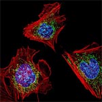
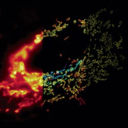
Category: Cells

Cool Image: Of Surfaces and Stem Cells

2 comments
Stem cells transform into neurons.

Stem cells grown on a soft surface begin to transform into neurons. Credit: Kiessling Lab, University of Wisconsin-Madison.

If you think this image looks like the fluorescent outline of a brain, you’re on the right track. The green threads show neurons that have just formed from unspecialized cells called stem cells.

Researchers led by Laura Kiessling Exit icon of the University of Wisconsin-Madison directed the stem cells to become neurons by changing the quality of the surface on which they grew. In experiments testing different gels used to grow stem cells in the lab, the scientists found that the stiffness of those gels influenced cell fate decisions.

When grown on a soft gel with a brain tissue-like surface, the stem cells began to transform into neurons. This happened without the addition of any of the proteins normally used to coax stem cells to specialize into different types of cells.

A better understanding of how stem cell fate is influenced by the mechanical properties of a surface could help researchers who are trying to harness the blank slate cells for tissue regeneration or other therapeutic uses.

This work also was funded by NIH’s National Institute of Biomedical Imaging and Bioengineering; National Heart, Lung, and Blood Institute; and National Institute of Neurological Disorders and Stroke.

Learn more:
University of Wisconsin-Madison News Release Exit icon

Nobel Prize for Powerful Microscopy Technology

0 comments
Fibroblasts
The cells shown here are fibroblasts, one of the most common cells in mammalian connective tissue. These particular cells were taken from a mouse. Scientists used them to test the power of a new microscopy technique that offers vivid views of the inside of a cell. The DNA within the nucleus (blue), mitochondria (green) and cellular skeleton (red) is clearly visible. Credit: Dylan Burnette and Jennifer Lippincott-Schwartz, Eunice Kennedy Shriver National Institute of Child Health and Human Development, National Institutes of Health.

William E. Moerner was at a conference in Brazil when he learned he’d be getting a Nobel Prize in chemistry. “I was incredibly excited and thrilled,” he said of his initial reaction.

An NIGMS grantee at Stanford University, Moerner received the honor for his role in achieving what was once thought impossible—developing super-resolution fluorescence microscopy, which is so powerful it allows researchers to see and track individual molecules in living organisms in real time.

Nobel recipients usually learn of the prize via a phone call from Stockholm, Sweden, in early October. For those in the United States, the call typically comes between 2:30 a.m. and 5:45 a.m.

Every year, the NIGMS communications office prepares for the Nobel Prize announcements in physiology or medicine and chemistry, the categories in which our grantees are most likely to be recognized. If the Institute played a significant role in funding the prize-winning research, we work quickly to provide information and context to reporters covering the story on tight deadlines. We issue a statement, identify an in-house expert on the research and arrange interviews with reporters. It’s all to help get the word out about the research and the taxpayers’ role in supporting it.

This year’s in-house expert, Cathy Lewis, shared her thoughts on the prize to Moerner in an NIGMS Feedback Loop post. You can also read this year’s statement and see a full list of NIGMS-supported Nobel laureates.

Outwitting Antibiotic Resistance

0 comments
Marine scene with fish and corals
The ocean is a rich source of microbes that could yield infection-fighting natural molecules. Credit: National Oceanic and Atmospheric Administration Exit icon.

Antibiotics save countless lives and are among the most commonly prescribed drugs. But the bacteria and other microbes they’re designed to eradicate can evolve ways to evade the drugs. This antibiotic resistance, which is on the rise due to an array of factors, can make certain infections difficult—and sometimes impossible—to treat.

Read the Inside Life Science article to learn how scientists are working to combat antibiotic resistance, from efforts to discover potential new antibiotics to studies seeking more effective ways of using existing ones.

Mighty Mitochondria

0 comments
Mitochondria from the heart muscle cell of a rat.
Mitochondria (red) from the heart muscle cell of a rat. Nearly all our cells have these structures. Credit: Thomas Deerinck, National Center for Microscopy and Imaging Research Exit icon.

Meet mitochondria: cellular compartments, or organelles, that are best known as the powerhouses that convert energy from the food we eat into energy that runs a range of biological processes.

As you can see in this close-up of mitochondria from a rat’s heart muscle cell, the organelles have an inner membrane that folds in many places (and that appears here as striations). This folding vastly increases the surface area for energy production. Nearly all our cells have mitochondria, but cells with higher energy demands have more. For instance, a skin cell has just a few hundred, while the cell pictured here has about 5,000.

Scientists are discovering there’s more to mitochondria than meets the eye, especially when it comes to understanding and treating disease.

Read more about mitochondria in this Inside Life Science article.

New Research Sheds Light on Drug-Induced Salivary Issues

0 comments
Open human mouth
Scientists have discovered a possible mechanism behind the bad taste and dry mouth caused by some drugs. Credit: Stock image.

The effects some medicines have on our salivary glands can at times extend beyond the fleeting flavor we experience upon ingesting them. Sometimes drugs cause a prolonged bad taste or dryness in the mouth, both of which can discourage people from taking medicines they need. Now, a research team led by Joanne Wang of the University of Washington has discovered a possible mechanism behind this phenomenon. Working primarily with mice and using a commonly prescribed antidiabetic drug known to impair taste, the scientists identified a protein in salivary gland cells that takes up the drug from the bloodstream and secretes it in saliva. Wang and her colleagues were also able to pinpoint a specific gene that, when removed, hindered this process. They hope their new insights will aid efforts to develop medicines that do not cause salivary issues.

This work also was funded by NIH’s Eunice Kennedy Shriver National Institute of Child Health and Human Development.

Learn more:
University of Washington News Release Exit icon

Cool Image: Training Cells to Devour Dying Neighbors

2 comments
A healthy cell that has ingested dying cells.

A healthy cell (green) that has ingested dying cells (purple). Credit: Toru Komatsu, University of Tokyo.

In this image, a healthy cell (green) has engulfed a number of dying cells (purple) just as a predator might ingest wounded or dying prey. A team of researchers is hoping to use this same strategy at the cellular level to help treat infection, neurodegenerative diseases or cancer.

Our bodies routinely use a process called phagocytosis to rid themselves of unhealthy cells. Takanari Inoue at Johns Hopkins University and collaborators at the University of Tokyo set out to investigate the molecular underpinnings of phagocytosis. Their goal was to endow laboratory-grown human cells with phagocytotic skills, namely the ability to recognize, swallow and digest dying cells. The scientists tried to do this by inserting into the cells two molecules known to play a role in phagocytosis.

The engineered cells accomplished the first two tasks—they recognized and surrounded dying cells. But they didn’t digest what they’d engulfed. Now the researchers are looking for a molecular trigger to get the engineered cells to complete this last task.

Eventually, the scientific team aims to build artificial cells that are programmed to target and destroy abnormal cells, such as those ravaged by bacteria, cancer or other diseases.

Learn more:
Johns Hopkins News Release Exit icon
How Cells Take Out the Trash Article from Inside Life Science
Cellular Suicide: An Essential Part of Life Article from Inside Life Science

Say Cheese

0 comments
Assorted cheeses
A biofilm of bacteria and fungi, commonly known as a rind, forms on the surface of traditionally aged cheeses. Credit: Elia Ben-Ari.

Biofilms—multispecies communities of microbes that live in and on us, and in the environment—are important for human health and the function of ecosystems. But studying these microbial metropolises can be challenging because many of the environments where they’re found are hard to replicate in the lab.

Enter cheese rinds. These biofilms of bacteria and fungi form on the surface of traditionally aged cheeses, and could serve as a system for understanding how microbial communities form and function. By sequencing DNA from the rinds of 137 artisan cheese varieties collected in 10 countries, Rachel Dutton and her colleagues at Harvard University identified three general types of microbial communities that live on their tasty study subjects. After individually culturing representatives of all the species found in the rind communities the scientists added them to a growth medium that included cheese curd. This approach allowed them to recreate the communities in the lab and use them to detect numerous bacterial-fungal interactions and patterns of community composition over time.

The scientists plan to use their lab-grown cheese rinds to study whether and how various microbes compete or cooperate as they form communities, as well as what molecules and mechanisms are involved. In addition to answering fundamental questions about microbial ecology, this cheesy research might ultimately yield insights that help fight infection-causing biofilms or lead to the discovery of new antibiotics.

Learn more:
Harvard University News Release Exit icon
Dutton Lab Exit icon

Restoring the Function of an Immune Receptor Involved in Crohn’s Disease

4 comments
Gut bacteria
Receptor proteins bind to bacterial cell wall fragments, initiating an immune response to remove bad gut bacteria. Credit: S. Melanie Lee, Caltech; Zbigniew Mikulski and Klaus Ley, La Jolla Institute for Allergy and Immunology.

Our bodies depend on a set of immune receptors to remove harmful bacteria and control the growth of helpful bacteria in our guts. Genetic changes that alter the function of the receptors can have an adverse effect and result in chronic inflammatory diseases like Crohn’s disease. Catherine Leimkuhler Grimes and Vishnu Mohanan of the University of Delaware researched a Crohn’s-associated immune receptor, NOD2, to figure out how it can lose the ability to respond properly to bacteria. In the process, they identified the involvement of a protective protein called HSP70. Increasing HSP70 levels in kidney, colon and white blood cells appeared to restore NOD2 function. This work represents a first step toward developing drugs to treat Crohn’s disease.

This work was funded in part by an Institutional Development Award (IDeA) Network of Biomedical Research Excellence (INBRE) grant.

Anesthesia and Brain Cells: A Temporary Disruption?

1 comment
Hippocampal neuron in culture.
Hippocampal neuron in culture. Dendrites are green, dendritic spines are red, and DNA in cell’s nucleus is blue. Credit: Shelley Halpain, University of California, San Diego.

Anesthetic drugs are vital to modern medicine, allowing patients to undergo even the longest and most invasive surgeries without consciousness or pain. Unfortunately, studies have raised the concern that exposing patients, particularly children and the elderly, to some anesthetics may increase risk of long-term cognitive and behavioral issues.

A scientific team led by Hugh Hemmings Exit icon of Weill Cornell Medical College and Shelley Halpain Exit icon of the University of California, San Diego, examined the effects of anesthesia on neurons isolated from juvenile rats. Given at doses and durations frequently used during surgery, the commonly administered general anesthetic isoflurane did in fact reduce the number and size of important structures within neurons called dendritic spines. Dendritic spines help pass information from neuron to neuron, and disruption of these structures can be associated with dysfunction in thinking and behavior.

Promisingly, the shrinkage observed by the researchers appeared to be temporary: After the researchers washed the anesthetic out of the cell cultures, the dendritic spines grew back. But because neurons in culture do not reproduce all aspects of intact neuronal networks, the scientists explain that the findings should be verified in more complex models. Other molecular mechanisms may also potentially contribute to late effects of anesthesia exposure.

This work also was funded by NIH’s National Institute of Mental Health.

Learn more:
University of California, San Diego News Release
Understanding Anesthesia from Inside Life Science

Meet Janet Iwasa

0 comments
Janet Iwasa
Credit: Janet Iwasa
Janet Iwasa
Fields: Cell biology and molecular animation
Works at: University of Utah
Raised in: Indiana and Maryland
Studied at: University of California, San Francisco, and Harvard Medical School
When not in the lab she’s: Keeping up with her two preschool-aged sons
Something she’s proud of that she’ll never try again: Baking a multi-tiered wedding cake, complete with sugar flowers, for a friend’s wedding.

Janet Iwasa wouldn’t have described herself as an artistic child. She didn’t carry around a sketch pad, pencils or paintbrushes. But she remembers accompanying her father, a scientist at the National Institutes of Health, to his lab on the weekends. She’d spend hours doodling in a drawing program on his old Macintosh computer while he worked on experiments.

“I always remember wanting to be a scientist, and that’s probably highly inspired by my dad,” says Iwasa. Her early affinity for art and technology set her on an unusual career path to become a molecular animator. A typical work day now finds her adapting computer programs originally designed to bring characters like Buzz Lightyear to life to help researchers probe complicated, dynamic interactions within cells.

Iwasa’s interest in animation was sparked when she was a graduate student in cell biology, studying a protein called actin, which helps cells to move and change shape. At the time, the only visual representations she had of actin networks were flat, two-dimensional drawings on paper. When she saw an animation of the dynamic movement of a molecule called kinesin, she thought, “Why are we relying on oversimplified, static illustrations [of molecules], when we can be doing something like this video?”

Within a year, she was taking an animation class at a local college. She quickly realized that she would need more intensive instruction to be able to animate complex biological processes. A few summers later, she flew to Hollywood for a 3-month training program in industry-standard animation technology.

The oldest student in that course—and the only woman—Iwasa immediately began thinking about how to adapt a standard animator’s toolkit to illustrate the inner life of cells. A technique used to create the effect of human hair blowing in the wind could also show the movement of an RNA molecule. A chunk of computer code used to make the facets of a soccer ball fall apart and come back together in a different order could be adapted to model virus assembly and disassembly.

Her Findings

Following her training, Iwasa spent 2 years as a National Science Foundation Discovery Corps fellow, producing the Exploring Life’s Origins exhibit with the Boston Museum of Science and the Szostak Lab at Massachusetts General Hospital/Harvard Medical School. As part of the multi-media exhibit, she created animations to illustrate how the simplest living organisms may have evolved on early Earth.

Since then, Iwasa has helped researchers model such complex actions as how cells ingest materials, how proteins are transported across a cell membrane, and how the motor protein dynein helps cells divide.

Screenshot from the video that shows how a protein called clathrin forms a cage-like container that cells use to engulf and ingest materials
Iwasa developed this video to show how a protein called clathrin forms a cage-like container that cells use to engulf and ingest materials.

Iwasa calls her animations “visual hypotheses”: The end results may be beautiful, but the process of animation itself is what encapsulates, clarifies and communicates the science.

“It’s really building the animated model that brings insights,” she says. “When you’re creating an animation, you’re really grappling with a lot of issues that don’t necessarily come up by any other means. In some cases, it might raise more questions, and make people go back and do some more experiments when they realize there might be something missing” in their theory of how a molecular process works.

Now she’s working with an NIH-funded research team at the University of Utah to develop a detailed animation of how HIV enters and exits human immune cells.

Abbreviated CHEETAH, the full name of the group is the Center for the Structural Biology of Cellular Host Elements in Egress, Trafficking, and Assembly of HIV.

“In the HIV life cycle, there are a number of events that aren’t really well understood, and people have different ideas of how things happen,” says Iwasa. She plans to animate the stages of viral infection in ways that reflect different proposals for how the process works, to give researchers a new way to visualize, communicate—and potentially harmonize—their hypotheses.

The full set of Iwasa’s HIV-related animations will be available online as they are completed, at https://scienceofhiv.org, with the first set launching in the fall of 2014.

Learn more:
Janet Iwasa’s TED Talk: How animations can help scientists test a hypothesis
Janet Iwasa’s 3D model of an HIV particle was a winner in the 2014 BioArt contest sponsored by Federation of American Societies for Experimental Biology
NIH Director’s blog post about Iwasa and her HIV video animation