Category: Chemistry, Biochemistry and Pharmacology

Nobel Prize for Powerful Microscopy Technology

0 comments
Fibroblasts
The cells shown here are fibroblasts, one of the most common cells in mammalian connective tissue. These particular cells were taken from a mouse. Scientists used them to test the power of a new microscopy technique that offers vivid views of the inside of a cell. The DNA within the nucleus (blue), mitochondria (green) and cellular skeleton (red) is clearly visible. Credit: Dylan Burnette and Jennifer Lippincott-Schwartz, Eunice Kennedy Shriver National Institute of Child Health and Human Development, National Institutes of Health.

William E. Moerner was at a conference in Brazil when he learned he’d be getting a Nobel Prize in chemistry. “I was incredibly excited and thrilled,” he said of his initial reaction.

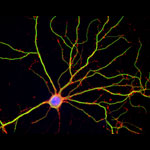
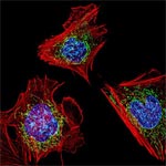
An NIGMS grantee at Stanford University, Moerner received the honor for his role in achieving what was once thought impossible—developing super-resolution fluorescence microscopy, which is so powerful it allows researchers to see and track individual molecules in living organisms in real time.

Nobel recipients usually learn of the prize via a phone call from Stockholm, Sweden, in early October. For those in the United States, the call typically comes between 2:30 a.m. and 5:45 a.m.

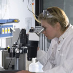
Every year, the NIGMS communications office prepares for the Nobel Prize announcements in physiology or medicine and chemistry, the categories in which our grantees are most likely to be recognized. If the Institute played a significant role in funding the prize-winning research, we work quickly to provide information and context to reporters covering the story on tight deadlines. We issue a statement, identify an in-house expert on the research and arrange interviews with reporters. It’s all to help get the word out about the research and the taxpayers’ role in supporting it.

This year’s in-house expert, Cathy Lewis, shared her thoughts on the prize to Moerner in an NIGMS Feedback Loop post. You can also read this year’s statement and see a full list of NIGMS-supported Nobel laureates.

Outwitting Antibiotic Resistance

0 comments
Marine scene with fish and corals
The ocean is a rich source of microbes that could yield infection-fighting natural molecules. Credit: National Oceanic and Atmospheric Administration Exit icon.

Antibiotics save countless lives and are among the most commonly prescribed drugs. But the bacteria and other microbes they’re designed to eradicate can evolve ways to evade the drugs. This antibiotic resistance, which is on the rise due to an array of factors, can make certain infections difficult—and sometimes impossible—to treat.

Read the Inside Life Science article to learn how scientists are working to combat antibiotic resistance, from efforts to discover potential new antibiotics to studies seeking more effective ways of using existing ones.

New Research Sheds Light on Drug-Induced Salivary Issues

0 comments
Open human mouth
Scientists have discovered a possible mechanism behind the bad taste and dry mouth caused by some drugs. Credit: Stock image.

The effects some medicines have on our salivary glands can at times extend beyond the fleeting flavor we experience upon ingesting them. Sometimes drugs cause a prolonged bad taste or dryness in the mouth, both of which can discourage people from taking medicines they need. Now, a research team led by Joanne Wang of the University of Washington has discovered a possible mechanism behind this phenomenon. Working primarily with mice and using a commonly prescribed antidiabetic drug known to impair taste, the scientists identified a protein in salivary gland cells that takes up the drug from the bloodstream and secretes it in saliva. Wang and her colleagues were also able to pinpoint a specific gene that, when removed, hindered this process. They hope their new insights will aid efforts to develop medicines that do not cause salivary issues.

This work also was funded by NIH’s Eunice Kennedy Shriver National Institute of Child Health and Human Development.

Learn more:
University of Washington News Release Exit icon

Aspirin’s Dual Action

0 comments
Aspirin
Aspirin can help reverse inflammation as well as prevent it from occurring. Credit: Stock image.

Ever wonder how aspirin knocks out aches? Scientists have known that medicine prevents an enzyme called cyclooxygenase from producing compounds linked to pain and inflammation, but they recently made another discovery about how aspirin works.

Edward Dennis and colleagues at the University of California, San Diego School of Medicine researched aspirin’s effect on macrophages–white blood cells that play a role in the body’s immune response to injury. They found that in addition to killing cyclooxygenase, aspirin causes the enzyme to make a product called 15-HETE. During infection and inflammation, 15-HETE can get converted by another enzyme into lipoxin, a compound that terminates and reverses inflammation.

Researchers will likely use lipoxin and similar compounds to develop new anti-inflammatory drugs.

Learn more:
University of California, San Diego News Release Exit icon
Dennis Lab (no longer available)

Anesthesia and Brain Cells: A Temporary Disruption?

1 comment
Hippocampal neuron in culture.
Hippocampal neuron in culture. Dendrites are green, dendritic spines are red, and DNA in cell’s nucleus is blue. Credit: Shelley Halpain, University of California, San Diego.

Anesthetic drugs are vital to modern medicine, allowing patients to undergo even the longest and most invasive surgeries without consciousness or pain. Unfortunately, studies have raised the concern that exposing patients, particularly children and the elderly, to some anesthetics may increase risk of long-term cognitive and behavioral issues.

A scientific team led by Hugh Hemmings Exit icon of Weill Cornell Medical College and Shelley Halpain Exit icon of the University of California, San Diego, examined the effects of anesthesia on neurons isolated from juvenile rats. Given at doses and durations frequently used during surgery, the commonly administered general anesthetic isoflurane did in fact reduce the number and size of important structures within neurons called dendritic spines. Dendritic spines help pass information from neuron to neuron, and disruption of these structures can be associated with dysfunction in thinking and behavior.

Promisingly, the shrinkage observed by the researchers appeared to be temporary: After the researchers washed the anesthetic out of the cell cultures, the dendritic spines grew back. But because neurons in culture do not reproduce all aspects of intact neuronal networks, the scientists explain that the findings should be verified in more complex models. Other molecular mechanisms may also potentially contribute to late effects of anesthesia exposure.

This work also was funded by NIH’s National Institute of Mental Health.

Learn more:
University of California, San Diego News Release
Understanding Anesthesia from Inside Life Science

The “Virtuous Cycle” of Technology and Science

0 comments
A scientist looking through a  microscope. Credit: Stock image.
Whether it’s a microscope, computer program or lab technique, technology is at the heart of biomedical research. Credit: Stock image.

Whether it’s a microscope, computer program or lab technique, technology is at the heart of biomedical research. Its central role is particularly clear from this month’s posts.

Some show how different tools led to basic discoveries with important health applications. For instance, a supercomputer unlocked the secrets of a drug-making enzyme, a software tool identified disease-causing variations among family members and high-powered microscopy revealed a mechanism allowing microtubules—and a cancer drug that targets them—to work.

Another theme featured in several posts is novel uses for established technologies. The scientists behind the cool image put a new spin on a long-standing imaging technology to gain surprising insights into how some brain cells dispose of old parts. Similarly, the finding related to sepsis demonstrates yet another application of a standard lab technique called polymerase chain reaction: assessing the immune state of people with this serious medical condition.

“We need tools to answer questions,” says NIGMS’ Doug Sheeley, who oversees biomedical technology research resource grants. “When we find the answers, we ask new questions that then require new or improved tools. It’s a virtuous cycle that keeps science moving forward.”

A Drug-Making Enzyme in Motion

0 comments
Mutated enzyme, LovD9. Credit: Silvia Osuna and Gonzalo Jiménez-Osés, University of California, Los Angeles.
The movement of this mutated enzyme, LovD9, facilitates rapid production of the cholesterol reducing-drug simvastatin. Credit: Silvia Osuna and Gonzalo Jiménez-Osés, University of California, Los Angeles.

LovD9, a mutated version of an enzyme extracted from mold growing in soil, produces the cholesterol-reducing drug simvastatin 1,000 times faster than its natural predecessor. But scientists didn’t understand why because the enzyme’s mutations are far from the active site, where the drug is actually made. Now they do.

Yi Tang of the University of California, Los Angeles (UCLA), in partnership with the pharmaceutical company Codexis, generated LovD9 by repeatedly inducing random mutations, each time selecting the mutated versions of the enzyme with the most promise for industrial simvastatin production.

Then, the team collaborated with UCLA colleagues Kendall Houk and Todd Yeates to unlock the secret of the enzyme’s speed. Using ANTON, a special-purpose supercomputer at the Pittsburgh Supercomputing Center, they simulated how different parts of the enzyme rotate and twist when synthesizing the drug. The scientists discovered that as LovD9 moves, it forms shapes that facilitate simvastatin production more often than the natural enzyme does.

With their better understanding of how mutations far from an active site may affect an enzyme’s motion, the researchers hope to one day directly engineer enzymes with precise mutations that enhance drug production.

Learn more:
University of California, Los Angeles News Release Exit icon
Houk Exit icon, Tang Exit icon and Yeates Exit icon Labs

Carbohydrates as Bacterial Camouflage: How Our Immune System Responds

1 comment
bacteria
Although invisible to our immune system’s antibodies, strains of a pneumonia-causing bacteria, Pseudomonas aeruginosa (orange), are easily detected by galectins. Credit: Centers for Disease Control and Prevention.

When harmful strains of bacteria invade our bodies, our immune system produces antibodies that identify the intruders by the specific carbohydrate structures coating them. Some strains, however, have coatings that mimic the carbohydrate structures found on our own cells, and this disguise allows them to evade detection by antibodies.

A team of scientists led by Richard Cummings of Emory University found that galectins, a class of proteins naturally produced by our bodies, can identify and kill these concealed bacteria without damaging our own mimicked cells. To make this discovery, the team used glass slides covered with more than 300 different carbohydrates extracted from the surface of bacterial cells. After testing the ability of galectins and antibodies to bind to specific carbohydrates on these slides, the researchers observed that the galectins easily detected the mammalian-like carbohydrates that the antibodies failed to recognize.

These findings provide a clearer understanding of the complementary roles played by galectins and antibodies in protecting us from a broad range of infections.

This work also was funded by NIH’s National Institute of Allergy and Infectious Diseases and National Heart, Lung, and Blood Institute.

Learn more:
Emory University News Release Exit icon

New Compound Improves Insulin Levels in Preliminary Studies

1 comment
compound
A new compound (chemical structure shown here) blocks the activity of an enzyme involved in glucose regulation.

The discovery of a compound that slows the natural degradation of insulin in mice opens up a new area of investigation in the search for drugs to treat diabetes. The research team, which included David Liu Exit icon and Alan Saghatelian Exit icon of Harvard University, Markus Seeliger of Stony Brook University School of Medicine, and Wei-Jen Tang Exit icon of the University of Chicago focused on insulin-degrading enzyme, or IDE. Using a method called DNA-templated synthesis, the scientists made 14,000 small molecules and found one that bound to the enzyme, suggesting it might modulate the enzyme’s activity. Work in test tubes and in animal models confirmed this—and showed that blocking IDE activity improved insulin levels and glucose tolerance. The researchers also learned that the enzyme is misnamed: In addition to insulin, it degrades two other hormones involved in glucose regulation.

NIGMS’ Peter Preusch says, “This is a very interesting fusion of chemical methods and biology that has uncovered new basic science findings about insulin processing with potential clinical impact.”

This work also was funded by NIH’s National Cancer Institute and the Office of the Director.

Learn more:
Harvard University News Article Exit icon
Chemistry of Health Booklet

Multitarget Drugs to Challenge Microbial Resistance

0 comments
A group of purple, rod-shaped bacterial cells rendered by computer at Centers for Disease Control and Prevention by Melissa Brower.
Computer-generated image of drug-resistant Mycobacterium tuberculosis bacteria. Credit: Melissa Brower, Centers for Disease Control and Prevention.

Drugs that target a single essential protein in a microbial invader can be effective treatments. But the genomes of pathogens—including bacteria, fungi and parasites—mutate rapidly, and resistance can develop if a mutation changes a target protein’s structure. Molecules that interfere with multiple microbial proteins at once have the potential to overcome the growing problem of antimicrobial drug resistance.

Researchers led by Eric Oldfield of the University of Illinois recently explored whether an experimental drug called SQ109, developed to treat tuberculosis (TB), could be tweaked to attack multiple enzymes, as well as to kill different types of microbes. The scientists succeeded in creating several multitarget analogs of SQ109 that were more effective than the original drug at killing their target pathogens in laboratory experiments. These analogs included one compound that was five times more potent against the bacterium that causes TB while also being less toxic to a human cell line tested.

This work was also funded by the National Cancer Institute; the National Heart, Lung, and Blood Institute; the National Institute of Allergy and Infectious Diseases and the NIH Office of the Director.

Learn more:
University of Illinois, Urbana-Champaign News Release exit icon