Category: Molecular Structures

A Drug-Making Enzyme in Motion

0 comments
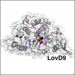
Mutated enzyme, LovD9. Credit: Silvia Osuna and Gonzalo Jiménez-Osés, University of California, Los Angeles.
The movement of this mutated enzyme, LovD9, facilitates rapid production of the cholesterol reducing-drug simvastatin. Credit: Silvia Osuna and Gonzalo Jiménez-Osés, University of California, Los Angeles.

LovD9, a mutated version of an enzyme extracted from mold growing in soil, produces the cholesterol-reducing drug simvastatin 1,000 times faster than its natural predecessor. But scientists didn’t understand why because the enzyme’s mutations are far from the active site, where the drug is actually made. Now they do.

Yi Tang of the University of California, Los Angeles (UCLA), in partnership with the pharmaceutical company Codexis, generated LovD9 by repeatedly inducing random mutations, each time selecting the mutated versions of the enzyme with the most promise for industrial simvastatin production.

Then, the team collaborated with UCLA colleagues Kendall Houk and Todd Yeates to unlock the secret of the enzyme’s speed. Using ANTON, a special-purpose supercomputer at the Pittsburgh Supercomputing Center, they simulated how different parts of the enzyme rotate and twist when synthesizing the drug. The scientists discovered that as LovD9 moves, it forms shapes that facilitate simvastatin production more often than the natural enzyme does.

With their better understanding of how mutations far from an active site may affect an enzyme’s motion, the researchers hope to one day directly engineer enzymes with precise mutations that enhance drug production.

Learn more:
University of California, Los Angeles News Release Exit icon
Houk Exit icon, Tang Exit icon and Yeates Exit icon Labs

Meet Rhiju Das

1 comment
Rhiju Das
Credit: Rhiju Das
Rhiju Das
Fields: Biophysics and biochemistry
Works at: Stanford University
Born and raised in: The greater Midwest (Texas, Indiana and Oklahoma)
Studied at: Harvard University, Stanford University
When he’s not in the lab he’s: Enjoying the California outdoors with his wife and 3-year-old daughter
If he could recommend one book about science to a lay reader, it would be: “The Eighth Day of Creation,” about the revolution in molecular biology in the 1940s and 50s.

At the turn of the 21st century, Rhiju Das saw a beautiful picture that changed his life. Then a student of particle physics with a focus on cosmology, he attended a lecture unveiling an image of the ribosome—the cellular machinery that assembles proteins in every living creature. Ribosomes are enormous, complicated machines made up of many proteins and nucleic acids similar to DNA. Deciphering the structure of a ribosome—the 3-D image Das saw—was such an impressive feat that the scientists who accomplished it won the 2009 Nobel Prize in chemistry.

Das, who had been looking for a way to apply his physics background to a research question he could study in a lab, had found his calling.

“It was an epiphany—it was just flabbergasting to me that a hundred thousand atoms could find their way into such a well-defined structure at atomic resolution. It was like miraculously a bunch of nuts and bolts had self-assembled into a Ferrari,” recounted Das. “That inspired me to drop everything and learn everything I could about nucleic acid structure.”

Das focuses on the nucleic acid known as RNA, which, in addition to forming part of the ribosome, plays many roles in the body. As is the case for most proteins, RNA folds into a 3-D shape that enables it to work properly.

Das is now the head of a lab at Stanford University that unravels how the structure and folding of RNA drives its function. He has taken a unique approach to uncovering the rules behind nucleic acid folding: harnessing the wisdom of the crowd.

Together with his collaborator, Adrien Treuille of Carnegie Mellon University, Das created an online, multiplayer video game called EteRNA Exit icon. More than a mere game, it does far more than entertain. With its tagline “Played by Humans, Scored by Nature,” it’s upending how scientists approach RNA structure discovery and design.

Das’ Findings

Treuille and Das launched EteRNA after working on another computer game called Foldit, which lets participants play with complex protein folding questions. Like Foldit, EteRNA asks players to assemble, twist and revise structures—this time of RNA—onscreen.

But EteRNA takes things a step further. Unlike Foldit, where the rewards are only game points, the winners of each round of EteRNA actually get to have their RNA designs synthesized in a wet lab at Stanford. Das and his colleagues then post the results—which designs resulted in a successful, functional RNAs and which didn’t—back online for the players to learn from.

In a paper published in the Proceedings of the National Academy of Sciences Exit icon, Das and his colleagues showed how effective this approach could be. The collective effort of the EteRNA participants—which now number over 100,000—was better and faster than several established computer programs at solving RNA design problems, and even came up with successful new structural rules never before proposed by scientists or computers.

“What was surprising to me was their speed,” said Das. “I had just assumed that it would take a year or so before players were really able to analyze experimental data, make conclusions and come up with robust rules. But it was one of the really shocking moments of my life when, about 2 months in, we plotted the performance of players against computers and they were out-designing the computers.”

“As far as I can tell, none of the top players are academic scientists,” he added. “But if you talk to them, the first thing they’ll tell you is not how many points they have in the game but how many times they’ve had a design synthesized. They’re just excited about seeing whether or not their hypotheses were correct or falsified. So I think the top players truly are scientists—just not academic ones. They get a huge kick out of the scientific method, and they’re good at it.”

To capture lessons learned through the crowd-sourcing approach, Das and his colleagues incorporated successful rules and features into a new algorithm for RNA structure discovery, called EteRNABot, which has performed better than older computer algorithms.

“We thought that maybe the players would react badly [to EteRNABot], that they would think they were going to be automated out of existence,” said Das. “But, as it turned out, it was exciting for them to have their old ideas put into an algorithm so they could move on to the next problems.”

You can try EteRNA for yourself at http://eternagame.org Exit icon. Das and Treuille are always looking for new players and soliciting feedback.
Cool Video: How a Microtubule Builds and Deconstructs

0 comments

A microtubule, part of the cell’s skeleton, builds and deconstructs. Credit: Eva Nogales lab, University of California, Berkeley.

In this animation, tubulin proteins snap into place like Lego blocks to build a microtubule, part of the cell’s skeleton. When construction ends, this long hollow cylinder falls to pieces from its top end. The breakdown is critical for many basic biological processes, including cell division, when rapidly shortening microtubules pull chromosomes into each daughter cell.

Until recently, scientists didn’t know exactly what drove microtubules to fall apart. A research team led by Eva Nogales of the Lawrence Berkeley National Laboratory and the University of California, Berkeley, now has an explanation.

Using high-powered microscopy, the scientists peered into the structure of a microtubule and found how a chemical reaction puts the stacking tubulin proteins under intense strain. The only thing keeping the proteins from springing apart is the pressure from the addition of more tubulin. So when assembly stops, the microtubule deconstructs.

The team also learned that Taxol, a common cancer drug, relieves this tension and allows microtubules to remain intact indefinitely. With microtubules frozen in place, a cancer cell cannot divide and eventually dies.

Because of this research, scientists now better understand both the success behind a common cancer drug and the molecular basis underlying the workings of microtubules.

Learn more:
University of California, Berkeley News Release Exit icon
Nogales Lab

Revealing the Human Proteome

0 comments
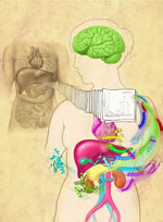
An artistic interpretation of the human proteome. Credit: Corinne Sandone and Jennifer Fairman, Johns Hopkins University.

Genes control the most basic functions of the cell, including what proteins to make and when. In 2003, the Human Genome Project created a draft map of our genes, and now researchers have completed a draft map of the human proteome—the set of all our proteins. The map, which includes proteins encoded by more than 17,000 genes as well as ones from regions of the genome previously thought to be non-coding, will help advance a broad range of research into human health and disease.

Read more about the proteome map in this NIH Research Matters article.

Learning How Mosquito-Borne Viruses Use Knot-like RNA to Cause Disease

0 comments
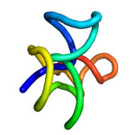
A knot-like structure in a section of RNA from a flavivirus
A knot-like structure in RNA enables flaviviruses to cause diseases like yellow fever, West Nile virus and dengue fever, which threaten roughly half the world’s population. Credit: Jeffrey Kieft.

Roughly half the world’s population is now at risk for mosquito-borne diseases other than malaria, such as yellow fever, West Nile virus and dengue fever. These three diseases are caused by flaviviruses, a type of virus that carries its genetic material as a single strand of RNA.

Flaviviruses have found a way not only to thwart our bodies’ normal defenses, but also to harness a human enzyme—paradoxically, one normally used to destroy RNA—to enhance their disease-causing abilities. A team of scientists led by Jeffrey Kieft at the University of Colorado at Denver found that flaviviruses accomplish both feats by bending and twisting a small part of their RNA into a knot-like structure.

The scientists set out to learn more about this unusual ability. First, they determined the detailed, three-dimensional architecture of the convoluted flaviviral RNA. Then, they examined several different variations of the RNA. In doing so, they pinpointed parts that are critical for forming the knot-like shape. If researchers can find a way to prevent the RNA from completing its potentially dangerous twist, they’ll be a step closer to developing a treatment for flaviviral diseases, which affect more than 100 million people worldwide.

This work also was supported by the National Institute of Allergy and Infectious Diseases and the National Cancer Institute.

Learn more:
University of Colorado News Release exit icon
Kieft Lab exit icon

Basic Research Fuels Medical Advances

0 comments
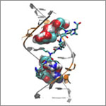
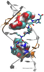
Genetic defect that causes myotonic dystrophy type 2 and used that information to design drug candidates to counteract the disease. Credit: Ilyas Yildirim, Northwestern University.
Scientists revealed a detailed image of the genetic change that causes myotonic dystrophy type 2 and used that information to design drug candidates to counteract the disease. Credit: Ilyas Yildirim, Northwestern University. View larger image

This image may look complicated, but it tells a fairly straightforward tale about basic research: Learning more about basic life processes can pave the way for medical and other advances.

In this example, researchers led by Matthew Disney of the Scripps Research Institute’s Florida campus focused on better understanding the structural underpinnings of myotonic dystrophy type 2, a relatively rare, inherited form of adult-onset muscular dystrophy. While this work is still in the preliminary stages, it may hold potential for someday treating the disorder.

Some 300,000 NIH-funded scientists are working on projects aimed at improving disease diagnosis, treatment and prevention, often through increasing understanding of basic life processes.

Read the complete Inside Life Science article.

Cool Image: Visualizing Viral Activity

0 comments
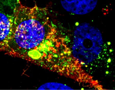
Viral RNA (red) in an RSV-infected cell. Credit: Eric Alonas and Philip Santangelo, Georgia Institute of Technology and Emory University. 

Viral RNA (red) in an RSV-infected cell. Credit: Eric Alonas and Philip Santangelo, Georgia Institute of Technology and Emory University.

What looks like a colorful pattern produced as light enters a kaleidoscope is an image of a cell infected with respiratory syncytial virus (RSV) illuminated by a new imaging technology. Although relatively harmless in most children, RSV can lead to bronchitis and pneumonia in others. Philip Santangelo of the Georgia Institute of Technology and Emory University, along with colleagues nationwide, used multiply-labeled tetravalent RNA imaging probes (MTRIPS) to observe the entry, assembly and replication of RSV inside a living cell. Once introduced into RSV-plagued cells, the MTRIPS latched onto the viral RNA (in the image, red) without altering the level of infectivity. This led to fluorescent RSV viral particles that let the researchers track the viral RNA in host cells and better understand what the virus was doing. The knowledge gained from this new technique might aid in the development of RSV antiviral drugs and possibly a vaccine. Scientists could also one day use the imaging approach to study other RNA viruses, such as the flu and Ebola.

Learn more:
Georgia Tech News Release Exit icon

Transporter Protein Dance Moves

2 comments
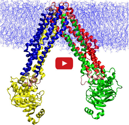
Animation depicts the changes that allow a protein transporter to do its job.

In this video, Emad Tajkhorshid of the University of Illinois at Urbana-Champaign explains the molecular dance of transporter proteins, molecules that move substances across the cell membrane.

In this video, Emad Tajkhorshid of the University of Illinois at Urbana-Champaign explains the molecular dance of ABC transporters, a family of molecular machines that utilize ATP to move substances across the cell membrane. Tajkhorshid and his team recently used computational methods to map the movements between two known structural models of MsbA, a bacterial version of a transporter in human cells that helps to export anti-cancer drugs. They then described the individual steps of the molecular motions during the transport cycle. Understanding the process at such a detailed level could suggest new targets for treating a range of diseases, including some drug-resistant cancers that often make more transporter proteins to kick out medications meant to kill them.

Learn more:
University of Illinois at Urbana-Champaign News Release Exit icon
Tajkhorshid Lab

Targeting Toxic RNA Molecules in Muscular Dystrophy

0 comments
Genetic defect that causes myotonic dystrophy type 2 and used that information to design drug candidates to counteract the disease. Credit: Ilyas Yildirim, Northwestern University.
Scientists revealed a detailed image of the genetic defect that causes myotonic dystrophy type 2 and used that information to design drug candidates to counteract the disease. Credit: Ilyas Yildirim, Northwestern University. View larger image

Myotonic dystrophy type 2 (DM2) is a relatively rare, inherited form of adult-onset muscular dystrophy that has no cure. It’s caused by a genetic defect in which a short series of nucleotides—the chemical units that spell out our genetic code—is repeated more times than normal. When the defective gene is transcribed, the resulting RNA repeat forms a hairpin-like structure that binds to and disables a protein called MBNL1.

Now, research led by Matthew Disney of The Scripps Research Institute (TSRI), Florida Campus, has revealed the detailed, three-dimensional structure of the RNA defect in DM2 and used this information to design small molecules that bind to the aberrant RNA. These designer molecules, even in small amounts, significantly improved disease-associated defects in a cellular model of DM2, and thus hold potential for reversing the disorder.

Drugs that target toxic RNA molecules associated with diseases such as DM2 are few and far between, as developing such compounds is technically challenging. The “bottom-up” approach that the scientists used to design potent new drug candidates, by first studying in detail how the RNA structure interacts with small molecules, is unconventional, noted Jessica Childs-Disney of TSRI, who was lead author of the paper with Ilyas Yildirim of Northwestern University. But it may serve as an effective strategy for pioneering the use of small molecules to manipulate disease-causing RNAs—a central focus of the Disney lab.

This work also was funded by NIH’s National Cancer Institute.

Learn more:
The Scripps Research Institute News Release Exit icon
Disney Lab

New Models Predict Where E. coli Strains Will Thrive

0 comments
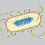
Illustration of E. coli. Credit: Janet Iwasa, University of Utah.
Illustration of E. coli. Credit: Janet Iwasa, University of Utah (image available under a Creative Commons Attribution-NonCommercial-ShareAlike license Exit icon). View larger image

Like plants and animals, different types of E. coli thrive in different environments. Now, scientists can even predict which environments—such as the bladder, stomach or blood—are most amenable to the growth of various strains, including pathogenic ones. A research team led by Bernhard Palsson Exit icon of the University of California, San Diego, accomplished this by using genome data to reconstruct the metabolic networks of 55 E. coli strains. The metabolic models, which identify differences in the ability to manufacture certain compounds and break down various nutrients, shed light on how certain E. coli strains become pathogenic and how to potentially control them. One approach could be depriving the deadly strains of the nutrients they need to survive in their niches. The researchers plan to use their new method to study other bacteria, such as those that cause staph infections.

This work also was funded by NIH’s National Cancer Institute.

Learn more:
University of California, San Diego News Release