Category: Molecular Structures

Structural Studies Demystify Membrane Protein

0 comments
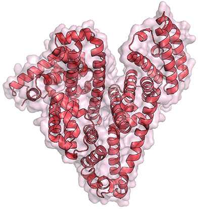
Animated structural model of TSPO.
Animated structural model of TSPO. Credit: Michigan State University.

Mitochondria have proteins that span their membranes to control the flow of messages and materials moving into and out of the organelle. One way scientists can learn more about how membrane proteins function—and how medicines might interact with them—is to determine their structures. But for a variety of reasons, obtaining the structures has been notoriously difficult.

Two structural studies have now shed light on the mysterious mitochondrial membrane protein TSPO. This protein plays a key role in transporting cholesterol and drugs into the cell’s mitochondria. While here, the cholesterol is converted to steroid hormones that are essential for numerous bodily functions. Although many researchers have been studying TSPO since the 1990s, they’ve remained uncertain about its mechanisms and how it truly functions. Continue reading “Structural Studies Demystify Membrane Protein”

Untangling a Trending Topic

0 comments

It’s not every day that we log into Facebook and Twitter to see conversations about denaturing proteins and the possibility of reducing biotechnology costs, but that changed last week when a story about “unboiling” eggs became a trending topic.

Since NIGMS partially funded the research advance Exit icon that led to the media scramble, we asked our scientific expert Jean Chin to tell us more about it.

What’s the advance?

Gregory Weiss of the University of California, Irvine, and his collaborators have designed a device that basically unties proteins that have been tangled together. Continue reading “Untangling a Trending Topic”

Meet Jennifer Doudna

3 comments
Jennifer Doudna
Credit: Jennifer Doudna
Jennifer Doudna
Fields: Biochemistry and structural biology
Studies: New genome editing tool called CRISPR
Works at: University of California, Berkeley
Raised in: Hilo, Hawaii
Studied at: Pomona College, Harvard University
Recent honors: Winner of the Lurie Prize in the Biomedical Sciences Exit icon, an annual award that recognizes outstanding achievement by promising scientists age 52 or younger
If she couldn’t be a scientist, she’d like to be: A papaya farmer or an architect

Jennifer Doudna likes to get her hands dirty. Literally. When she’s not in her laboratory, she can often be found amid glossy green leaves and brightly colored fruit in her Berkeley garden. She recently harvested her first three strawberry guavas.

Coaxing tropical fruit plants from her childhood home in Hawaii to grow in Northern California is more than just a hobby—it’s an intellectual challenge.

“I like solving puzzles, I like the process of figuring things out, and I enjoy working with my hands,” says Doudna. “Those things were what really drew me to science in the beginning.”

Since she was a graduate student, Doudna’s professional puzzle has been RNA, a type of genetic material inside our cells. Recently, there has been an explosion of discoveries about the many roles RNA molecules play in the body. Doudna’s work probes into how RNA molecules work, what 3-D shapes they form and how their structures drive their functions.

“I’ve been fascinated by understanding RNA at a mechanistic level,” Doudna says.

While teasing out answers to these fundamental questions, Doudna’s lab has played a leading role in a discovery that is upending the field of genetic engineering, with exciting implications for human health.

Her Findings

The discovery started with bacteriophages—viruses that infect bacteria, just like the common cold infects humans. About 10 years ago, researchers using high-powered computing to sift through bacterial genomes began to find mysterious repetitive gene sequences that matched those from viruses known to infect the bacteria. The researchers named these sequences “clustered regularly interspaced short palindromic repeats,” or CRISPRs for short.

Over the next few years, scientists came to understand that these CRISPR sequences are part of something not previously thought to exist—an adaptive bacterial immune system, which remembers viruses fought off before and raises a response to fight them when exposed again. CRISPRs were this immune system’s reference library, holding records of viral exposure.

Somehow, bacteria with a CRISPR-based immune system (there are three types now known to scientists) use these records to command certain proteins to recognize and chop up DNA from returning viruses.

Wanting to know more about this process, Doudna’s team picked one protein in a CRISPR-based defense system to study. This protein, called Cas9, had been identified by other researchers as being essential for protection against viral invasion.

To their delight, Doudna’s group had hit the jackpot. Cas9 turned out to be the system’s scalpel. Once CRISPR identifies a DNA sequence from the invading virus, Cas9 slices the sequence out of the viral genome, destroying the virus’s ability to copy itself.

Doudna’s lab and their European collaborators also identified the other key components of the CRISPR-Cas9 system—two RNA molecules that guide Cas9 to the piece of viral DNA identified by CRISPR.

Even more importantly, the researchers showed that the two guide RNAs could be manipulated in the lab to create a tool that both recognizes any specified DNA sequence and carries Cas9 there to make its cut.

“That was really where we made the connection between the basic, curiosity-driven research that we were doing and recognizing that we had in our hands something that could be a very powerful technology for genome editing,” remembers Doudna.

She was right. After publication of their 2012 paper, the field of CRISPR-guided genetic manipulation exploded. Labs around the world now use the tool Doudna’s team developed to cut target gene sequences in organisms ranging from plants to humans. The technique is already replacing more time-consuming, less-reliable methods of creating ‘knock-out’ model organisms (those missing a specific gene) for laboratory research. CRISPR-based editing even allows more than one gene to be knocked out at the same time, something that was not possible with previous genome-editing techniques.

The ability of CRISPR systems to recognize DNA sequences with extraordinary precision also holds potential for human therapeutics. For example, a paper from another laboratory published early this year showed that, in a mouse model, CRISPR-based editing could cut out and replace a defective gene responsible for a type of muscular dystrophy. Researchers are testing similar CRISPR-based techniques in models of human diseases ranging from cystic fibrosis to blood disorders.

Doudna is a co-founder of two biotechnology companies hoping to harness the potential of CRISPR-based genome editing. Although the technology holds great promise, she acknowledges that much work needs to be done before CRISPR can be considered safe for human trials. Major challenges include assuring that no off-target cuts are made in the genome and finding a safe way to deliver the editing system to living tissues.

She is also excited to continue working with her research team, advancing the basic understanding of the CRISPR-based system.

“I’m very interested in seeing what we can contribute to the whole question about how you deliver a technology like this, how you can use it therapeutically in an organism. That’s an area where we hope that our biochemical understanding of this system will be able to contribute,” she concludes.
A Crisper View of the CRISPR Gene-Editing Mechanism

0 comments
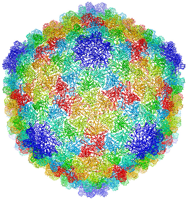
Structural model of the Cascade surveillance machine
Structural model of the Cascade surveillance machine. Credit: Ryan Jackson, Montana State University. Click for larger image

To dismantle the viruses that infect them, bacteria have evolved an immune system that identifies invading viral DNA and signals for its destruction. This gene-editing system is called CRISPR, and it’s being harnessed as a tool for modifying human genes associated with disease.

Taking another important step toward this potential application, researchers now know the structure of a key CRISPR component: a multi-subunit surveillance machine called Cascade that identifies the viral DNA. Shaped like a sea horse, Cascade is composed of 11 proteins and CRISPR-related RNA. A research team led by Blake Wiedenheft of Montana State University used X-ray crystallography and computational analysis to determine Cascade’s configuration. In a complementary study, Scott Bailey of Johns Hopkins University and his colleagues determined the structure of the complex bound to a viral DNA target.

Like blueprints, these structural models help scientists understand how Cascade assembles into an efficient surveillance machine and, more broadly, how the CRISPR system functions and how to adapt it as a tool for basic and clinical research.

Learn more:
Montana State University News Release Exit icon
Johns Hopkins University News Release Exit icon

Intercepting Amyloid-Forming Proteins

0 comments
Structure of a protein involved in disease-associated amyloid fibrils.
A molecule targets the intermediary structure of a protein involved in disease-associated amyloid fibrils. Credit: University of Washington.

Alzheimer’s disease, type 2 diabetes and many other illnesses are linked to the buildup of proteins whose structures have changed into shapes that enable the formation of cell-entangling threads called amyloid fibrils. About 10 years ago, researchers led by Valerie Daggett of the University of Washington used computer simulations to suggest that such proteins, on their way to creating fibrils, form an intermediary structure called an alpha sheet that’s even more toxic to cells than fibrils. Now Daggett’s team has experimentally investigated this possibility. The scientists made alpha sheet molecules expected to bind to amyloid-forming proteins in the computationally predicted intermediate state. When they tested the molecules on two amyloid disease-related proteins, they observed a substantial reduction in fibril formation. The work is still very preliminary, but it highlights a potential new avenue for treating a range of amyloid-related diseases.

This work also was funded by NIH’s National Institute of Allergy and Infectious Diseases.

Learn more:
University of Washington News Release Exit icon
Daggett Lab Exit icon
Monster Mash: Protein Folding Gone Wrong Article from Inside Life Science

A Data Bank Built for Discovery

0 comments
Dynein, a motor protein. Credit: David S. Goodsell, The Scripps Research Institute and the RCSB PDB.
The PDB archive holds structural data for dynein, a motor protein, and more than 100,000 other molecules. Credit: David S. Goodsell, The Scripps Research Institute and the RCSB PDB. Click for larger image

Meet dynein, the August Molecule of the Month presented by the Research Collaboratory for Structural Bioinformatics Protein Data Bank (RCSB PDB). This motor protein travels along the cables of our cellular skeleton, delivering cargo throughout the cell. The structure of dynein’s stalk enables it to bind to regular grooves along its path.

Dynein’s shape is just one of more than 100,000 structures that scientists have deposited in the PDB archive, a freely available digital repository. Because understanding a protein’s shape helps researchers better understand its function, the structural information in the PDB can lead to additional scientific advancements. For example, scientists use the structure of HIV protease, a protein that helps the virus replicate in the body, to develop drugs that fit snugly into the protein’s center, shutting it down. And they use the shape of RNA polymerase to learn how this protein fits together with smaller ones to read our genetic code.

The PDB has doubled in size over the last 6 years. As the collection continues to grow, so does our potential for drug discovery and our understanding of basic life processes.

Learn more:
Molecule of the Month Archive from RCSB PDB

How Heat-Loving Organisms Are Helping Advance Medicine

2 comments
Hot spring. Credit: Stock image.
Icelandic hot springs are the natural habitat of Rhodothermus marinus, one of many species of thermophiles that the Gennis Lab studies to better understand membrane proteins. Credit: Stock image.

As the temperature climbs, most humans look for ways to cool down fast. But for some species of microorganisms, a midsummer heat wave isn’t nearly hot enough. These heat lovers, known as thermophiles, thrive at temperatures of 113°F or more. They’re often found in hot springs, geysers and even home water heaters.

Like humans and other organisms, thermophiles rely on proteins to maintain normal cell function. While our protein molecules break down under intense heat, a thermophile’s proteins actually work more efficiently. They also tend to be more stable at room temperature than our own. An NIH-funded research team is taking advantage of this property of thermophiles to better understand a group of human proteins commonly targeted by today’s medicines.

Read more about the team’s thermophile research in this Inside Life Science article.

The “Virtuous Cycle” of Technology and Science

0 comments
A scientist looking through a  microscope. Credit: Stock image.
Whether it’s a microscope, computer program or lab technique, technology is at the heart of biomedical research. Credit: Stock image.

Whether it’s a microscope, computer program or lab technique, technology is at the heart of biomedical research. Its central role is particularly clear from this month’s posts.

Some show how different tools led to basic discoveries with important health applications. For instance, a supercomputer unlocked the secrets of a drug-making enzyme, a software tool identified disease-causing variations among family members and high-powered microscopy revealed a mechanism allowing microtubules—and a cancer drug that targets them—to work.

Another theme featured in several posts is novel uses for established technologies. The scientists behind the cool image put a new spin on a long-standing imaging technology to gain surprising insights into how some brain cells dispose of old parts. Similarly, the finding related to sepsis demonstrates yet another application of a standard lab technique called polymerase chain reaction: assessing the immune state of people with this serious medical condition.

“We need tools to answer questions,” says NIGMS’ Doug Sheeley, who oversees biomedical technology research resource grants. “When we find the answers, we ask new questions that then require new or improved tools. It’s a virtuous cycle that keeps science moving forward.”

A Drug-Making Enzyme in Motion

0 comments
Mutated enzyme, LovD9. Credit: Silvia Osuna and Gonzalo Jiménez-Osés, University of California, Los Angeles.
The movement of this mutated enzyme, LovD9, facilitates rapid production of the cholesterol reducing-drug simvastatin. Credit: Silvia Osuna and Gonzalo Jiménez-Osés, University of California, Los Angeles.

LovD9, a mutated version of an enzyme extracted from mold growing in soil, produces the cholesterol-reducing drug simvastatin 1,000 times faster than its natural predecessor. But scientists didn’t understand why because the enzyme’s mutations are far from the active site, where the drug is actually made. Now they do.

Yi Tang of the University of California, Los Angeles (UCLA), in partnership with the pharmaceutical company Codexis, generated LovD9 by repeatedly inducing random mutations, each time selecting the mutated versions of the enzyme with the most promise for industrial simvastatin production.

Then, the team collaborated with UCLA colleagues Kendall Houk and Todd Yeates to unlock the secret of the enzyme’s speed. Using ANTON, a special-purpose supercomputer at the Pittsburgh Supercomputing Center, they simulated how different parts of the enzyme rotate and twist when synthesizing the drug. The scientists discovered that as LovD9 moves, it forms shapes that facilitate simvastatin production more often than the natural enzyme does.

With their better understanding of how mutations far from an active site may affect an enzyme’s motion, the researchers hope to one day directly engineer enzymes with precise mutations that enhance drug production.

Learn more:
University of California, Los Angeles News Release Exit icon
Houk Exit icon, Tang Exit icon and Yeates Exit icon Labs

Meet Rhiju Das

1 comment
Rhiju Das
Credit: Rhiju Das
Rhiju Das
Fields: Biophysics and biochemistry
Works at: Stanford University
Born and raised in: The greater Midwest (Texas, Indiana and Oklahoma)
Studied at: Harvard University, Stanford University
When he’s not in the lab he’s: Enjoying the California outdoors with his wife and 3-year-old daughter
If he could recommend one book about science to a lay reader, it would be: “The Eighth Day of Creation,” about the revolution in molecular biology in the 1940s and 50s.

At the turn of the 21st century, Rhiju Das saw a beautiful picture that changed his life. Then a student of particle physics with a focus on cosmology, he attended a lecture unveiling an image of the ribosome—the cellular machinery that assembles proteins in every living creature. Ribosomes are enormous, complicated machines made up of many proteins and nucleic acids similar to DNA. Deciphering the structure of a ribosome—the 3-D image Das saw—was such an impressive feat that the scientists who accomplished it won the 2009 Nobel Prize in chemistry.

Das, who had been looking for a way to apply his physics background to a research question he could study in a lab, had found his calling.

“It was an epiphany—it was just flabbergasting to me that a hundred thousand atoms could find their way into such a well-defined structure at atomic resolution. It was like miraculously a bunch of nuts and bolts had self-assembled into a Ferrari,” recounted Das. “That inspired me to drop everything and learn everything I could about nucleic acid structure.”

Das focuses on the nucleic acid known as RNA, which, in addition to forming part of the ribosome, plays many roles in the body. As is the case for most proteins, RNA folds into a 3-D shape that enables it to work properly.

Das is now the head of a lab at Stanford University that unravels how the structure and folding of RNA drives its function. He has taken a unique approach to uncovering the rules behind nucleic acid folding: harnessing the wisdom of the crowd.

Together with his collaborator, Adrien Treuille of Carnegie Mellon University, Das created an online, multiplayer video game called EteRNA Exit icon. More than a mere game, it does far more than entertain. With its tagline “Played by Humans, Scored by Nature,” it’s upending how scientists approach RNA structure discovery and design.

Das’ Findings

Treuille and Das launched EteRNA after working on another computer game called Foldit, which lets participants play with complex protein folding questions. Like Foldit, EteRNA asks players to assemble, twist and revise structures—this time of RNA—onscreen.

But EteRNA takes things a step further. Unlike Foldit, where the rewards are only game points, the winners of each round of EteRNA actually get to have their RNA designs synthesized in a wet lab at Stanford. Das and his colleagues then post the results—which designs resulted in a successful, functional RNAs and which didn’t—back online for the players to learn from.

In a paper published in the Proceedings of the National Academy of Sciences Exit icon, Das and his colleagues showed how effective this approach could be. The collective effort of the EteRNA participants—which now number over 100,000—was better and faster than several established computer programs at solving RNA design problems, and even came up with successful new structural rules never before proposed by scientists or computers.

“What was surprising to me was their speed,” said Das. “I had just assumed that it would take a year or so before players were really able to analyze experimental data, make conclusions and come up with robust rules. But it was one of the really shocking moments of my life when, about 2 months in, we plotted the performance of players against computers and they were out-designing the computers.”

“As far as I can tell, none of the top players are academic scientists,” he added. “But if you talk to them, the first thing they’ll tell you is not how many points they have in the game but how many times they’ve had a design synthesized. They’re just excited about seeing whether or not their hypotheses were correct or falsified. So I think the top players truly are scientists—just not academic ones. They get a huge kick out of the scientific method, and they’re good at it.”

To capture lessons learned through the crowd-sourcing approach, Das and his colleagues incorporated successful rules and features into a new algorithm for RNA structure discovery, called EteRNABot, which has performed better than older computer algorithms.

“We thought that maybe the players would react badly [to EteRNABot], that they would think they were going to be automated out of existence,” said Das. “But, as it turned out, it was exciting for them to have their old ideas put into an algorithm so they could move on to the next problems.”

You can try EteRNA for yourself at http://eternagame.org Exit icon. Das and Treuille are always looking for new players and soliciting feedback.