Category: Molecular Structures

Fall 2017 Issue of Findings Magazine

0 comments

It’s back! Check out the new issue of Findings magazine.

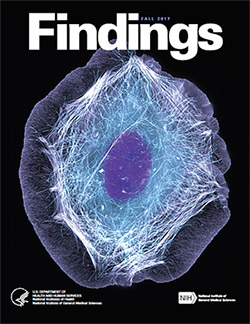
Findings Magazine Cover for Fall 2017.Findings presents cutting-edge research from scientists in diverse biomedical fields. The articles are aimed at high school students with the goal of making science—and the people who do it—interesting and exciting, and to inspire young readers to pursue careers in biomedical research. In addition to putting a face on science, Findings offers activities such as quizzes and crossword puzzles and, in its online version, video interviews with scientists.

The Fall 2017 issue profiles Yale University biologist Enrique De La Cruz, who studies how actin—a protein chain that supports cell structure—breaks so easily. Also profiled is University of California, Berkeley, biologist Rebecca Heald and her study of developmental factors that control an animal’s size.

This issue also features:

  • A virtual reality program designed to help burn patients manage pain
  • The promise of gene therapy for glaucoma
  • The many ways scientists categorize the biological world using “omics”
  • What researchers know—and don’t know—about how general anesthetics work
  • How animation helps researchers visualize interactions between biological molecules
  • How cells use sugary outer coatings to distinguish friend from foe
  • What makes our tissues stiff, squishy, solid, or see-through (hint: its initials are ECM)
  • How super-powerful microscopes are revealing views of biology never possible before

View Findings online, or order a print copy (classroom sets of up to 30 copies are available for educators).

Protein Alphabet: A Picture Is Worth One Letter

0 comments

It’s back-to-school time. That means learning lots of new facts and figures. In science, terms tend to be several syllables, sometimes with a Latin word thrown into the mix. As a result, many are referred to by their acronyms, such as DNA—short for deoxyribonucleic acid. This makes them easier both to remember and to say.

Researcher Mark Howarth Exit icon of Oxford University, has taken this a step further. Searching through information stored in the NIGMS-funded Protein Data Bank Exit icon, he curated a 3-D protein alphabet. It’s a set of 26 protein crystal structures that look like they were fashioned from bits of rainbow-colored curly ribbon. This 3-D alphabet helps us see what different protein strands look like, and explains terms and concepts relating to protein structure and function.

Proteins are molecules that play important roles in virtually every activity in the body. They form hair and fingernails, carry oxygen in the blood, enable muscle movement and much more. Although proteins are made of long strands of small molecules called amino acids, they do not remain as a straight chain. Some proteins are composed of multiple amino acid strands that wind together in the completed protein. The strands twist, bend and fold into a specific shape, and the protein’s structure enables it to perform its task. For instance, the “Y” shape of antibodies helps these immune system proteins bind to foreign molecules such as bacteria or viruses while also drawing in other immune system molecules.

Continue reading “Protein Alphabet: A Picture Is Worth One Letter”

Happy Birthday, BioBeat

0 comments

This month, our blog that highlights NIGMS-funded research turns four years old! For each candle, we thought we’d illuminate an aspect of the blog to offer you, our reader, an insider’s view.

Who are we?

Over the years, the editorial team has included onsite science writers, office interns, staff scientists and guest authors from universities. Kathryn, who’s a regular contributor, writes entirely from her home office. Chris, who has a Ph.D. in neuroscience and now manages the blog, used to do research in a lab. Alisa has worked in NIGMS’ Bethesda-based office the longest: 22 years! She and I remember when we first launched Biomedical Beat as an e-newsletter in 2005. You can read more about each of the writers on the contributors page and if you know someone who’s considering a career in science communications, tell them to drop us a line.

How do we come up with the stories?

We get our story ideas from a range of sources. For instance, newspaper articles about an experimental pest control strategy in Florida and California prompted us to write about NIGMS-funded studies exploring the basic science of the technique. A beautiful visual from a grantee’s institution inspired a short post on tissue regeneration research. And an ongoing conversation with NIGMS scientific staff about the important role of research organisms in biological studies sparked the idea for a playful profile of one such science superstar.

A big change in our storytelling has been shifting the focus from a single finding to broader progress in a lab or field. So instead of reporting on a study just published in a scientific journal, we may write about the scientist’s career path or showcase a collection of recent findings in that particular field. These approaches help us demonstrate that scientific understanding usually progresses through the slow and steady work undertaken by many labs.

What are our favorite posts?

I polled the writers on posts they liked, and the list is really long! Here are the top picks.


Four Ways Inheritance Is More Complex Than Mendel Knew
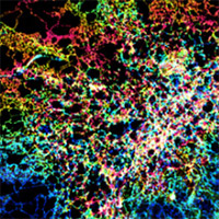

The Endoplasmic Reticulum: Networking in the Cell


Interview With a Scientist: Janet Iwasa, Molecular Animator


From Basic Research to Bioelectric Medicine
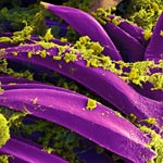

An Insider’s Look at Life: Magnified, an Airport Exhibit of Stunning Microscopy Images

What are your favorite posts?

We regularly review data about the number of times a blog post has been viewed to identify the ones that interest readers the most. That information also helps guide our decisions about other topics to feature on the blog. The Cool Image posts are among the most popular! Below are some other chart-topping posts.


Our Complicated Relationship With Viruses
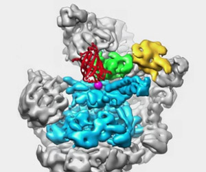

The Proteasome: The Cells Trash Processor in Action
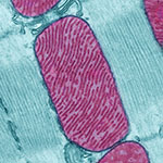

Demystifying General Anesthetics


Meet Sarkis Mazmanian and the Bacteria That Keep Us Healthy


5 Reasons Biologists Love Math

We always like hearing from readers! If there’s a basic biomedical research topic you’d like us to write about, or if you have feedback on a story or the blog in general, please leave your suggestions in the comment field below.
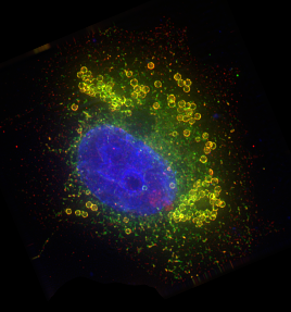
Researchers Score Goal with New Atomic-Scale Model of Salmonella-Infecting Virus

1 comment

An atomic-scale model of a virus that infects the Salmonella bacterium. Credit: C. Hryc and the Chiu Lab, Baylor College of Medicine.

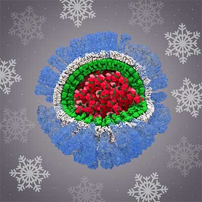
This sphere could be a prototype design for the 2018 World Cup official match soccer ball, but you won’t see it dribbled around any soccer fields. The image is actually an atomic-scale model of a virus that infects the Salmonella bacterium. Like a soccer ball, both are approximately spherical shapes created by a combination of hexagonal (six-sided) and pentagonal (five-sided) units. Wah Chiu, a biochemist at Baylor College of Medicine, and his colleagues used new computational methods to construct the model from more than 20,000 cryo-electron microscopy (cryo-EM) images. Cryo-EM is a sophisticated technique that uses electron beams for visualizing frozen samples of proteins and other biological specimens.

The researchers’ model, published in a recent issue of PNAS, shows the virus’ protein shell, or capsid, that encloses the virus’ genetic material. Each color shows capsid proteins having the same interactions with their neighbors. The fine resolution allowed researchers to identify the protein interactions essential to building a stable shell. They developed a new approach to checking the accuracy and reliability of the virus model and reporting what parts are the most certain. The approach could be used to evaluate other complex biological structures, potentially leading to better quality models and new avenues for drug design and development.

This research is funded in part by NIH under grants R01GM079429, P01GM063210, P41GM103832, PN2EY016525, T15LM007093.

Viral Views: New Insights on Infection Strategies

0 comments

The following images show a few ways in which cutting-edge research tools are giving us clearer views of viruses—and possible ways to disarm them. The examples, which highlight work involving HIV and the coronavirus, were funded in part by our Biomedical Technology Research Resources program.

Uncloaking HIV’s Camouflage

HIV capsid with (right, red) and without (left) a camouflaging human protein.
HIV capsid with (right, red) and without (left) a camouflaging human protein. Credit: Juan R. Perilla, Klaus Schulten and the Theoretical and Computational Biophysics Group, University of Illinois at Urbana-Champaign.

To sneak past our immune defenses and infect human cells, HIV uses a time-honored strategy—disguise. The virus’ genome is enclosed in a protein shell called a capsid (on left) that’s easily recognized and destroyed by the human immune system. To evade this fate, the chrysalis-shaped capsid cloaks itself with a human protein known as cyclophilin A (in red, on right). Camouflaged as human, the virus gains safe passage into and through a human cell to deposit its genetic material in the nucleus and start taking control of cellular machinery.

Biomedical and technical experts teamed up to generate these HIV models at near-atomic resolution. First, structural biologists at the Pittsburgh Center for HIV Protein Interactions Exit icon used a technique called cryo-electron microscopy (cryo-EM) to get information on the shape of an HIV capsid as well as the capsid-forming proteins’ connections to each other and to cyclophilin A. Then experts at the Resource for Macromolecular Modeling and Bioinformatics fed the cryo-EM data into their visualization and simulation programs to computationally model the physical interactions among every single atom of the capsid and the cyclophilin A protein. The work revealed a previously unknown site where cyclophilin A binds to the capsid, offering new insights on the biology of HIV infection. Continue reading “Viral Views: New Insights on Infection Strategies”

Finding Adventure: Blake Wiedenheft’s Path to Gene Editing

1 comment
Blake Wiedenheft
Blake Wiedenheft
Grew up in: Fort Peck, Montana
Fields: Microbiology, biochemistry, structural biology
Job site: Montana State University
Secret talent: Being a generalist; enjoying many different subjects and activities
When not in the lab, he’s: Running, biking, skiing or playing scrabble with his grandmother

Scientific discoveries are often stories of adventure. This is the realization that set Blake Wiedenheft on a path toward one of the hottest areas in biology.

His story begins in Montana, where he grew up and now lives. Always exploring different interests, Wiedenheft decided in his final semester at Montana State University (MSU) in Bozeman to volunteer for Mark Young, a scientist who studies plant viruses. Even though he majored in biology, Wiedenheft had spent little time in a lab and hadn’t even considered research as a career option. Continue reading “Finding Adventure: Blake Wiedenheft’s Path to Gene Editing”

A Heart-Shaped Protein

1 comment
Structure of the serum albumin protein

The structure of the serum albumin protein is shaped like a heart. Credit: Wladek Minor, University of Virginia.

From cookies and candies to balloons and cards, heart-shaped items abound this time of year. They’re even in our blood. It turns out that the most abundant protein molecule in blood plasma—serum albumin (SA)—is shaped very much like a heart. Continue reading “A Heart-Shaped Protein”

Cryo-Electron Microscopy Reveals Molecules in Ever Greater Detail

0 comments

The molecular visualization technique known as cryo-electron microscopy (cryo-EM) was recently named the “2015 Method of the Year” by the journal Nature Methods. In a recent blog post, NIH Director Francis Collins explains how the technology works and just how rapidly it has advanced. He writes, “Today’s cryo-EM is so powerful that researchers can almost make out individual atoms!” He also notes, “These dramatic advances serve as a reminder of the ways in which basic technological innovation can open new realms of scientific possibility.”

We fund many scientists who are developing and applying cryo-EM to bring the details of biological structures into unprecedented focus. Here are two examples of their work and its potential impact. Continue reading “Cryo-Electron Microscopy Reveals Molecules in Ever Greater Detail”

Seeing Telomerase’s ‘Whiskers’ and ‘Toes’

1 comment
Telomerase and its components.

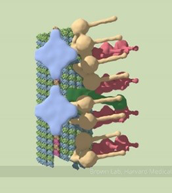
The image here is the “front view” of telomerase, with the enzyme’s components shown in greater detail than ever before. Credit: UCLA Department of Chemistry and Biochemistry.

Like the features of a cat in a dark alley, those of an important enzyme called telomerase have been elusive. Using a combination of imaging techniques, a research team led by Juli Feigon Exit icon of the University of California, Los Angeles, has now captured the clearest view ever of the enzyme.

Telomerase maintains the DNA at the ends of our chromosomes, known as telomeres, which act like the plastic tips on the ends of shoelaces. In the absence of telomerase activity, telomeres get shorter each time our cells divide. Eventually, the telomeres become so short that the cells stop dividing or die. On the other hand, cells with abnormally high levels of telomerase activity can constantly rebuild their protective chromosomal caps. Telomerase is particularly active within cancer cells. Continue reading “Seeing Telomerase’s ‘Whiskers’ and ‘Toes’”

Unusual DNA Form May Help Virus Withstand Extreme Conditions

0 comments
A, B and Z DNA.
DNA comes in three forms: A, B and Z. Credit: A-DNA, B-DNA and Z-DNA by Zephyris (Richard Wheeler) under CC BY-SA 3.0.

DNA researcher Rosalind Franklin Exit icon first described an unusual form of DNA called the A-form in the early 1950s (Franklin, who died in 1958, would have turned 95 next month). New research on a heat- and acid-loving virus has revealed surprising information about this DNA form, which is one of three known forms of DNA: A, B and Z.

“Many people have felt that this A-form of DNA is only found in the laboratory under very non-biological conditions, when DNA is dehydrated or dry,” says Edward Egelman Exit icon in a University of Virginia news release Exit icon about the recent study. But considered with earlier studies on bacteria by other researchers, the new findings suggest that the A-form “appears to be a general mechanism in biology for protecting DNA.” Continue reading “Unusual DNA Form May Help Virus Withstand Extreme Conditions”