Category: Molecular Structures

Transporter Protein Dance Moves

2 comments
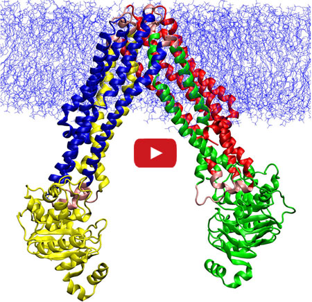
Animation depicts the changes that allow a protein transporter to do its job.

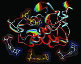
In this video, Emad Tajkhorshid of the University of Illinois at Urbana-Champaign explains the molecular dance of transporter proteins, molecules that move substances across the cell membrane.

In this video, Emad Tajkhorshid of the University of Illinois at Urbana-Champaign explains the molecular dance of ABC transporters, a family of molecular machines that utilize ATP to move substances across the cell membrane. Tajkhorshid and his team recently used computational methods to map the movements between two known structural models of MsbA, a bacterial version of a transporter in human cells that helps to export anti-cancer drugs. They then described the individual steps of the molecular motions during the transport cycle. Understanding the process at such a detailed level could suggest new targets for treating a range of diseases, including some drug-resistant cancers that often make more transporter proteins to kick out medications meant to kill them.

Learn more:
University of Illinois at Urbana-Champaign News Release Exit icon
Tajkhorshid Lab

Targeting Toxic RNA Molecules in Muscular Dystrophy

0 comments
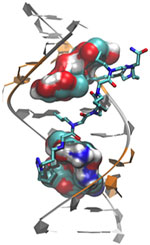
Genetic defect that causes myotonic dystrophy type 2 and used that information to design drug candidates to counteract the disease. Credit: Ilyas Yildirim, Northwestern University.
Scientists revealed a detailed image of the genetic defect that causes myotonic dystrophy type 2 and used that information to design drug candidates to counteract the disease. Credit: Ilyas Yildirim, Northwestern University. View larger image

Myotonic dystrophy type 2 (DM2) is a relatively rare, inherited form of adult-onset muscular dystrophy that has no cure. It’s caused by a genetic defect in which a short series of nucleotides—the chemical units that spell out our genetic code—is repeated more times than normal. When the defective gene is transcribed, the resulting RNA repeat forms a hairpin-like structure that binds to and disables a protein called MBNL1.

Now, research led by Matthew Disney of The Scripps Research Institute (TSRI), Florida Campus, has revealed the detailed, three-dimensional structure of the RNA defect in DM2 and used this information to design small molecules that bind to the aberrant RNA. These designer molecules, even in small amounts, significantly improved disease-associated defects in a cellular model of DM2, and thus hold potential for reversing the disorder.

Drugs that target toxic RNA molecules associated with diseases such as DM2 are few and far between, as developing such compounds is technically challenging. The “bottom-up” approach that the scientists used to design potent new drug candidates, by first studying in detail how the RNA structure interacts with small molecules, is unconventional, noted Jessica Childs-Disney of TSRI, who was lead author of the paper with Ilyas Yildirim of Northwestern University. But it may serve as an effective strategy for pioneering the use of small molecules to manipulate disease-causing RNAs—a central focus of the Disney lab.

This work also was funded by NIH’s National Cancer Institute.

Learn more:
The Scripps Research Institute News Release Exit icon
Disney Lab

New Models Predict Where E. coli Strains Will Thrive

0 comments
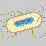
Illustration of E. coli. Credit: Janet Iwasa, University of Utah.
Illustration of E. coli. Credit: Janet Iwasa, University of Utah (image available under a Creative Commons Attribution-NonCommercial-ShareAlike license Exit icon). View larger image

Like plants and animals, different types of E. coli thrive in different environments. Now, scientists can even predict which environments—such as the bladder, stomach or blood—are most amenable to the growth of various strains, including pathogenic ones. A research team led by Bernhard Palsson Exit icon of the University of California, San Diego, accomplished this by using genome data to reconstruct the metabolic networks of 55 E. coli strains. The metabolic models, which identify differences in the ability to manufacture certain compounds and break down various nutrients, shed light on how certain E. coli strains become pathogenic and how to potentially control them. One approach could be depriving the deadly strains of the nutrients they need to survive in their niches. The researchers plan to use their new method to study other bacteria, such as those that cause staph infections.

This work also was funded by NIH’s National Cancer Institute.

Learn more:
University of California, San Diego News Release

Detailing Key Structures of HIV

1 comment
Structure of Env. Credit: The Scripps Research Institute.
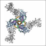
This model shows a birds-eye view of the structure of Env, a protein on the HIV surface that enables it to infect cells, bound to neutralizing antibodies. Credit: The Scripps Research Institute.

In a statement on World AIDS Day 2013, NIH leaders wrote, “In the 25 years that have passed since the first annual commemoration of World AIDS Day, extraordinary scientific progress has been made in the fight against HIV/AIDS. That progress has turned an HIV diagnosis from an almost-certain death sentence to what is now for many, a manageable medical condition and nearly normal lifespan. We have come far, yet not far enough.”

One area of progress is in understanding the structural biology of HIV, the virus that causes AIDS. Capturing details about the virus’ shape has helped scientists better understand how HIV operates and pinpoint its Achilles’ heels. Recently, scientists got a closer look at two key pieces of the virus.

In one study, researchers developed the most detailed picture yet of Env, a three-segment protein on the HIV surface that allows the virus to infect cells. The work illuminated the complex process by which the protein assembles, undergoes radical shape changes during infection and interacts with neutralizing antibodies, which can block many strains of HIV from infecting human cells. The findings also may guide the development of HIV vaccines.

In another study, researchers created the best image yet of the cocoon-like container, or capsid, that carries HIV’s genome. Capsids have been difficult to study because individual imaging techniques had not produced high enough detail. By combining several cutting-edge imaging methods, the scientists pieced together the individual polygonal units of the capsid like a jigsaw puzzle to determine its structure in detail. Now that they know how HIV’s inner vessel looks, the research team is searching for its cracks—potential targets for drug development.

Learn more:
Imaging HIV’S Inner Shell Article from Inside Life Science
Key HIV Protein Structure Revealed Article from NIH Research Matters
Interactive HIV Structural Model

Fitting a Piece of the Protein-Function Puzzle

0 comments
Comparison of the predicted binding of the substrate to the active site of HpbD (blue) with the binding sites determined experimentally by crystallography (magenta). Credit: Matt Jacobson, University of California, San Francisco; Steve Almo, Albert Einstein College of Medicine.
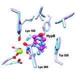
The image shows a comparison of the predicted binding of the substrate to the active site of HpbD (blue) with the binding sites determined experimentally by crystallography (magenta). Credit: Matt Jacobson, University of California, San Francisco; Steve Almo, Albert Einstein College of Medicine.

Sequencing the genomes of almost 7,000 organisms has identified more than 40 million proteins. But how do we figure out what all these proteins do? New results from an initiative led by John Gerlt of the University of Illinois suggest a possible method for identifying the functions of unknown enzymes, proteins that speed chemical reactions within cells. Using high-powered computing, the research team modeled how the structure of a mystery bacterial enzyme, HpbD, might fit like a puzzle piece into thousands of proteins in known metabolic pathways. Since an enzyme acts on other molecules, finding its target or substrate can shed light on its function. The new method narrowed HpbD’s candidate substrate down from more than 87,000 to only four. Follow-up lab work led to the actual substrate, tHypB, and determined the enzyme’s biological role. This combination of computational and experimental methods shows promise for uncovering the functions of many more proteins.

Learn more:

University of Illinois at Urbana-Champaign News Release
Gerlt Lab
Enzyme Function Initiative

Cell Biology Advances and Computational Techniques Earn Nobels for NIGMS Grantees

0 comments
The winners of the 2013 Nobel Prize in physiology or medicine discovered that cells import and export materials using fluid-filled sacs called vesicles. Credit: Judith Stoffer.

Every October, a few scientists receive a call from Sweden that changes their lives. From that day forward, they will be known as Nobel laureates. This year, five of the new Nobelists have received funding from NIGMS.

In physiology or medicine, NIGMS grantees James Rothman (Yale University) and Randy Schekman (University of California, Berkeley) were honored “for their discoveries of machinery regulating vesicle traffic, a major transport system in our cells.” They share the prize with Thomas C. Südhof of Stanford University.

Rothman and Schekman started out working separately and in different systems—Schekman in yeast and Rothman in reconstituted mammalian cells—and their conclusions validated each others’. It’s yet another example of the power of investigator-initiated research and the value of model systems.

Highly accurate molecular models like this one are based on computational techniques developed by the winners of the 2013 Nobel Prize in chemistry. Credit: Rommie E. Amaro, University of California, San Diego.

The three Nobelists in chemistry are NIGMS grantees Martin Karplus (Harvard University), Michael Levitt (Stanford University) and Arieh Warshel (University of Southern California), who developed “multiscale models for complex chemical systems.” They used computational techniques to obtain, for the first time, detailed structural information about proteins and other large molecules. Because of that work, scientists around the world are now able to access, with a few keystrokes, highly accurate models of nearly 100,000 molecular structures. Studying these structures has advanced our understanding of countless diseases, pharmaceuticals and basic biological processes.

Learn more:

NIGMS Nobel Prize News Announcement
NIGMS Nobelists Fact Sheet

Meet Galina Lepesheva

2 comments
Galina Lepesheva
Galina Lepesheva
Field: Biochemistry
Works at: Vanderbilt University, Nashville, TN
Born, raised and studied in: Belarus
To unwind, she: Reads, travels, spends time with her family

Galina Lepesheva knows that kissing bugs are anything but romantic. When the lights get low, these blood-sucking insects begin feasting—and defecating—on the faces of their sleeping victims. Their feces are often infected with a protozoan (a single-celled, eukaryotic parasite) called Trypanosoma cruzi that causes Chagas disease. Lepesheva has developed a compound that might be an effective treatment for Chagas. She has also tested the substance, called VNI, as a treatment for two related diseases—African sleeping sickness and leishmaniasis.

“This particular research is mainly driven by one notion: Why should people suffer from these terrible illnesses if there could be a relatively easy solution?” she says.

Lepesheva’s Findings

Currently, most cases of Chagas disease occur in rural parts of Mexico, Central America and South America. According to some estimates, up to 1 million people in the U.S. could have Chagas disease, and most of them don’t realize it. If left untreated, the infection is lifelong and can be deadly.

The initial, acute stage of the disease is usually mild and lasts 4 to 8 weeks. Then the disease goes dormant for a decade or two. In about one in three people, Chagas re-emerges in its life-threatening, chronic stage, which can affect the heart, digestive system or both. Once chronic Chagas disease develops, about 60 percent of people die from it within 2 years.

The Centers for Disease Control and Prevention (CDC) has targeted Chagas disease as one of five “neglected parasitic infections,” indicating that it warrants special public health action.

“Chagas disease does not attract much attention from pharmaceutical companies,” Lepesheva says. Right now, there are only two medicines to treat it. They are only available by special request from the CDC, aren’t always effective and can cause severe side effects.

Lepesheva’s research focuses on a particular enzyme, CYP51, that is the target of some anti-fungal medicines. If CYP51 can also act as an effective drug target for the parasites that cause Chagas, her work might help meet an important public health need.

CYP51 is found in all kingdoms of life. It helps produce molecules called sterols, which are essential for the development and viability of eukaryotic cells. Lepesheva and her colleagues are studying VNI and related compounds to examine whether they can block the activity of CYP51 in human pathogens such as protozoa, but do no harm to the enzyme in mammals. In other words, her goal is to cripple disease-causing organisms without creating side effects in infected humans or other mammals.

Lepesheva has tested the effectiveness of VNI on Chagas-infected mice. Remarkably, it has worked 100 percent of the time, curing both the acute and chronic stages of the disease. It acts by preventing the protozoan from establishing itself in the host’s body. If it is similarly effective in humans, VNI could become the first reliable treatment for Chagas disease.
NIH Director Blogs About NIGMS-Funded Research

0 comments
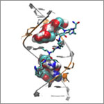
Antifolate drugs (bottom) work by blocking the folate receptor (top). Credit: Charles Dann III, Indiana University.
Caption: Antifolate drugs (bottom) work by blocking the folate receptor (top), starving cancer cells of an essential vitamin. Credit: Charles Dann III, Indiana University.

Within the last few weeks, NIH Director Francis Collins has blogged about several findings that NIGMS helped fund: the identification of a genetic link between hair color and melanoma risk and the solving of human folate receptor structures, which may aid the design of drugs for cancer and inflammatory diseases like rheumatoid arthritis and Crohn’s disease. Both advances are excellent examples of the value and impact of basic research. Want more examples? Check out Curiosity Creates Cures!

New Door Opens in the Effort to Stave off Mosquito-Borne Diseases

1 comment
Mosquito net Pyrethroids are used in mosquito nets distributed around the globe. Credit: Kurt Stepnitz, Michigan State University.

In the past decade, mosquitoes in many countries have become increasingly resistant to pyrethroid insecticides used to fend off mosquito-borne diseases such as malaria and dengue fever. Now, Ke Dong of Michigan State University and her colleagues have discovered a second pyrethroid-docking site in the molecular doorways, or channels, that control the flow of sodium into cells. Pyrethroids paralyze and kill mosquitoes and other insects by propping open the door and causing the pests to overdose on sodium, a critical regulator of nerve function. By providing new insights on pyrethroid action at the molecular level—and how mutations in the dual docking sites cause resistance—the findings open avenues to better monitoring and management of insecticide resistance.

Learn more:
Michigan State University News Release Exit icon