Category: Tools and Techniques

Simulating the Potential Spread of Measles

0 comments
Try out FRED Measles:

  1. Go to http://fred.publichealth.
    pitt.edu/measles
    Exit icon
  2. Select “Get Started”
  3. Pick a state and city
  4. Play both simulations

To help the public better understand how measles can spread, a team of infectious disease computer modelers at the University of Pittsburgh has launched a free, mobile-friendly tool that lets users simulate measles outbreaks in cities across the country.

The tool is part of the Pitt team’s Framework for Reconstructing Epidemiological Dynamics, or FRED, that it previously developed to simulate flu epidemics. FRED is based on anonymized U.S. census data that captures demographic and geographic distributions of different communities. It also incorporates details about the simulated disease, such as how contagious it is.

Screenshot of the FRED simulation.

A free, mobile-friendly tool lets users simulate potential measles outbreaks in cities across the country. Credit: University of Pittsburgh Graduate School of Public Health.

Continue reading “Simulating the Potential Spread of Measles”

E. Coli Bacteria as Medical Sensors and Hard Drives?

1 comment
E.Coli
Modified E. coli bacteria can serve as sensors and data storage devices for environmental and medical monitoring. Credit: Centers for Disease Control and Prevention. View larger image

E. coli bacteria help us digest our food, produce vitamin K and have served as a model organism in research for decades. Now, they might one day be harnessed as environmental or medical sensors and long-term data storage devices Exit icon.

MIT researchers Timothy Lu and Fahim Farzadfard modified the DNA of E. coli cells so that the cells could be deployed to detect a signal (for example, a small molecule, a drug or the presence of light) in their surroundings. To create the modified E. coli, the scientists inserted into the bacteria a custom-designed genetic tool.

When exposed to the specified signal, the tool triggers a series of biochemical processes that work together to introduce a single mutation at a specific site in the E. coli’s DNA. This genetic change serves to record exposure to the signal, and it’s passed on to subsequent generations of bacteria, providing a continued record of exposure to the signal. In essence, the modified bacteria act like a hard drive, storing biochemical memory for long periods of time. The memory can be retrieved by sequencing the bacteria or through a number of other laboratory techniques.

Continue reading “E. Coli Bacteria as Medical Sensors and Hard Drives?”

Forecasting Infectious Disease Spread with Web Data

0 comments

Just as you might turn to Twitter or Facebook for a pulse on what’s happening around you, researchers involved in an infectious disease computational modeling project are turning to anonymized social media and other publicly available Web data to improve their ability to forecast emerging outbreaks and develop tools that can help health officials as they respond.

Mining Wikipedia Data

Screen shot of the Wikipedia site
Incorporating real-time, anonymized data from Wikipedia and other novel sources of information is aiding efforts to forecast and respond to emerging outbreaks. Credit: Stock image.

“When it comes to infectious disease forecasting, getting ahead of the curve is problematic because data from official public health sources is retrospective,” says Irene Eckstrand of the National Institutes of Health, which funds the project, called Models of Infectious Disease Agent Study (MIDAS). “Incorporating real-time, anonymized data from social media and other Web sources into disease modeling tools may be helpful, but it also presents challenges.”

To help evaluate the Web’s potential for improving infectious disease forecasting efforts, MIDAS researcher Sara Del Valle of Los Alamos National Laboratory conducted proof-of-concept experiments involving data that Wikipedia releases hourly to any interested party. Del Valle’s research group built models based on the page view histories of disease-related Wikipedia pages in seven languages. The scientists tested the new models against their other models, which rely on official health data reported from countries using those languages. By comparing the outcomes of the different modeling approaches, the Los Alamos team concluded that the Wikipedia-based modeling results for flu and dengue fever performed better than those for other diseases. Continue reading “Forecasting Infectious Disease Spread with Web Data”

Field Focus: Asking Our Expert About Modeling Ebola

0 comments
Irene Eckstrand
NIGMS’ Irene Eckstrand answers questions about modeling Ebola. Credit: National Institute of General Medical Sciences.

Ebola is the focus of many NIH-supported research efforts, from analyzing the genetics of virus samples to evaluating the safety and effectiveness of treatments and vaccines. Researchers involved in our Models of Infectious Disease Agent Study, or MIDAS, have been using computational methods to forecast the potential course of the outbreak and the impact of various intervention strategies.

Wondering how their work is going, I recently asked our modeling expert Irene Eckstrand a few questions.

How useful are the forecasts?

Forecasts give us a range of possible outcomes. In addition to being a useful public health tool to prepare for an outbreak, they’re an important research tool to test assumptions about how a disease may spread. When we compare the predicted and actual outcomes, we can confirm assumptions, such as the groups of individuals who are more likely to spread the infection to others. Continually doing this helps refine the models and ensure that their forecasts are as accurate as possible.

What are some of the challenges the modelers face?

Ebola virus
Ebola virus particles (green) attached to and budding from a cell (blue). Credit: National Institute of Allergy and Infectious Diseases.

We need data to build and test models. The data available from this outbreak have been more limited than in most previous outbreaks of Ebola simply because the public health systems are overwhelmed with sick people, and recording information is a secondary priority.

Another issue with forecasting future trends is incorporating information about the deployment of resources and the implementation of interventions that actually slowed the outbreak. We also need to incorporate changes in people’s behavior. If people think an outbreak is leveling off, they may relax the precautions they’ve been taking—and that could lead to another spike in the disease.

What other Ebola-related projects are the MIDAS modelers working on?

The MIDAS researchers are:

  • Modeling logistical factors such as the number and placement of treatment beds and staffing needs.
  • Tracking potential transmission within and between communities and at hospitals and funerals.
  • Developing a method to estimate the amount of underreporting of case data.
  • Applying models of “tipping points” to look for evidence that the disease curve is slowing.
  • Estimating the number of people who are infected but not symptomatic.
  • Creating new resources for Ebola modelers, including standards for using infectious disease data.
  • Calculating the risk of importation of cases for a wide variety of countries based on travel networks.

How are the modelers working together?

The MIDAS modelers conference call 1-2 times a week to discuss results, modeling strategies, data sources and questions amenable to modeling. They also participate in discussions with government and other academic groups, so there’s a sizable number of modelers working on a wide variety of public health, logistical and basic research questions.

If you’re interested in learning more about Ebola, Irene recommends a video overview of the 2014 outbreak from Penn State University Exit icon and a slide presentation on the myths and realities of the disease from Nigeria’s Kaduna State University Exit icon.

Intercepting Amyloid-Forming Proteins

0 comments
Structure of a protein involved in disease-associated amyloid fibrils.
A molecule targets the intermediary structure of a protein involved in disease-associated amyloid fibrils. Credit: University of Washington.

Alzheimer’s disease, type 2 diabetes and many other illnesses are linked to the buildup of proteins whose structures have changed into shapes that enable the formation of cell-entangling threads called amyloid fibrils. About 10 years ago, researchers led by Valerie Daggett of the University of Washington used computer simulations to suggest that such proteins, on their way to creating fibrils, form an intermediary structure called an alpha sheet that’s even more toxic to cells than fibrils. Now Daggett’s team has experimentally investigated this possibility. The scientists made alpha sheet molecules expected to bind to amyloid-forming proteins in the computationally predicted intermediate state. When they tested the molecules on two amyloid disease-related proteins, they observed a substantial reduction in fibril formation. The work is still very preliminary, but it highlights a potential new avenue for treating a range of amyloid-related diseases.

This work also was funded by NIH’s National Institute of Allergy and Infectious Diseases.

Learn more:
University of Washington News Release Exit icon
Daggett Lab Exit icon
Monster Mash: Protein Folding Gone Wrong Article from Inside Life Science

Meet Janet Iwasa

0 comments
Janet Iwasa
Credit: Janet Iwasa
Janet Iwasa
Fields: Cell biology and molecular animation
Works at: University of Utah
Raised in: Indiana and Maryland
Studied at: University of California, San Francisco, and Harvard Medical School
When not in the lab she’s: Keeping up with her two preschool-aged sons
Something she’s proud of that she’ll never try again: Baking a multi-tiered wedding cake, complete with sugar flowers, for a friend’s wedding.

Janet Iwasa wouldn’t have described herself as an artistic child. She didn’t carry around a sketch pad, pencils or paintbrushes. But she remembers accompanying her father, a scientist at the National Institutes of Health, to his lab on the weekends. She’d spend hours doodling in a drawing program on his old Macintosh computer while he worked on experiments.

“I always remember wanting to be a scientist, and that’s probably highly inspired by my dad,” says Iwasa. Her early affinity for art and technology set her on an unusual career path to become a molecular animator. A typical work day now finds her adapting computer programs originally designed to bring characters like Buzz Lightyear to life to help researchers probe complicated, dynamic interactions within cells.

Iwasa’s interest in animation was sparked when she was a graduate student in cell biology, studying a protein called actin, which helps cells to move and change shape. At the time, the only visual representations she had of actin networks were flat, two-dimensional drawings on paper. When she saw an animation of the dynamic movement of a molecule called kinesin, she thought, “Why are we relying on oversimplified, static illustrations [of molecules], when we can be doing something like this video?”

Within a year, she was taking an animation class at a local college. She quickly realized that she would need more intensive instruction to be able to animate complex biological processes. A few summers later, she flew to Hollywood for a 3-month training program in industry-standard animation technology.

The oldest student in that course—and the only woman—Iwasa immediately began thinking about how to adapt a standard animator’s toolkit to illustrate the inner life of cells. A technique used to create the effect of human hair blowing in the wind could also show the movement of an RNA molecule. A chunk of computer code used to make the facets of a soccer ball fall apart and come back together in a different order could be adapted to model virus assembly and disassembly.

Her Findings

Following her training, Iwasa spent 2 years as a National Science Foundation Discovery Corps fellow, producing the Exploring Life’s Origins exhibit with the Boston Museum of Science and the Szostak Lab at Massachusetts General Hospital/Harvard Medical School. As part of the multi-media exhibit, she created animations to illustrate how the simplest living organisms may have evolved on early Earth.

Since then, Iwasa has helped researchers model such complex actions as how cells ingest materials, how proteins are transported across a cell membrane, and how the motor protein dynein helps cells divide.

Screenshot from the video that shows how a protein called clathrin forms a cage-like container that cells use to engulf and ingest materials
Iwasa developed this video to show how a protein called clathrin forms a cage-like container that cells use to engulf and ingest materials.

Iwasa calls her animations “visual hypotheses”: The end results may be beautiful, but the process of animation itself is what encapsulates, clarifies and communicates the science.

“It’s really building the animated model that brings insights,” she says. “When you’re creating an animation, you’re really grappling with a lot of issues that don’t necessarily come up by any other means. In some cases, it might raise more questions, and make people go back and do some more experiments when they realize there might be something missing” in their theory of how a molecular process works.

Now she’s working with an NIH-funded research team at the University of Utah to develop a detailed animation of how HIV enters and exits human immune cells.

Abbreviated CHEETAH, the full name of the group is the Center for the Structural Biology of Cellular Host Elements in Egress, Trafficking, and Assembly of HIV.

“In the HIV life cycle, there are a number of events that aren’t really well understood, and people have different ideas of how things happen,” says Iwasa. She plans to animate the stages of viral infection in ways that reflect different proposals for how the process works, to give researchers a new way to visualize, communicate—and potentially harmonize—their hypotheses.

The full set of Iwasa’s HIV-related animations will be available online as they are completed, at https://scienceofhiv.org, with the first set launching in the fall of 2014.

Learn more:
Janet Iwasa’s TED Talk: How animations can help scientists test a hypothesis
Janet Iwasa’s 3D model of an HIV particle was a winner in the 2014 BioArt contest sponsored by Federation of American Societies for Experimental Biology
NIH Director’s blog post about Iwasa and her HIV video animation

The “Virtuous Cycle” of Technology and Science

0 comments
A scientist looking through a  microscope. Credit: Stock image.
Whether it’s a microscope, computer program or lab technique, technology is at the heart of biomedical research. Credit: Stock image.

Whether it’s a microscope, computer program or lab technique, technology is at the heart of biomedical research. Its central role is particularly clear from this month’s posts.

Some show how different tools led to basic discoveries with important health applications. For instance, a supercomputer unlocked the secrets of a drug-making enzyme, a software tool identified disease-causing variations among family members and high-powered microscopy revealed a mechanism allowing microtubules—and a cancer drug that targets them—to work.

Another theme featured in several posts is novel uses for established technologies. The scientists behind the cool image put a new spin on a long-standing imaging technology to gain surprising insights into how some brain cells dispose of old parts. Similarly, the finding related to sepsis demonstrates yet another application of a standard lab technique called polymerase chain reaction: assessing the immune state of people with this serious medical condition.

“We need tools to answer questions,” says NIGMS’ Doug Sheeley, who oversees biomedical technology research resource grants. “When we find the answers, we ask new questions that then require new or improved tools. It’s a virtuous cycle that keeps science moving forward.”

Raking the Family Tree for Disease-Causing Variations

1 comment
Silhouettes of people with nucleic acid sequences and a stethoscope.
A new software tool analyzes disease-causing genetic variations within a family. Credit: NIH’s National Human Genome Research Institute.

Changes in your DNA sequence occur randomly and rarely. But when they do happen, they can increase your risk of developing common, complex diseases, such as cancer. One way to identify disease-causing variations is to study the genomes of family members, since the changes typically are passed down to subsequent generations.

To rake through a family tree for genetic variations with the highest probabilities of causing a disease, researchers combined several commonly-used statistical methods into a new software tool called pVAAST. The scientific team, which included Mark Yandell and Lynn Jorde of the University of Utah and Chad Huff of the University of Texas MD Anderson Cancer Center, used the tool to identify the genetic causes of a chronic intestinal inflammation disease and of developmental defects affecting the heart, face and limbs.

The results confirmed previously identified genetic variations for the developmental diseases and pinpointed a previously unknown variation for the intestinal inflammation. Together, the findings confirm the ability of the tool to detect disease-causing genetic changes within a family. Another research team has already used the software tool to discover rare genetic changes associated with family cases of breast cancer. These studies are likely just the beginning for studying genetic patterns of diseases than run in a family.

This work also was funded by NIH’s National Institute of Diabetes and Digestive and Kidney Diseases; National Cancer Institute; National Human Genome Research Institute; National Heart, Lung, and Blood Institute; and National Institute of Mental Health.

Learn more:
University of Utah News Release (no longer available)
Yandell Exit icon, Jorde and Huff Exit icon Labs

Meet Rhiju Das

1 comment
Rhiju Das
Credit: Rhiju Das
Rhiju Das
Fields: Biophysics and biochemistry
Works at: Stanford University
Born and raised in: The greater Midwest (Texas, Indiana and Oklahoma)
Studied at: Harvard University, Stanford University
When he’s not in the lab he’s: Enjoying the California outdoors with his wife and 3-year-old daughter
If he could recommend one book about science to a lay reader, it would be: “The Eighth Day of Creation,” about the revolution in molecular biology in the 1940s and 50s.

At the turn of the 21st century, Rhiju Das saw a beautiful picture that changed his life. Then a student of particle physics with a focus on cosmology, he attended a lecture unveiling an image of the ribosome—the cellular machinery that assembles proteins in every living creature. Ribosomes are enormous, complicated machines made up of many proteins and nucleic acids similar to DNA. Deciphering the structure of a ribosome—the 3-D image Das saw—was such an impressive feat that the scientists who accomplished it won the 2009 Nobel Prize in chemistry.

Das, who had been looking for a way to apply his physics background to a research question he could study in a lab, had found his calling.

“It was an epiphany—it was just flabbergasting to me that a hundred thousand atoms could find their way into such a well-defined structure at atomic resolution. It was like miraculously a bunch of nuts and bolts had self-assembled into a Ferrari,” recounted Das. “That inspired me to drop everything and learn everything I could about nucleic acid structure.”

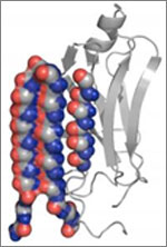
Das focuses on the nucleic acid known as RNA, which, in addition to forming part of the ribosome, plays many roles in the body. As is the case for most proteins, RNA folds into a 3-D shape that enables it to work properly.

Das is now the head of a lab at Stanford University that unravels how the structure and folding of RNA drives its function. He has taken a unique approach to uncovering the rules behind nucleic acid folding: harnessing the wisdom of the crowd.

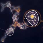
Together with his collaborator, Adrien Treuille of Carnegie Mellon University, Das created an online, multiplayer video game called EteRNA Exit icon. More than a mere game, it does far more than entertain. With its tagline “Played by Humans, Scored by Nature,” it’s upending how scientists approach RNA structure discovery and design.

Das’ Findings

Treuille and Das launched EteRNA after working on another computer game called Foldit, which lets participants play with complex protein folding questions. Like Foldit, EteRNA asks players to assemble, twist and revise structures—this time of RNA—onscreen.

But EteRNA takes things a step further. Unlike Foldit, where the rewards are only game points, the winners of each round of EteRNA actually get to have their RNA designs synthesized in a wet lab at Stanford. Das and his colleagues then post the results—which designs resulted in a successful, functional RNAs and which didn’t—back online for the players to learn from.

In a paper published in the Proceedings of the National Academy of Sciences Exit icon, Das and his colleagues showed how effective this approach could be. The collective effort of the EteRNA participants—which now number over 100,000—was better and faster than several established computer programs at solving RNA design problems, and even came up with successful new structural rules never before proposed by scientists or computers.

“What was surprising to me was their speed,” said Das. “I had just assumed that it would take a year or so before players were really able to analyze experimental data, make conclusions and come up with robust rules. But it was one of the really shocking moments of my life when, about 2 months in, we plotted the performance of players against computers and they were out-designing the computers.”

“As far as I can tell, none of the top players are academic scientists,” he added. “But if you talk to them, the first thing they’ll tell you is not how many points they have in the game but how many times they’ve had a design synthesized. They’re just excited about seeing whether or not their hypotheses were correct or falsified. So I think the top players truly are scientists—just not academic ones. They get a huge kick out of the scientific method, and they’re good at it.”

To capture lessons learned through the crowd-sourcing approach, Das and his colleagues incorporated successful rules and features into a new algorithm for RNA structure discovery, called EteRNABot, which has performed better than older computer algorithms.

“We thought that maybe the players would react badly [to EteRNABot], that they would think they were going to be automated out of existence,” said Das. “But, as it turned out, it was exciting for them to have their old ideas put into an algorithm so they could move on to the next problems.”

You can try EteRNA for yourself at http://eternagame.org Exit icon. Das and Treuille are always looking for new players and soliciting feedback.

Knowing Networks

1 comment
Artist's rendition of a network diagram. Credit: Allison Kudla, Institute for Systems Biology.
Artist’s rendition of a network diagram. Credit: Allison Kudla, Institute for Systems Biology.

Networks—both real and virtual—are everywhere, from our social media circles to the power grid that delivers electricity. The interactions of genes, proteins and other molecules in a cell are examples of networks, too.

Scientists working in a field called systems biology study and chart living networks to learn how the individual parts work together to make a functioning whole and what happens when these complex, dynamic systems go awry. For example, the network diagram here depicts yeast cells (superimposed circles) and the biochemical “chatter” between them (lines) that tells the cells to gather together in clumps. This clumping helps them survive stressful conditions like a shortage of nutrients.

Network diagrams provide more than just hub-and-spoke pictures. They can yield information that helps us better understand—and potentially influence—complex phenomena that affect our health.

Read more about network analysis and systems biology in this Inside Life Science article.