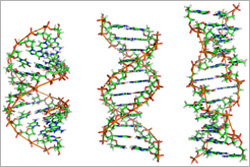
DNA researcher Rosalind Franklin first described an unusual form of DNA called the A-form in the early 1950s (Franklin, who died in 1958, would have turned 95 next month). New research on a heat- and acid-loving virus has revealed surprising information about this DNA form, which is one of three known forms of DNA: A, B and Z.
“Many people have felt that this A-form of DNA is only found in the laboratory under very non-biological conditions, when DNA is dehydrated or dry,” says Edward Egelman in a University of Virginia news release about the recent study. But considered with earlier studies on bacteria by other researchers, the new findings suggest that the A-form “appears to be a general mechanism in biology for protecting DNA.”
A new study suggests that an antibiotic regimen half as long as the standard course could be just as effective in treating intra-abdominal infections and preventing sepsis. Credit: Stock image.
When treating infections, the most critical actions are to quash the infection at its site of origin and prevent it from spreading. If allowed to spread to the bloodstream, an infection could result in body-wide inflammation known as sepsis that can cause organ failure and death.
Intra-abdominal infections, most often caused by gut bacteria, can lead to painful inflammation and present a high risk for sepsis. These infections, which include appendicitis, are some of the most common illnesses around the world.
A standard treatment regimen includes surgically removing the original infection and then prescribing antibiotics to keep the infection from coming back and to prevent sepsis. Currently, doctors administer antibiotics until 2 days after the symptoms disappear, for a total of up to 2 weeks.
Like many other researchers, University of Virginia’s Robert Sawyer wondered if treating intra-abdominal infections with shorter antibiotic courses could be just as effective as the standard treatment. To find out, he and a team of researchers from around the country designed the Study to Optimize Peritoneal Infection Therapy (STOP-IT). Continue reading “Preventing Sepsis in Half the Time”
We asked the heads of our scientific divisions to tell us about some of the big questions in fundamental biomedical science that researchers are investigating with NIGMS support. This article is the first in an occasional series that will explore these questions and explain how pursuing the answers could advance understanding of important biological processes.
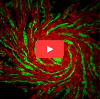
This video shows different strains of amoeba cells in red and green. As cells move toward one another, they use two sets of proteins to recognize others from the same strain. When close relatives meet, their proteins match and the cells join together to form a multicellular structure. When cells from different strains meet, their proteins don’t match, so they can’t aggregate. Credit: Shigenori Hirose, Baylor College of Medicine.
Cells are faced with many decisions: When’s the best time to produce a new protein? To grow and split into two? To treat another cell as an invader? Scientists are working to understand how cells make these and many other decisions, and how these decisions contribute to health and disease.
An active area of research on cell decisions focuses on allorecognition, the ability of an organism to distinguish its own cells from those of another. Immune cells use a system called the major histocompatibility complex (MHC) to identify which cells belong to the body and which are foreign. The particular set of MHC proteins on the outer surface of a cell helps immune cells decide whether it does not belong and should be attacked.
Last month, we shared some facts about the microbes that inhabit us. Here’s another: From head to toe, our skin bacteria coexist with chemicals in hygiene products, fibers from clothes and proteins shed by dead or dying skin cells.
These images highlight the complex composition of our body’s largest organ. They show the association between microbial diversity (top images) and skin chemistry (middle images). The different colors note the abundance of a certain bacterium or molecule—red is high, and blue is low. The skin maps remind NIH Director Francis Collins of a 60’s rock album cover.
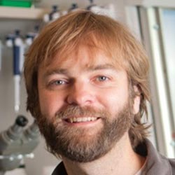
Nels Elde, Ph.D. Fields: Evolutionary genetics, virology, microbiology, cell biology Works at: University of Utah, Salt Lake City When not in the lab, he’s: Gardening, supervising pets, procuring firewood Hobbies: Canoeing, skiing, participating in facial hair competitions
“I really look at my job as an adventure,” says Nels Elde. “The ability to follow your nose through different fields is what motivates me.”
Elde has used that approach to weave evolutionary genetics, bacteriology, virology, genomics and cell biology into his work. While a graduate student at the University of Chicago and postdoctoral researcher at the Fred Hutchinson Cancer Research Center in Seattle, he became interested in how interactions between pathogens (like viruses and bacteria) and their hosts (like humans) drive the evolution of both parties. He now works in Salt Lake City, where, as an avid outdoorsman, he draws inspiration from the wild landscape.
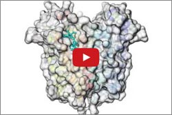
Animated structural model of TSPO. Credit: Michigan State University.
Mitochondria have proteins that span their membranes to control the flow of messages and materials moving into and out of the organelle. One way scientists can learn more about how membrane proteins function—and how medicines might interact with them—is to determine their structures. But for a variety of reasons, obtaining the structures has been notoriously difficult.
Two structural studies have now shed light on the mysterious mitochondrial membrane protein TSPO. This protein plays a key role in transporting cholesterol and drugs into the cell’s mitochondria. While here, the cholesterol is converted to steroid hormones that are essential for numerous bodily functions. Although many researchers have been studying TSPO since the 1990s, they’ve remained uncertain about its mechanisms and how it truly functions. Continue reading “Structural Studies Demystify Membrane Protein”
Trillions of microorganisms inhabit us—inside and out. Scientists are surveying these microbial metropolises to learn more about their role in health. Microbiologists Darren Sledjeski of NIGMS and Andrew Goodman of Yale University share a few details of what researchers have learned so far.
Researchers are surveying the microbes that inhabit us to learn more about their role in health. Credit: Andrew Goodman, Yale University.
The majority of the microbes that inhabit us are bacteria. The rest of the microbial menagerie is fungi and viruses, including ones that infect the bacteria! Collectively, our resident microorganisms are referred to as the human microbiota, and their genomes are called the human microbiome.
Our bodies harbor more bacterial cells than human ones. Even so, the microbiota accounts for less than 3 percent of a person’s body mass. That’s because our cells are up to 10,000 times bigger in volume than bacterial cells.
Your collection of bacteria has more genes than you do. Scientists estimate that the genomes of gut bacteria contain 100-fold or more genes than our own genomes. For this reason, the human microbiome is sometimes called our second genome.
Most of our microbes are harmless, and some are helpful. For example, harmless microbes on the skin keep infectious microbes from occupying that space. Microbes in the colon break down lactose and other complex carbohydrates that our bodies can’t naturally digest.
Different microbes occupy different parts of the body. Some skin bacteria prefer the oily nooks near the nose, while others like the dry terrain of the forearm. Bacteria don’t all fare well in the same environment and have adapted to live in certain niches. The NIGMS Findings Magazine article Body Bacteria: Exploring the Skin’s Microbial Metropolis shows what types of bacteria colonize where.
Are we more microbial than human? Richard Losick, a microbiologist at Harvard University, explores that question in this video lecture produced by iBiology.
Each person’s microbiota is unique. The demographics of microbiota differ among individuals. Diet is one reason. Also, while a type of microbe might be part of one person’s normal microbial flora, it might not be part of another’s, and could potentially make that person sick.
Host-microbial interactions are universal. Microbial communities may vary from person to person, but everyone’s got them, including other creatures. For this reason, researchers can use model organisms to tease apart the complexities of host-microbial interactions and develop broad principles for understanding them. The mouse is the most widely used animal model for microbiome studies.
The role of microbiota in our health isn’t entirely clear. While it’s now well accepted that the microbial communities that inhabit us are actively involved in a range of conditions—from asthma to obesity—research studies have not yet pinpointed why or how. In other words, the results may suggest that the presence of a bacterial community is associated with a disease, but they don’t show cause and effect.
Most of our microbes have not been grown in the lab. Microbes require a certain mix of nutrients and other microbes to survive, making it challenging to replicate their natural environments in a petri dish. New culturing techniques are enabling scientists to study previously uncultivated microbes.
The impact of probiotic and prebiotic products isn’t clear. Fundamental knowledge gaps remain regarding how these products may work and what effects they might have on host-microbial interactions. A new NIH effort to stimulate research in this area is under way.
There’s even more we don’t know! Additional areas of research include studying the functions of microbial genes and the effects of gut microbes on medicines. The more we learn from these and other studies, the more we’ll understand how our normal microbiota interacts with us and how to apply that knowledge to promote our health.
NIGMS’ Bob Lees answers questions about green chemistry. Credit: National Institute of General Medical Sciences.
Chemists funded by NIGMS are working to develop “greener” processes for discovering, developing and manufacturing medicines and other molecules with therapeutic potential, as well as compounds used in biomedical research. One of our scientific experts, organic chemist Bob Lees, recently spoke to me about some of these efforts.
What is green chemistry?
Green chemistry is the design of chemical processes and products that are more environmentally friendly. Among the 12 guiding principles of green chemistry are producing less waste, including fewer toxic byproducts; using more sustainable (renewable) or biodegradable materials; and saving energy.
Cells are the ultimate smart material. They can sense the demands being placed on them during critical life processes and then respond by strengthening, remodeling or self-repairing, for instance. To do this, cells use “mechanosensory” systems similar to the cruise control that lets a car’s engine adjust its power output when going up or down hills.
Researchers are uncovering new details on cells’ molecular cruise control systems. By learning more about the inner workings of these systems, scientists hope ultimately to devise ways to tinker with them for therapeutic purposes.
Cell Fusion
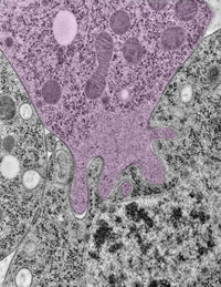
To examine how cells fine-tune their architecture and force output during the merging or fusion of cells, Elizabeth Chen and Douglas Robinson of Johns Hopkins University teamed up with Daniel Fletcher of the University of California, Berkeley. Cell fusion is critical to many developmental and physiological processes, including fertilization, placenta formation, immune response, and skeletal muscle development and regeneration.
Fingerlike protrusions of one cell (pink) invade another cell prior to cell fusion. Credit: Shuo Li. Used with permission from Developmental Cell.
Using the fruit fly Drosophila melanogaster as a model system, Chen’s research group previously found that when two muscle cells merge during muscle development, fingerlike protrusions of one cell invade the territory of the other cell to promote fusion. In the new study, led by Chen, the researchers showed that cell fusion depends on the ability of the “receiving” cell to put up resistance against the invading cells.
In fusing fruit fly cells, the scientists saw that in areas where the invading cells drilled in, the receiving cells quickly stiffened their cell skeletons, effectively pushing back. This mechanosensory response allows the outer membranes of the two cells to be pushed together and later fuse, Chen explains.
The team then explored the mechanisms underlying the stiffening response. They found that a protein called myosin II, which is part of the cell skeleton, senses the pushing force from the invading cell. Myosin II swarms to the fusion site and binds with fibers just beneath the cell membrane to put up the necessary resistance.
Hunting for the cause of a disease can be like tracing a river back to its many sources. Myriad factors, large and small, may contribute to a condition. One approach to the search focuses on the massive amounts of genomic and other biological data that scientists are gathering in the course of their studies. To examine this data and look for meaningful patterns and other clues, scientists turn to bioinformatics, a field focused on the development of analytical methods and software tools.
Here are a few examples of how National Institutes of Health-funded scientists are using bioinformatics to dig deeply into data and learn more about the development of diseases, including Huntington’s, preeclampsia and asthma.
Huntington’s Disease
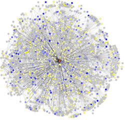
Researchers have mapped a network of 2,141 proteins that all interact either directly or through one other protein with huntingtin (red), the protein associated with Huntington’s disease. Credit: Cendrine Tourette, Buck Institute for Research on Aging, J Biol Chem 2014 Mar 7;289(10):6709-26.
The cause of Huntington’s disease, a degenerative neurological disorder with no known cure, may appear simple. It begins with a change in a single gene that alters the shape and functioning of the huntingtin protein. But this protein, whether in its normal or altered form, does not act alone. It interacts with other proteins, which in turn interact with others.
A research team led by Robert Hughes of the Buck Institute for Research on Aging set out to understand how this ripple effect contributes to the breakdown in normal cellular function associated with Huntington’s disease. The scientists used experimental and computational approaches to map a network of 2,141 proteins that interact with the huntingtin protein either directly or through one other protein. They found that many of these proteins were involved in cell movement and intercellular communication. Understanding how the huntingtin protein leads to mistakes in these cellular processes could help scientists pursue new approaches to developing treatments. Continue reading “Digging Deeply Into Data for the Causes of Disease”