Capitalizing on Cellular Conversations

0 comments
Fat cells
Fat cells such as these listen for incoming signals like FGF21, which tells them to burn more fat. Credit: David Gregory and Debbie Marshall. All rights reserved by Wellcome Images.

Living things are chatty creatures. Even when they’re not making actual sounds, organisms constantly communicate using chemical signals that course through their systems. In multicellular organisms like people, brain cells might call, “I’m in trouble!” signaling others to help mount a protective response. Single-celled organisms like bacteria may broadcast, “We have to stick together to survive!” so they can coordinate certain activities that they can’t carry out solo. In addition to sending out signals, cells have to receive information. To help them do this, they use molecular “ears” called receptors on their surfaces. When a chemical messenger attaches to a receptor, it tells the cell what’s going on and causes a response.

Scientists are following the dialogue, learning how cell signals affect health and disease. They’re also starting to take part in the cellular conversations, inserting their own comments with the goal of developing therapies that set a diseased system right.

Continue reading this new Inside Life Science article

Nanoparticles Developed to Stick to Damaged Blood Vessels, Deliver Drugs

0 comments
Artery with fat deposits and a formed clot. Credit: Stock image.
Artery with fat deposits and a formed clot. Credit: Stock image. View larger image

Heart disease is the leading cause of death for both men and women in the United States, according to the Centers for Disease Control and Prevention. One treatment challenge is developing non-invasive ways to direct medication to damaged or clogged arteries, which can block blood flow and increase the risk for heart attack and stroke. A team led by Naren Vyavahare at Clemson University has engineered extremely tiny particles—nanoparticles—that offer a promising step forward.

Healthy arteries have elastic fibers that make the arteries flexible. But, in most cardiovascular diseases, the fibers get damaged. The new nanoparticles, which can deliver drugs, attach only to damaged fibers and could enable site-specific drug delivery to minimize off-target side effects. The nanoparticles also allow drugs to be released over longer periods of time, potentially increasing the drugs’ effectiveness. The new biomaterial was tested in rodent models for studying vascular disease, so it is still in the early stages of development.

This work also was funded by NIH’s National Heart, Lung, and Blood Institute.

Learning More About Our Partners in Digestion

0 comments
Bacteroides ovatus
Bacteroides ovatus. Credit: Eric Martens, University of Michigan Medical School.

After eating, we don’t do all the work of digestion on our own. Trillions of gut bacteria help us break food down into the simple building blocks our cells need to function. New research from an international team co-led by Eric Martens of the University of Michigan Medical School has uncovered how a strain of beneficial gut bacteria, Bacteroides ovatus, digests complex carbohydrates called xyloglucans that are found in fruits and vegetables. The researchers traced the microorganism’s digestive ability to a single piece of the genome. They also examined a publicly available set of genomic data, which included information from both humans and their resident bacteria, and found that more than 90 percent of 250 adults harbored at least one Bacteroides strain with xyloglucan-digesting capabilities. These results underscore the importance of the bacteria to human health and nutrition.

This work also was funded by the National Institute of Diabetes and Digestive and Kidney Diseases.

Learn more:
University of Michigan News Release
University of Michigan Host Microbiome Initiative
Gut Reactions and Other Findings About Our Resident Microbes from Inside Life Science
Body Bacteria from Findings Magazine

Anti-Clotting Drugs: The Next Generation

1 comment
Form of heparin
Scientists created a tailor-made form of the anti-clotting drug heparin that offers several advantages.
View larger image

The low molecular weight (LMW) form of the drug heparin is commonly used to prevent unwanted blood clots that can lead to heart attacks and strokes. It’s usually derived from pig intestines and normally cleared from the human body by the kidneys. In individuals with impaired kidney function, the drug can build up in the circulation and cause excessive bleeding. Impurities and the risk of contamination are also concerns with pig-derived heparin.

Now, Robert Linhardt of Rensselaer Polytechnic Institute and Jian Liu of the University of North Carolina at Chapel Hill have created a synthetic, tailor-made form of LMW heparin that offers several advantages over the animal-derived version, including alleviating the risk of contamination from natural sources. Studies in the test tube and in mice showed that the activity of this customized heparin molecule is easily reversible in cases of overdose or uncontrolled bleeding. And, since it is cleared from the body by the liver rather than the kidneys, this form of heparin would be safer for people with impaired kidney function. Additional research, including testing in humans, will be needed before this new version of LMW heparin can be considered for medical use.

This work also was funded by NIH’s National Heart, Lung, and Blood Institute.

Learn more:

Rensselaer Polytechnic Institute News Release Exit icon
University of North Carolina at Chapel Hill News Release
Linhardt Labs
Liu Lab

Resetting Our Clocks: New Details About How the Body Tells Time

0 comments
VIP in time-keeping brain cells
Boosting doses of a molecule called VIP (green) in time-keeping brain cells (blue) helped mice adjust quickly to major shifts in light-dark cycles. Credit: Cristina Mazuski in the lab of Erik Herzog, Washington University in St. Louis.

Springing clocks forward by an hour this Sunday, traveling across time zones, staring at a computer screen late at night or working the third shift are just a few examples of activities that can disrupt our daily, or circadian, rhythms. These roughly 24-hour cycles influence our physiology and behavior, and they’re driven by our body’s network of tiny timekeepers. If our daily routines fall out of sync with our body clocks, sleep, metabolic and other disorders can result.

Researchers funded by the National Institutes of Health have spent decades piecing together the molecular mechanisms of our biological clocks. Now, they’re building on that basic knowledge to better understand the intricate relationship among these clocks, circadian rhythms and physiology—and ultimately, find ways to manipulate the moving parts to improve our modern-day lives.

Continue reading this new Inside Life Science article

Meet Ravi Iyengar

0 comments
Ravi Iyengar
Ravi Iyengar
Fields: Systems pharmacology and systems biology
Works at: Mount Sinai School of Medicine, New York, NY
Favorite sports team: Yankees
Favorite subject in high school: Math
Recently read book: The Signal and the Noise by Nate Silver
Credit: Pedro Martinez, Systems Biology Center New York

Ravi Iyengar, a professor at Mount Sinai School of Medicine, stood in an empty lecture hall, primed to tell thousands of students about systems biology, a holistic approach to studying fundamental life processes. To prepare for this moment, he had spent 4 months reading hundreds of scientific papers and distilling the research into understandable nuggets. But that day, his only student was a videographer.

Together, they recorded 15 different lectures about systems biology—many related to Iyengar’s own research—that thousands of people would stream or download as part of a MOOC, or massive open online course.

Trained in biochemistry, Iyengar built his research career around studying molecules and developing a list of all the parts that help nerve, kidney and skin cells to function. As he obtained more information, he realized he needed to know how all the components worked together. To achieve this comprehensive understanding, Iyengar turned to computational techniques and mathematical analyses—cornerstones of systems biology.

For more than a decade, he has been using and developing systems biology approaches to explore a range of biomedical questions, from very basic to translational ones with immediate relevance to human health.

Iyengar’s Findings

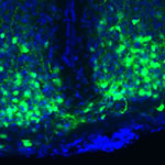
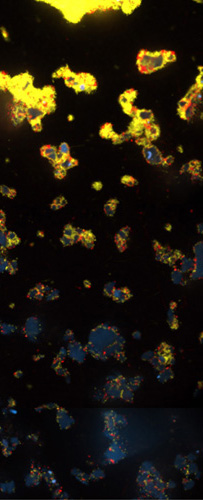
In his earlier work, Iyengar used mathematical analyses to show that molecules within cells connect with one another to form switches that produce cellular memory. This may allow, for instance, an immune cell to remember a foreign object and secrete an antibody. In recent work, he and his team developed a mathematical model showing that the shape of a cell influences the flow of information across the membrane, possibly contributing to disease states and offering a way to study and identify them under the microscope. In another study, they analyzed a database of drug side effects to find combinations of medications that produce fewer adverse reactions and then created a cell biology interaction network that explains why a certain drug pair had this beneficial outcome. The approach could point to other combinations of FDA-approved drugs that reduce serious side effects and thereby guide clinical practice.

“Systems biology is a powerful way to explore important biological and medical questions, and it’s relevant to many fields of science,” said Iyengar. But he added that the majority of educational institutions, including liberal arts and community colleges, don’t have systems biology courses. So, Iyengar teamed with colleagues to create a series of MOOCs.

The first course, offered last summer and taught by Iyengar, presented all the facets of systems biology. The syllabus included lessons on genomics and bioinformatics, fields that have contributed to systems biology; gathering and integrating data; and the use of modeling in drug development.

“My goal was for the students to get the general gestalt of systems biology,” explained Iyengar, who directs an NIH-funded center focused on the systems-level study of medicine and therapeutics.

In total, more than 12,000 participants watched at least one video lecture, 3,000 submitted one or more of the weekly quizzes and 1,800 took a mid-term or final exam. The online discussions forum included nearly 400 topics with about 5,000 posts. The students, most enrolled in a graduate program or working full-time, had some training in the biological, biomedical, computer and information sciences.

“The stats tell me that many people are in fields adjacent to systems biology and don’t have access to more traditional systems biology courses,” concluded Iyengar. “Through the MOOC, we can reach them in a substantial way.”

The second course, which covers network analysis, wrapped up in early December, and the third course, which covers dynamical modeling methods, began in January. Iyengar plans to offer the intro course again in late March.

Learn more:
MOOC Systems Biology Courses Exit icon

Cool Image: Denying Microbial Moochers

0 comments
V. cholerae and V. cholerae


Productive V. cholerae (yellow) and exploitive V. cholerae (red). Credit: Carey Nadell, Princeton University.

What looks like an abstract oil painting is actually an image of several cholera-causing V. cholerae bacterial communities. These communities, called biofilms, include productive and exploitive microbial members. The industrious bacteria (yellow) tend to thrive in denser biofilms (top) while moochers (red) thrive in weaker biofilms (bottom). In an effort to understand this phenomenon, Princeton University researchers led by Bonnie Bassler discovered two ways the freeloaders are denied food. They found that some V. cholerae cover themselves with a thick coating to prevent nutritious carbon- and nitrogen-containing molecules from drifting over to the scroungers. In addition, the natural flow of fluids over biofilms can wash away any leftovers. Encouraging such bacterial fairness could boost the efficient breakdown of organic materials into useful products, such as biofuels. On the other hand, counteracting it could lead to better treatment of illnesses, like cholera, by starving the most productive bacteria and thereby weakening the infection.

Learn more:
Princeton University News Release Exit icon


Visualizing Vessels

0 comments
Blood vessels in a mouse retina
Blood vessels in a mouse retina visualized using cutting-edge imaging technology. Credit: Tom Deerinck and Mark Ellisman, NCMIR.

For poets and lovers, the eyes are the windows of the soul. For scientists and doctors, blood vessels at the back of the eye are windows into many diseases.

Blood vessel abnormalities can indicate a variety of serious conditions such as atherosclerosis (hardening of the arteries), heart attacks and strokes. But most vessels are buried beneath skin and other tissues, making them difficult to examine without surgery.

There’s one exception—in the eye. Unlike anywhere else in the body, larger vessels on the retina at the back of the eye are directly visible through the pupil, requiring essentially only light and magnifying lenses to view.

These vessels are used to diagnose glaucoma and diabetic eye disease. Because they display characteristic changes in people with high blood pressure, some researchers hope retinal vessels might one day help predict an impending stroke, congestive heart failure or other diseases stemming from dangerously high blood pressure.

The medical importance of retinal vessels piqued the interest of scientists funded by the National Institutes of Health at the National Center for Microscopy and Imaging Research (NCMIR) at the University of California, San Diego, who captured this micrograph image of mouse retinal vessels.

Continue reading this new Inside Life Science article

Dendrites Show Ability to Regenerate After Injury

0 comments
Dendrites
Cutting off the dendrites from nerve cells in fruit flies revealed that they can regenerate. Credit: Melissa Rolls, Penn State University.

When a bone breaks, it might slice axons—the part of nerve cells that sends information to other cells—and potentially cause loss of mobility or feeling. Prior research had shown that a damaged nerve cell could repair such an injury through the regrowth of axons. Scientists at Penn State University wondered if dendrites—the part of nerve cells that receive information from other nerve cells—could also regenerate. To find out, Melissa Rolls and her team cut off the dendrites from nerve cells in fruit flies. Instead of dying, as was expected, the cells regrew dendrites. The research also revealed that dendrite regeneration happens independently of axon regeneration, leading investigators to believe there are two separate regeneration pathways: one for axons and one for dendrites. Learning more about this new dendrite regrowth pathway might one day lead to new approaches for healing injured nerve cells, including those damaged after a stroke.

Learn more:
Penn State News Release Exit icon
Rolls Lab

Epilepsy Drug Improves Health in Animal Model of Obesity

2 comments
Liver cells of obese mice treated with valproic acid (right) and untreated obese mice (left).
Liver cells (magenta) of obese mice treated with valproic acid (right) had much less fat accumulation (white) than those of untreated obese mice (left). Credit: Lindsay B. Avery and Namandjé N. Bumpus, Johns Hopkins University. View larger image

With more than 90 million Americans affected by obesity, developing medications to help combat weight gain and its associated diseases has become a priority. In a study using obese mice, a team led by Namandjé Bumpus of Johns Hopkins University recently showed that a commonly prescribed epilepsy drug, valproic acid, reduced fat accumulation in the liver and lowered elevated blood sugar levels like those associated with type 2 diabetes. Body weight also stabilized in mice given the drug, whereas untreated mice continued to gain weight. Additional experiments in mouse and human liver cells suggested that the byproducts of valproic acid produced as the body breaks down the drug, rather than valproic acid itself, were responsible for the observed effects. These byproducts achieved the same effects in cells at one-fortieth the concentration of valproic acid, making them promising candidates for further drug development.

Learn more:
Johns Hopkins University News Release Exit icon
Bumpus Laboratory