Tag: Bacteria

New Models Predict Where E. coli Strains Will Thrive

0 comments
Illustration of E. coli. Credit: Janet Iwasa, University of Utah.
Illustration of E. coli. Credit: Janet Iwasa, University of Utah (image available under a Creative Commons Attribution-NonCommercial-ShareAlike license Exit icon). View larger image

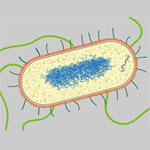
Like plants and animals, different types of E. coli thrive in different environments. Now, scientists can even predict which environments—such as the bladder, stomach or blood—are most amenable to the growth of various strains, including pathogenic ones. A research team led by Bernhard Palsson Exit icon of the University of California, San Diego, accomplished this by using genome data to reconstruct the metabolic networks of 55 E. coli strains. The metabolic models, which identify differences in the ability to manufacture certain compounds and break down various nutrients, shed light on how certain E. coli strains become pathogenic and how to potentially control them. One approach could be depriving the deadly strains of the nutrients they need to survive in their niches. The researchers plan to use their new method to study other bacteria, such as those that cause staph infections.

This work also was funded by NIH’s National Cancer Institute.

Learn more:
University of California, San Diego News Release

Cool Video: How Bee Venom Toxin Kills Cells

0 comments
Credit: Huey Huang, Rice University.

Credit: Huey Huang, Rice University.

A new video, starring the toxin in bee venom, might help scientists design new drugs to combat bacterial infections. The video, by Rice University biophysicist Huey Huang Exit icon, condenses 6.5 minutes into less than a minute to show how the toxin, called melittin, destroys an animal or bacterial cell.

What looks like a red balloon is an artificial cell filled with red dye. Melittin molecules are colored green and float on the cell’s surface like twigs on a pond. As melittin accumulates on the cell’s membrane, the membrane expands to accommodate it. In the video, the membrane stretches into a column on the left.

When melittin levels reach a critical threshold, countless pinhole leaks burst open in the membrane. The cell’s vital fluids—red dye in the video—leak out through these pores. Within minutes, the cell collapses.

Many organisms use such a pore-forming technique to kill attacking bacterial cells. This research reveals molecular-level details of the strategy, bringing pharmaceutical scientists a step closer to harnessing it in the design of new antibiotics.

How Some Bacteria Colonize the Gut

1 comment
A section of mouse colon with gut bacteria (center, in green). Credit: S. Melanie Lee, Caltech; Zbigniew Mikulski and Klaus Ley, La Jolla Institute for Allergy and Immunology.
A section of mouse colon with gut bacteria (center, in green) residing within a protective pocket. Credit: S. Melanie Lee, Caltech; Zbigniew Mikulski and Klaus Ley, La Jolla Institute for Allergy and Immunology.

Have you ever felt that your gut was trying to tell you something? The guts of germ-free mice have told scientists a few new things about our resident microorganisms. By studying a genus of bacteria called Bacteriodes that live in the gastrointestinal tract, Sarkis Mazmanian of the California Institute of Technology discovered how Bacteriodes species stake their claim in a mouse’s gut. Mazmanian and his colleagues introduced different species of Bacteriodes into germ-free mice to learn how the microbes competed and found that most of them peacefully co-existed. However, when microbes of a species that was already present were introduced, they couldn’t take up residence. Further investigation revealed that Bacteriodes species, due to a set of specific genes, can live in tiny pockets or “crypts” of the colon, where they are sheltered from antibiotics and infectious microbes passing through. Understanding how these microorganisms colonize could help devise ways to correct for abnormal changes in bacterial communities that are associated with disorders like inflammatory bowel disease.

This work also was funded by NIH’s National Institute of Diabetes and Digestive and Kidney Diseases.

Learn more:

California Institute of Technology News Release
Mazmanian Lab
Mazmanian Video Interview Exit icon