Tag: Infectious Diseases

What Zombie Ants Are Teaching Us About Fungal Infections: Q & A with Entomologists David Hughes and Maridel Fredericksen

0 comments

I can still remember that giddy feeling I had seven years ago, when I first read about the “zombie ant.” The story was gruesome and fascinating, and it was everywhere. Even friends and family who aren’t so interested in science knew the basics: in a tropical forest somewhere there’s a fungus that infects an ant and somehow takes control of the ant’s brain, forcing it to leave its colony, crawl up a big leaf, bite down and wait for the sweet relief of death. A grotesque stalk then sprouts from the poor creature’s head, from which fungal spores rain down to infect a new batch of ants.

A fungal fruiting body erupts through the head of a carpenter ant infected by a parasitic fungus in Thailand. Credit: David Hughes, Penn State University.

The problem is, it doesn’t happen quite like that. David Hughes, the Penn State University entomologist who reported his extensive field observations of the fungus/ant interactions in BMC Ecology Exit icon, which caused much excitement back in 2011, has continued to study the fungus, Ophiocordyceps unliateralis, and its carpenter ant host, Camponotus leonardi.

In late 2017, Hughes and his colleagues published an article in PNAS Exit icon in which they used sophisticated microscopy and image-processing techniques to describe in great detail how the fungus invades various parts of the ant’s body including muscles in its legs and head.

Although Hughes’s earlier BMC Ecology paper showed fungus in the head of an ant, the new study reveals that the fungus never actually enters the brain.

To me, the new finding somehow made the fungus’ control over the ant even more baffling. What exactly was going on?

To find out, I spoke with Hughes and his graduate student Maridel Fredericksen.

Continue reading “What Zombie Ants Are Teaching Us About Fungal Infections: Q & A with Entomologists David Hughes and Maridel Fredericksen”

Flipping the Switch on Controlling Disease-Carrying Insects

4 comments
Illustration of some of the jobs that the ER performs in the cell.

This image shows a mosquito egg. Wolbachia bacteria, which infect many species of insects including mosquitos, move from one generation to the next inside insect eggs. Credit: Wikimedia Commons, Mogana Das Murtey and Patchamuthu Ramasamy, Universiti Sains, Malaysia.

Suppressing insects that spread disease is an essential public health effort, and scientists are testing a possible new tool to use in this challenging arena. They’re harnessing a microbe capable of controlling insects’ reproductive processes.

The microbes, called Wolbachia, live inside the cells of about two-thirds of insect species worldwide, and they can manipulate the host’s reproductive cells in ways that boost their own survival. Scientists think they can use Wolbachia’s methods to reduce populations of insects that spread disease among humans.

A Switch to Control Fertility

Wolbachia have evolved complex ways to control insect reproduction so as to infect increasing numbers of an insect species—such as those prolific disease-spreaders, mosquitos. One method Wolbachia uses is called cytoplasmic incompatibility, or CI. The end result of CI, basically, is that the sperm of infected male insects cause sterility in uninfected females.

Wolbachia that have infected male insects can insert proteins that produce a kind of infertility switch into the host’s sperm. When the sperm later fuses with an egg from an uninfected female, the switch is triggered and renders the egg sterile. If the female is already infected, her eggs will contain Wolbachia, which can turn off the switch and allow the egg to develop. This trick ensures that more Wolbachia-infected insects will survive and continue to reproduce, while uninfected ones will be less successful.

Already, some states Exit icon and countries Exit icon are releasing Wolbachia-infected male mosquitoes into wild mosquito populations that carry disease-causing viruses to test this strategy for insect control. Males carrying a Wolbachia strain that strongly induces infertility in uninfected females should reduce the numbers of mosquito eggs that mature, leading to fewer mosquitos. Continue reading “Flipping the Switch on Controlling Disease-Carrying Insects”

New Technology May Help Reduce Serious and Costly Post-Surgical Infections—Using Nothing but Air

7 comments

According to a recent estimate, implant infections following hip and knee replacement surgeries in the U.S. may number 65,000 by 2020, with the associated healthcare costs exceeding $1 billion. A new small, high-tech device could have a significant impact on improving health outcomes and reducing cost for these types of surgeries. The device, Air Barrier System (ABS), attaches on top of the surgical drape and gently emits HEPA-filtered air over the incision site. By creating a “cocoon” of clean air, the device prevents airborne particles—including the bacteria that can cause healthcare-associated infections—from entering the wound.

Air Barrier System
The Air Barrier System creates a “cocoon” of clean air (gray area with size indicated) over a surgical site to remove airborne contaminants and reduce the risk of infection in patients who are receiving an artificial hip, a blood vessel graft, a titanium plate in the spine or other implants.

Scientists recently analyzed the effectiveness of the ABS device in a clinical study—funded by NIGMS—involving nearly 300 patients. Each patient needed an implant, such as an artificial hip, a blood vessel graft in the leg or a titanium plate in the spine. Because implant operations involve inserting foreign materials permanently into the body, they present an even higher risk of infection than many other surgeries, and implant infections can cause life-long problems.

The researchers focused on one of the most common causes of implant infections—the air in the operating room. Although operating rooms are much cleaner than almost any other non-hospital setting, it’s nearly impossible to sterilize the entire room. Instead, the scientists focused on reducing contaminants directly over the surgical site. They theorized that if the air around the wound was cleaner, the number of implant infections might go down. Continue reading “New Technology May Help Reduce Serious and Costly Post-Surgical Infections—Using Nothing but Air”

Bit by the Research Bug: Priscilla’s Growth as a Scientist

2 comments

This is the third post in a new series highlighting NIGMS’ efforts toward developing a robust, diverse and well-trained scientific workforce.

Priscilla Del Valle
Credit: Christa Reynolds.
Priscilla Del Valle
Academic Institution: The University of Texas at El Paso
Major: Microbiology
Minors: Sociology and Biomedical Engineering
Mentor: Charles Spencer
Favorite Book: The Immortal Life of Henrietta Lacks, by Rebecca Skloot
Favorite Food: Tacos
Favorite music: Pop
Hobbies: Reading and drinking coffee

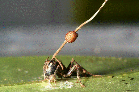
It’s not every day that you’ll hear someone say, “I learned more about parasites, and I thought, ‘This is so cool!’” But it’s also not every day that you’ll meet an undergraduate researcher like 21-year-old Priscilla Del Valle.

BUILD and the Diversity Program Consortium

The Diversity Program Consortium (DPC) aims to enhance diversity in the biomedical research workforce through improved recruitment, training and mentoring nationwide. It comprises three integrated programs—Building Infrastructure Leading to Diversity (BUILD), which implements activities at student, faculty and institutional levels; the National Research Mentoring Network (NRMN), which provides mentoring and career development opportunities for scientists at all levels; and the Coordination and Evaluation Center (CEC), which is responsible for evaluating and coordinating DPC activities.

Ten undergraduate institutions across the United States have received BUILD grants, and together, they serve a diverse population. Each BUILD site has developed a unique program intended to engage and prepare students for success in the biomedical sciences and maximize opportunities for research training and faculty development. BUILD programs include everything from curricular redesign, lab renovations, faculty training and research grants, to student career development, mentoring and research-intensive summer programs.

Del Valle’s interest in studying infectious diseases and parasites is motivating her to pursue an M.D./Ph.D. focusing on immunology and pathogenic microorganisms. Currently, Del Valle is a junior at The University of Texas at El Paso (UTEP)’s BUILDing SCHOLARS Center. BUILDing SCHOLARS, which stands for “Building Infrastructure Leading to Diversity Southwest Consortium of Health-Oriented Education Leaders and Research Scholars,” focuses on providing undergraduate students interested in the biomedical sciences with academic, financial and professional development opportunities. Del Valle is one of the first cohort of students selected to take part in this training opportunity.

BUILD scholars receive individual support through this training model, and Del Valle says she likes “the way that they [BUILDing SCHOLARS] take care of us and the workshops and opportunities that we have.”

Born in El Paso, Texas, Del Valle moved to Saltillo, Mexico, where she spent most of her childhood. Shortly after graduating from high school, she returned to El Paso to start undergraduate courses at El Paso Community College (EPCC), to pursue an M.D. Del Valle explains that in Mexico, unlike in the United States, careers in medical research are not really emphasized in the student community or in society, so she did not have firsthand experience with research.

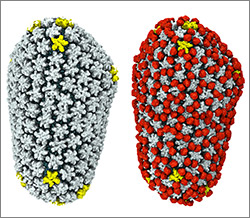
Del Valle discovered her passion for research when she was assigned a project on malaria as part of an EPCC course. She was fascinated by the parasite that causes malaria. “It impressed me how something so little could infect a person so harshly,” she says. Continue reading “Bit by the Research Bug: Priscilla’s Growth as a Scientist”

Online Virus Tracking Tool Nextstrain Wins Inaugural Open Science Prize

0 comments
Nextstrain’s analysis of the genomes from Zika virus obtained in 25 countries over the past few years. 
Credit: Trevor Bedford and Richard Neher, nextstrain.org.

Over the past decade, scientists and clinicians have eagerly deposited their burgeoning biomedical data into publicly accessible databases. However, a lack of computational tools for sharing and synthesizing the data has prevented this wealth of information from being fully utilized.

In an attempt to unleash the power of open-access data, the National Institutes of Health, in collaboration with the Howard Hughes Medical Institute and Britain’s Wellcome Trust, launched the Open Science Prize. Last week, after a multi-stage public voting process, the inaugural award was announced. The winner of the grand prize—and $230,000—is a prototype computational tool called nextstrain Exit icon that tracks the spread of emerging viruses such as Ebola and Zika. This tool could be especially valuable in revealing the transmission patterns and geographic spread of new outbreaks before vaccines are available, such as during the 2013-2016 Ebola epidemic and the current Zika epidemic.

An international team of scientists—led by NIGMS grantee Trevor Bedford of the Fred Hutchinson Cancer Research Center, Seattle, and Richard Neher Exit icon of Biozentrum at the University of Basel, Switzerland—developed nextstrain as an open-access system capable of sharing and analyzing viral genomes. The system mines viral genome sequence data that researchers have made publicly available online. nextstrain then rapidly determines the evolutionary relationships among all the viruses in its database and displays the results of its analyses on an interactive public website.

The image here shows nextstrain’s analysis of the genomes from Zika virus obtained in 25 countries over the past few years. Plotting the relatedness of these viral strains on a timeline provides investigators a sense of how the virus has spread and evolved, and which strains are genetically similar. Researchers can upload genome sequences of newly discovered viral strains—in this case Zika—and find out in short order how their new strain relates to previously discovered strains, which could potentially impact treatment decisions.

Nearly 100 interdisciplinary teams comprising 450 innovators from 45 nations competed for the Open Science Prize. More than 3,500 people from six continents voted online for the winner. Other finalists for the prize focused on brain maps, gene discovery, air-quality monitoring, neuroimaging and drug discovery.

nextstrain was funded in part by NIH under grant U54GM111274.

Student Researcher Finds New Clues About Flu with Old Data

1 comment

Do you like to find new uses for old things? Like weaving old shirts into a rug, repurposing bottles into candle holders or turning packing crates into tables? Katie Gostic, a University of California, Los Angeles (UCLA) graduate student, likes finding new uses for old data. She channeled this interest when she analyzed existing data to study whether childhood exposure to flu affects a person’s future immunity to the disease.

Katie Gostic
Gostic conducted research for the flu project during the summer of 2015 when she was visiting her boyfriend, a tropical biologist, in Alamos, Sonora, Mexico. Credit: Charlie de la Rosa.

As an undergraduate student at Princeton University, Gostic was originally pursuing a degree in engineering. Her focus shifted to biology after taking an infectious disease modeling class. Gostic’s background in math and programming allows her to take large, complex pre-existing data sets and reanalyze them using new tools and methods. The result: Information that wasn’t accessible when the data were first collected.

Now a graduate researcher in the ecology and evolutionary biology lab of James Lloyd-Smith Exit icon, Gostic studies infectious diseases. The lab builds mathematical models to investigate zoonotic diseases—diseases that animals can transmit to humans but that humans don’t frequently spread between each other. Examples include diseases caused by Leptospira, a type of bacteria that infects household pets and many other animals, and monkeypox, a virus whose transmission to humans is increasing since the eradication of smallpox. The lab also studies bird flus, a category of flu viruses that infect birds and other animals and only occasionally jump to people. A very small number of cases of human-to-human transmission of bird flus have been recorded. However, if a bird flu virus mutated in a way that allowed it to spread among humans, it could cause a pandemic. Continue reading “Student Researcher Finds New Clues About Flu with Old Data”

Viral Views: New Insights on Infection Strategies

0 comments

The following images show a few ways in which cutting-edge research tools are giving us clearer views of viruses—and possible ways to disarm them. The examples, which highlight work involving HIV and the coronavirus, were funded in part by our Biomedical Technology Research Resources program.

Uncloaking HIV’s Camouflage

HIV capsid with (right, red) and without (left) a camouflaging human protein.
HIV capsid with (right, red) and without (left) a camouflaging human protein. Credit: Juan R. Perilla, Klaus Schulten and the Theoretical and Computational Biophysics Group, University of Illinois at Urbana-Champaign.

To sneak past our immune defenses and infect human cells, HIV uses a time-honored strategy—disguise. The virus’ genome is enclosed in a protein shell called a capsid (on left) that’s easily recognized and destroyed by the human immune system. To evade this fate, the chrysalis-shaped capsid cloaks itself with a human protein known as cyclophilin A (in red, on right). Camouflaged as human, the virus gains safe passage into and through a human cell to deposit its genetic material in the nucleus and start taking control of cellular machinery.

Biomedical and technical experts teamed up to generate these HIV models at near-atomic resolution. First, structural biologists at the Pittsburgh Center for HIV Protein Interactions Exit icon used a technique called cryo-electron microscopy (cryo-EM) to get information on the shape of an HIV capsid as well as the capsid-forming proteins’ connections to each other and to cyclophilin A. Then experts at the Resource for Macromolecular Modeling and Bioinformatics fed the cryo-EM data into their visualization and simulation programs to computationally model the physical interactions among every single atom of the capsid and the cyclophilin A protein. The work revealed a previously unknown site where cyclophilin A binds to the capsid, offering new insights on the biology of HIV infection. Continue reading “Viral Views: New Insights on Infection Strategies”

El Niño Season Temperatures Linked to Dengue Epidemics

1 comment
Screen shot from a video showing dengue incidence in Southeast Asia.
Incidence of dengue fever across Southeast Asia, 1993-2010. Note increasing incidence (red) starting about June 1997, which corresponds to a period of higher temperatures driven by a strong El Niño season. At the end of the El Niño event, in January 1999, dengue incidence is much lower (green). Credit: Wilbert van Panhuis, University of Pittsburgh.

Weather forecasters are already warning about an intense El Niño season that’s expected to alter precipitation levels and temperatures worldwide. El Niño seasons, characterized by warmer Pacific Ocean water along the equator, may impact the spread of some infectious diseases transmitted by mosquitoes.

In a study published last month in the Proceedings of the National Academy of Sciences, researchers reported a link between intense dengue fever epidemics in Southeast Asia and the high temperatures that a previous El Niño weather event brought to that region.

Dengue fever, a viral infection transmitted by the Aedes mosquito, can cause life-threatening high fever, severe joint pain and bleeding. Infection rates soar every two to five years. Interested in understanding why, an international team of researchers collected and analyzed incidence reports including 3.5 million dengue fever cases across eight Southeast Asian countries spanning an 18-year period. The study is part of Project Tycho, an effort to study disease transmission dynamics by mining historical data and making that data freely available to others. Continue reading “El Niño Season Temperatures Linked to Dengue Epidemics”

Designing Drugs That Kill Invasive Fungi Without Harming Humans

0 comments
Top to bottom: Cryptococcus, Candida, Aspergillus, Pneumocystis
Invasive fungal infections kill more than 1 million people worldwide every year. Almost all of these deaths are due to fungi in one of these four groups. Credit: Centers for Disease Control and Prevention.

Invasive fungal infections—the kind that infect the bloodstream, lung and brain—are inordinately deadly. A big part of the problem is the lack of drugs that are both effective against the fungi and nontoxic to humans.

The situation might change in the future though, thanks to the work of a multidisciplinary research team led by chemist Martin Burke at the University of Illinois. For years, the team has focused on an antifungal agent called amphotericin B (AmB for short). Although impressively lethal to fungi, AmB is also notoriously toxic to human cells.

Most recently, the research team chemically modified the drug to create compounds that kill fungi, but don’t disrupt human cells. The scientists explain it all in the latest issue of Nature Chemical Biology.

Invasive fungal infections are so intractable because most antifungal drugs aren’t completely effective. Plus, fungi have a tendency to develop resistance to them. AmB is a notable exception. Isolated 50 years ago from Venezuelan dirt, AmB has evaded resistance and remains highly effective. Unfortunately, it causes side effects so debilitating that some doctors call it “ampho-terrible.” At high doses, it is fatal.

For decades, scientists believed that AmB molecules kill fungal cells by forming membrane-piercing pores, or ion channels, through which the cells’ innards leak out. Last year, Burke’s group overturned this well-established concept using evidence from nuclear magnetic resonance, chemistry and cell-based experiments. The researchers showed that AmB molecules assemble outside cells into lattice-like structures. These structures act as powerful sponges, sucking vital lipid molecules, called ergosterol, right out of the fungal cell membrane, destroying the cell. Continue reading “Designing Drugs That Kill Invasive Fungi Without Harming Humans”

Preventing Sepsis in Half the Time

0 comments
Human digestive system
A new study suggests that an antibiotic regimen half as long as the standard course could be just as effective in treating intra-abdominal infections and preventing sepsis. Credit: Stock image.

When treating infections, the most critical actions are to quash the infection at its site of origin and prevent it from spreading. If allowed to spread to the bloodstream, an infection could result in body-wide inflammation known as sepsis that can cause organ failure and death.

Intra-abdominal infections, most often caused by gut bacteria, can lead to painful inflammation and present a high risk for sepsis. These infections, which include appendicitis, are some of the most common illnesses around the world.

A standard treatment regimen includes surgically removing the original infection and then prescribing antibiotics to keep the infection from coming back and to prevent sepsis. Currently, doctors administer antibiotics until 2 days after the symptoms disappear, for a total of up to 2 weeks.

Like many other researchers, University of Virginia’s Robert Sawyer Exit icon wondered if treating intra-abdominal infections with shorter antibiotic courses could be just as effective as the standard treatment. To find out, he and a team of researchers from around the country designed the Study to Optimize Peritoneal Infection Therapy (STOP-IT). Continue reading “Preventing Sepsis in Half the Time”