Tag: Medicines

A Medicine’s Life Inside the Body

0 comments
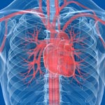
Heart
Most often, the bloodstream is the vehicle for carrying medicines throughout the body. Credit: Stock image.

Pharmacology is the scientific field that studies how the body reacts to medicines and how medicines affect the body. Scientists funded by the National Institutes of Health are interested in many aspects of pharmacology, including one called pharmacokinetics, which deals with understanding the entire cycle of a medicine’s life inside the body.

Knowing more about each of the four main stages of pharmacokinetics—absorption, distribution, metabolism and excretion—aids the design of medicines that are more effective and that produce fewer side effects.

Read more about a medicine’s life inside the body in this Inside Life Science article.

New Life for Toxic Antibiotics?

0 comments
Pills and a bottle
Researchers found that the antibiotic trovafloxacin cuts off a channel for communication between cells and interferes with a cell-death process. Credit: Stock image.

Many compounds that show promise as new antibiotics for treating bacterial infections never make it to the clinic because they turn out to be toxic to humans as well as to bacteria. A research team led by Kodi Ravichandran of the University of Virginia recently gained insights into why one such antibiotic, trovafloxacin, harms human cells. They found that the compound cuts off a channel for communication between cells, which in turn interferes with how dying cells are broken down and recycled by the body. Roughly 200 billion cells in the human body die and are replaced every day as part of a routine cleanup process, and interference in this process by trovafloxacin may have contributed to the serious liver damage seen in some patients in clinical trials of the drug. Understanding how trovafloxacin causes toxicity in people may help researchers re-engineer this and related compounds to make them safe and effective for use in fighting bacterial infections.

Learn more:
Ravichandran Lab

Nanoparticles Developed to Stick to Damaged Blood Vessels, Deliver Drugs

0 comments
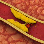
Artery with fat deposits and a formed clot. Credit: Stock image.
Artery with fat deposits and a formed clot. Credit: Stock image. View larger image

Heart disease is the leading cause of death for both men and women in the United States, according to the Centers for Disease Control and Prevention. One treatment challenge is developing non-invasive ways to direct medication to damaged or clogged arteries, which can block blood flow and increase the risk for heart attack and stroke. A team led by Naren Vyavahare at Clemson University has engineered extremely tiny particles—nanoparticles—that offer a promising step forward.

Healthy arteries have elastic fibers that make the arteries flexible. But, in most cardiovascular diseases, the fibers get damaged. The new nanoparticles, which can deliver drugs, attach only to damaged fibers and could enable site-specific drug delivery to minimize off-target side effects. The nanoparticles also allow drugs to be released over longer periods of time, potentially increasing the drugs’ effectiveness. The new biomaterial was tested in rodent models for studying vascular disease, so it is still in the early stages of development.

This work also was funded by NIH’s National Heart, Lung, and Blood Institute.

Anti-Clotting Drugs: The Next Generation

1 comment
Form of heparin
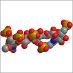
Scientists created a tailor-made form of the anti-clotting drug heparin that offers several advantages.
View larger image

The low molecular weight (LMW) form of the drug heparin is commonly used to prevent unwanted blood clots that can lead to heart attacks and strokes. It’s usually derived from pig intestines and normally cleared from the human body by the kidneys. In individuals with impaired kidney function, the drug can build up in the circulation and cause excessive bleeding. Impurities and the risk of contamination are also concerns with pig-derived heparin.

Now, Robert Linhardt of Rensselaer Polytechnic Institute and Jian Liu of the University of North Carolina at Chapel Hill have created a synthetic, tailor-made form of LMW heparin that offers several advantages over the animal-derived version, including alleviating the risk of contamination from natural sources. Studies in the test tube and in mice showed that the activity of this customized heparin molecule is easily reversible in cases of overdose or uncontrolled bleeding. And, since it is cleared from the body by the liver rather than the kidneys, this form of heparin would be safer for people with impaired kidney function. Additional research, including testing in humans, will be needed before this new version of LMW heparin can be considered for medical use.

This work also was funded by NIH’s National Heart, Lung, and Blood Institute.

Learn more:

Rensselaer Polytechnic Institute News Release Exit icon
University of North Carolina at Chapel Hill News Release
Linhardt Labs
Liu Lab

Meet Ravi Iyengar

0 comments
Ravi Iyengar
Ravi Iyengar
Fields: Systems pharmacology and systems biology
Works at: Mount Sinai School of Medicine, New York, NY
Favorite sports team: Yankees
Favorite subject in high school: Math
Recently read book: The Signal and the Noise by Nate Silver
Credit: Pedro Martinez, Systems Biology Center New York

Ravi Iyengar, a professor at Mount Sinai School of Medicine, stood in an empty lecture hall, primed to tell thousands of students about systems biology, a holistic approach to studying fundamental life processes. To prepare for this moment, he had spent 4 months reading hundreds of scientific papers and distilling the research into understandable nuggets. But that day, his only student was a videographer.

Together, they recorded 15 different lectures about systems biology—many related to Iyengar’s own research—that thousands of people would stream or download as part of a MOOC, or massive open online course.

Trained in biochemistry, Iyengar built his research career around studying molecules and developing a list of all the parts that help nerve, kidney and skin cells to function. As he obtained more information, he realized he needed to know how all the components worked together. To achieve this comprehensive understanding, Iyengar turned to computational techniques and mathematical analyses—cornerstones of systems biology.

For more than a decade, he has been using and developing systems biology approaches to explore a range of biomedical questions, from very basic to translational ones with immediate relevance to human health.

Iyengar’s Findings

In his earlier work, Iyengar used mathematical analyses to show that molecules within cells connect with one another to form switches that produce cellular memory. This may allow, for instance, an immune cell to remember a foreign object and secrete an antibody. In recent work, he and his team developed a mathematical model showing that the shape of a cell influences the flow of information across the membrane, possibly contributing to disease states and offering a way to study and identify them under the microscope. In another study, they analyzed a database of drug side effects to find combinations of medications that produce fewer adverse reactions and then created a cell biology interaction network that explains why a certain drug pair had this beneficial outcome. The approach could point to other combinations of FDA-approved drugs that reduce serious side effects and thereby guide clinical practice.

“Systems biology is a powerful way to explore important biological and medical questions, and it’s relevant to many fields of science,” said Iyengar. But he added that the majority of educational institutions, including liberal arts and community colleges, don’t have systems biology courses. So, Iyengar teamed with colleagues to create a series of MOOCs.

The first course, offered last summer and taught by Iyengar, presented all the facets of systems biology. The syllabus included lessons on genomics and bioinformatics, fields that have contributed to systems biology; gathering and integrating data; and the use of modeling in drug development.

“My goal was for the students to get the general gestalt of systems biology,” explained Iyengar, who directs an NIH-funded center focused on the systems-level study of medicine and therapeutics.

In total, more than 12,000 participants watched at least one video lecture, 3,000 submitted one or more of the weekly quizzes and 1,800 took a mid-term or final exam. The online discussions forum included nearly 400 topics with about 5,000 posts. The students, most enrolled in a graduate program or working full-time, had some training in the biological, biomedical, computer and information sciences.

“The stats tell me that many people are in fields adjacent to systems biology and don’t have access to more traditional systems biology courses,” concluded Iyengar. “Through the MOOC, we can reach them in a substantial way.”

The second course, which covers network analysis, wrapped up in early December, and the third course, which covers dynamical modeling methods, began in January. Iyengar plans to offer the intro course again in late March.

Learn more:
MOOC Systems Biology Courses Exit icon

Epilepsy Drug Improves Health in Animal Model of Obesity

2 comments
Liver cells of obese mice treated with valproic acid (right) and untreated obese mice (left).
Liver cells (magenta) of obese mice treated with valproic acid (right) had much less fat accumulation (white) than those of untreated obese mice (left). Credit: Lindsay B. Avery and Namandjé N. Bumpus, Johns Hopkins University. View larger image

With more than 90 million Americans affected by obesity, developing medications to help combat weight gain and its associated diseases has become a priority. In a study using obese mice, a team led by Namandjé Bumpus of Johns Hopkins University recently showed that a commonly prescribed epilepsy drug, valproic acid, reduced fat accumulation in the liver and lowered elevated blood sugar levels like those associated with type 2 diabetes. Body weight also stabilized in mice given the drug, whereas untreated mice continued to gain weight. Additional experiments in mouse and human liver cells suggested that the byproducts of valproic acid produced as the body breaks down the drug, rather than valproic acid itself, were responsible for the observed effects. These byproducts achieved the same effects in cells at one-fortieth the concentration of valproic acid, making them promising candidates for further drug development.

Learn more:
Johns Hopkins University News Release Exit icon
Bumpus Laboratory

An Experimental Contact Lens to Prevent Glaucoma-Induced Blindness

3 comments
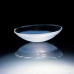
Contact lens. Credit: Peter Mallen, Massachusetts Eye and Ear Laboratory/Kohane Laboratory, Boston Children's Hospital.
An experimental contact lens design releases a glaucoma medicine at a steady rate for up to a month. Credit: Peter Mallen, Massachusetts Eye and Ear Laboratory/Kohane Laboratory, Boston Children’s Hospital.

Like a miniature donut stuffed inside a tiny pita pocket, a common glaucoma medicine held within a biomaterial ring is sandwiched inside this contact lens. In laboratory experiments, the lens, which can also correct vision, releases the eyesight-saving medication at a steady rate for up to a month. Its construction offers numerous potential clinical advantages over the standard glaucoma treatment and may have additional applications, such as delivering anti-inflammatory drugs or antibiotics to the eye. Led by Daniel Kohane and Joseph Ciolino at Harvard Medical School, the researchers who developed the lens are now gearing up to test its effectiveness in additional laboratory studies. They hope a Phase I clinical trial to evaluate the safety and ability of the lens to reduce pressure in the human eye could begin in about a year.

This work also was funded by NIH’s National Eye Institute.

Learn more
:
NEI Glaucoma Awareness Month Resources

Targeting Toxic RNA Molecules in Muscular Dystrophy

0 comments
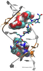
Genetic defect that causes myotonic dystrophy type 2 and used that information to design drug candidates to counteract the disease. Credit: Ilyas Yildirim, Northwestern University.
Scientists revealed a detailed image of the genetic defect that causes myotonic dystrophy type 2 and used that information to design drug candidates to counteract the disease. Credit: Ilyas Yildirim, Northwestern University. View larger image

Myotonic dystrophy type 2 (DM2) is a relatively rare, inherited form of adult-onset muscular dystrophy that has no cure. It’s caused by a genetic defect in which a short series of nucleotides—the chemical units that spell out our genetic code—is repeated more times than normal. When the defective gene is transcribed, the resulting RNA repeat forms a hairpin-like structure that binds to and disables a protein called MBNL1.

Now, research led by Matthew Disney of The Scripps Research Institute (TSRI), Florida Campus, has revealed the detailed, three-dimensional structure of the RNA defect in DM2 and used this information to design small molecules that bind to the aberrant RNA. These designer molecules, even in small amounts, significantly improved disease-associated defects in a cellular model of DM2, and thus hold potential for reversing the disorder.

Drugs that target toxic RNA molecules associated with diseases such as DM2 are few and far between, as developing such compounds is technically challenging. The “bottom-up” approach that the scientists used to design potent new drug candidates, by first studying in detail how the RNA structure interacts with small molecules, is unconventional, noted Jessica Childs-Disney of TSRI, who was lead author of the paper with Ilyas Yildirim of Northwestern University. But it may serve as an effective strategy for pioneering the use of small molecules to manipulate disease-causing RNAs—a central focus of the Disney lab.

This work also was funded by NIH’s National Cancer Institute.

Learn more:
The Scripps Research Institute News Release Exit icon
Disney Lab

Meet Galina Lepesheva

2 comments
Galina Lepesheva
Galina Lepesheva
Field: Biochemistry
Works at: Vanderbilt University, Nashville, TN
Born, raised and studied in: Belarus
To unwind, she: Reads, travels, spends time with her family

Galina Lepesheva knows that kissing bugs are anything but romantic. When the lights get low, these blood-sucking insects begin feasting—and defecating—on the faces of their sleeping victims. Their feces are often infected with a protozoan (a single-celled, eukaryotic parasite) called Trypanosoma cruzi that causes Chagas disease. Lepesheva has developed a compound that might be an effective treatment for Chagas. She has also tested the substance, called VNI, as a treatment for two related diseases—African sleeping sickness and leishmaniasis.

“This particular research is mainly driven by one notion: Why should people suffer from these terrible illnesses if there could be a relatively easy solution?” she says.

Lepesheva’s Findings

Currently, most cases of Chagas disease occur in rural parts of Mexico, Central America and South America. According to some estimates, up to 1 million people in the U.S. could have Chagas disease, and most of them don’t realize it. If left untreated, the infection is lifelong and can be deadly.

The initial, acute stage of the disease is usually mild and lasts 4 to 8 weeks. Then the disease goes dormant for a decade or two. In about one in three people, Chagas re-emerges in its life-threatening, chronic stage, which can affect the heart, digestive system or both. Once chronic Chagas disease develops, about 60 percent of people die from it within 2 years.

The Centers for Disease Control and Prevention (CDC) has targeted Chagas disease as one of five “neglected parasitic infections,” indicating that it warrants special public health action.

“Chagas disease does not attract much attention from pharmaceutical companies,” Lepesheva says. Right now, there are only two medicines to treat it. They are only available by special request from the CDC, aren’t always effective and can cause severe side effects.

Lepesheva’s research focuses on a particular enzyme, CYP51, that is the target of some anti-fungal medicines. If CYP51 can also act as an effective drug target for the parasites that cause Chagas, her work might help meet an important public health need.

CYP51 is found in all kingdoms of life. It helps produce molecules called sterols, which are essential for the development and viability of eukaryotic cells. Lepesheva and her colleagues are studying VNI and related compounds to examine whether they can block the activity of CYP51 in human pathogens such as protozoa, but do no harm to the enzyme in mammals. In other words, her goal is to cripple disease-causing organisms without creating side effects in infected humans or other mammals.

Lepesheva has tested the effectiveness of VNI on Chagas-infected mice. Remarkably, it has worked 100 percent of the time, curing both the acute and chronic stages of the disease. It acts by preventing the protozoan from establishing itself in the host’s body. If it is similarly effective in humans, VNI could become the first reliable treatment for Chagas disease.