Year: 2016

The Cell’s Mailroom

1 comment
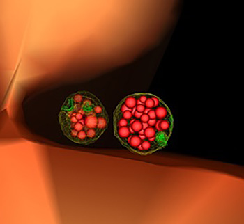
Yeast cell showing two mature, or “late” endosomes that are filled with small vesicles.

This illustration of the inside of a yeast cell shows two mature, or “late” endosomes (green-ringed structures) that are filled with small vesicles (red bubbles). Endosomes are cellular containers that can carry many types of cargo, including cellular waste, which they typically dump into vacuoles (orange). Credit: Matthew West and Greg Odorizzi, University of Colorado, Boulder.

In large offices, mailroom workers read the labels on incoming letters and packages to sort and deliver them and dispose of junk mail. In cells, these tasks—as well as importing food and other materials—fall to small cellular sacs called endosomes. Acting as mailroom staff, endosomes sort and deliver nutrients and building blocks, like amino acids, fat and sugars, to their proper destinations, and send cellular junk, like damaged proteins, to trash processors, such as vacuoles or lysosomes. Continue reading “The Cell’s Mailroom”

A World Without Pain

6 comments

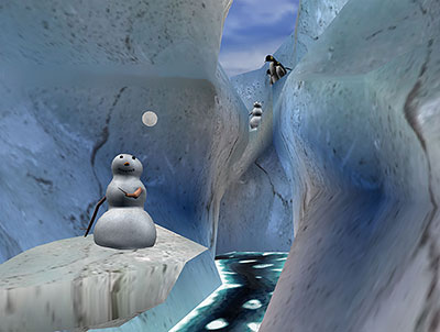
In an immersive virtual reality environment called “Snow World,” burn patients distract themselves from their pain by tossing snow balls, building snowmen and interacting with penguins. Credit: Ari Hollander and Howard Rose, copyright Hunter Hoffman, UW, www.vrpain.com Exit icon.

You glide across an icy canyon where you meet smiling snowmen, waddling penguins and a glistening river that winds forever. You toss snowballs, hear them smash against igloos, then watch them explode in vibrant colors.

Back in the real world, a dentist digs around your mouth to remove an impacted tooth, a procedure that really, really hurts. Could experiencing a “virtual” world distract you from the pain? NIGMS grantees David Patterson Exit icon and Hunter Hoffman Exit icon show it can.

Patterson, a psychologist at the University of Washington (UW) in Seattle, and Hoffman, a UW cognitive psychologist, helped create the virtual reality program “Snow World” in an effort to reduce excessive pain experienced by burn patients. However, the researchers expect Snow World to help alleviate all kinds of pain, including pain experienced during dental procedures. Continue reading “A World Without Pain”

Cool Images: An Independence Day-Inspired Collection

1 comment

@media screen and (max-width: 480px) {
.halloween-out {
width: 96.5%;
}
.halloween-out .halloween-in img {
padding-top: 42px;
padding-right: 12px;
}
}

@media screen and (max-width: 320px) {
.halloween-out {
width: 96%;
}
}

In case you missed the fireworks this weekend, we’ve put together a collection of firework-like images from basic research studies.

Viral Electricity
Viral Electricity Image
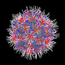
This patriotic Koosh ball is an adeno-associated virus. Most people will come into contact with the virus at some point in their lives, and they’ll probably never know it. Even though it doesn’t cause disease—in fact, because it doesn’t cause disease—this virus is scientifically important. Researchers hope to harness the virus’ ability to enter cells and hijack genes and to use it to to deliver gene therapy. This image, created with the software DelPhi, shows which parts of the virus are positively charged (blue) and which parts are negatively charged (red). The charge of a molecule—like the charge of this virus—influences the way it behaves. In addition to helping researchers understand how viruses might enter cells, images like this one could help them understand how molecules interact with each other as well as drugs.
Continue reading “Cool Images: An Independence Day-Inspired Collection”

Demystifying General Anesthetics

0 comments

When Margaret Sedensky, now of Seattle Children’s Research Institute, started as an anesthesiology resident, she wasn’t entirely clear on how anesthetics worked. “I didn’t know, but I figured someone did,” she says. “I asked the senior resident. I asked the attending. I asked the chair. Nobody knew.”

For many years, doctors called general anesthetics a “modern mystery.” Even though they safely administered anesthetics to millions of Americans, they didn’t know exactly how the drugs produced the different states of general anesthesia. These states include unconsciousness, immobility, analgesia (lack of pain) and amnesia (lack of memory).

Stock image of a symphony.
Like the instruments that make up an orchestra, many molecular targets may contribute to an anesthetic producing the desired effect. Credit: Stock image.

Understanding anesthetics has been challenging for a number of reasons. Unlike many drugs that act on a limited number of proteins in the body, anesthetics interact with seemingly countless proteins and other molecules. Additionally, some anesthesiologists believe that anesthetics may work through a number of different molecular pathways. This means no single molecular target may be required for an anesthetic to work, or no single molecular target can do the job without the help of others.

“It’s like a symphony,” says Roderic Eckenhoff of the University of Pennsylvania Perelman School of Medicine, who has studied anesthesia for decades. “Each molecular target is an instrument, and you need all of them to produce Beethoven’s 5th.” Continue reading “Demystifying General Anesthetics”
CRISPR Serves Up More than DNA

0 comments
Marine bacterium Marinomonas mediterranea
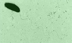
The marine bacterium Marinomonas mediterranea uses a CRISPR system to spot invading RNAs and store a memory of the invasion event in its genome. Research team member Antonio Sanchez-Amat was the first to isolate and characterize this bacterial species. Credit: Antonio Sanchez-Amat, University of Murcia.

A new study has added another twist to the CRISPR story. As we’ve highlighted in several recent posts, CRISPR is an immune system in bacteria that recognizes and destroys viral DNA and other invading DNA elements, such as transposons. Scientists have adapted CRISPR into an indispensable gene-editing tool now widely used in both basic and applied research.

Many previously described CRISPR systems detect and cut viral DNA, insert the DNA pieces into the bacterial genome and then use them as molecular “mug shots” to flag and destroy the virus if it attacks again. But various viruses use RNA, not DNA, as genetic material. Although research has shown that some CRISPR systems also can target RNA, how these systems can archive harmful RNA encounters in the bacterial genome was unknown. Continue reading “CRISPR Serves Up More than DNA”

Viral Views: New Insights on Infection Strategies

0 comments

The following images show a few ways in which cutting-edge research tools are giving us clearer views of viruses—and possible ways to disarm them. The examples, which highlight work involving HIV and the coronavirus, were funded in part by our Biomedical Technology Research Resources program.

Uncloaking HIV’s Camouflage

HIV capsid with (right, red) and without (left) a camouflaging human protein.
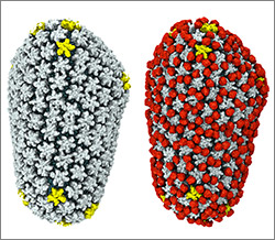
HIV capsid with (right, red) and without (left) a camouflaging human protein. Credit: Juan R. Perilla, Klaus Schulten and the Theoretical and Computational Biophysics Group, University of Illinois at Urbana-Champaign.

To sneak past our immune defenses and infect human cells, HIV uses a time-honored strategy—disguise. The virus’ genome is enclosed in a protein shell called a capsid (on left) that’s easily recognized and destroyed by the human immune system. To evade this fate, the chrysalis-shaped capsid cloaks itself with a human protein known as cyclophilin A (in red, on right). Camouflaged as human, the virus gains safe passage into and through a human cell to deposit its genetic material in the nucleus and start taking control of cellular machinery.

Biomedical and technical experts teamed up to generate these HIV models at near-atomic resolution. First, structural biologists at the Pittsburgh Center for HIV Protein Interactions Exit icon used a technique called cryo-electron microscopy (cryo-EM) to get information on the shape of an HIV capsid as well as the capsid-forming proteins’ connections to each other and to cyclophilin A. Then experts at the Resource for Macromolecular Modeling and Bioinformatics fed the cryo-EM data into their visualization and simulation programs to computationally model the physical interactions among every single atom of the capsid and the cyclophilin A protein. The work revealed a previously unknown site where cyclophilin A binds to the capsid, offering new insights on the biology of HIV infection. Continue reading “Viral Views: New Insights on Infection Strategies”

Cool Video: Watching Bacteria Turn Virulent

1 comment

Researchers created an apparatus to study quorum sensing, a communication system that allows some bacteria to cause dangerous infections. Their findings suggest that blocking bacterial communication might lead to a new way to combat such infections. Credit: Minyoung Kevin Kim and Bonnie Bassler, Princeton University.

If you’ve ever felt a slimy coating on your teeth, scrubbed grime from around a sink drain or noticed something growing between the tiles of a shower, you’ve encountered a biofilm. Made up of communities of bacteria and other microorganisms, biofilms thrive where they can remain moist and relatively undisturbed. As they enlarge, biofilms can block narrow passages like medical stents, airways, pipes or intestines. Continue reading “Cool Video: Watching Bacteria Turn Virulent”

Ticks, Mice and Microbes—Studying Disease Spread

0 comments
Maria Diuk-Wasser
Credit: Oscar Gonzalez (Diuk-Wasser’s husband)
Maria Diuk-Wasser
Hometown: Buenos Aires, Argentina
Childhood dream job: Veterinarian
Hobbies: Hiking and gardening with her son (age 10) and daughter (age 7)
Favorite music: Salsa
Worksite: Lab at Columbia University and forests in coastal New England

Maria Diuk-Wasser grew up on the 10th floor of an apartment building in the middle of a bustling city. With no forests or meadows nearby, she read book after book about the natural world and surrounded herself with houseplants.

“I yearned for nature,” Diuk-Wasser says. “But my parents couldn’t provide it. They’re city people. They didn’t know anything about hiking or camping.”

These days, Diuk-Wasser still spends a lot of time in a city—she’s a professor at Columbia University in New York, the most populous city in the U.S.—but she also gets plenty of time in the woods. She hikes for hours through coastal New England forests, some of the loveliest in the country, searching for what many consider less-than-lovely inhabitants: mice and ticks. Continue reading “Ticks, Mice and Microbes—Studying Disease Spread”

Visualizing Skin Regeneration in Real Time

2 comments
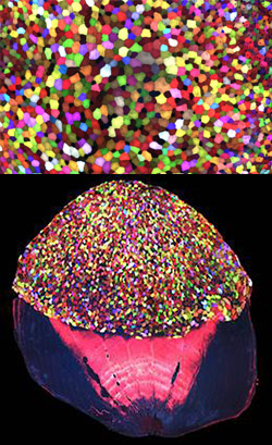
Top: Colorful skin cells on a zebrafish . Bottom: Cells from the outer surface of the scale.
More than 70 Skinbow colors distinguish hundreds of live cells from a tiny bit (0.0003348 square inches) of skin on the tail fin of an adult zebrafish. The bottom image shows the cells on the outer surface of a scale. Credit: Chen-Hui Chen, Duke University.

Zebrafish, blue-and-white-striped fish that are about 1.5 inches long, can regrow injured or lost fins. This feature makes the small fish a useful model organism for scientists who study tissue regeneration.

To better understand how zebrafish skin recovers after a scrape or amputation, researchers led by Kenneth Poss of Duke University tracked thousands of skin cells in real time. They found that lifespans of individual skin cells on the surface were 8 to 9 days on average and that the entire skin surface turned over in 20 days.

The scientists used an imaging technique they developed called “Skinbow,” which essentially shows the fish’s outer layer of skin cells in a spectrum of colors when viewed under a microscope. Skinbow is based on a technique created to study nerve cells in mice, another model organism.

The research team’s color-coded experiments revealed several unexpected cellular responses during tissue repair and replacement. The scientists plan to incorporate additional imaging techniques to generate an even more detailed picture of the tissue regeneration process.

The NIH director showcased the Skinbow technique and these images on his blog, writing: “You can see more than 70 detectable Skinbow colors that make individual cells as visually distinct from one another as jellybeans in a jar.”

This work was funded in part by NIH under grant R01GM074057.

The Proteasome: The Cell’s Trash Processor in Action

2 comments

Our cells are constantly removing and recycling molecular waste. On the occasion of Earth Day, we put together this narrated animation to show you one way cells process their trash. The video features the proteasome, a cellular machine that breaks down damaged or unwanted proteins into bits that the cell can re-use to make new proteins. For this reason, the proteasome is as much a recycling plant as it is a garbage disposal.

For more details about the proteasome and other cellular disposal systems, check out our article How Cells Take Out the Trash.