Year: 2016

Newly Identified Cell Wall Construction Workers: A Novel Antibiotic Target?

0 comments
SEDS

A family of proteins abbreviated SEDS (bright, pink) help build bacterial cell walls, so they are a potential target for new antibiotic drugs. Credit: Rudner lab, Harvard Medical School.

Scientists have identified a new family of proteins that, like the targets of penicillin, help bacteria build their cell walls. The finding might reveal a new strategy for treating a range of bacterial diseases.

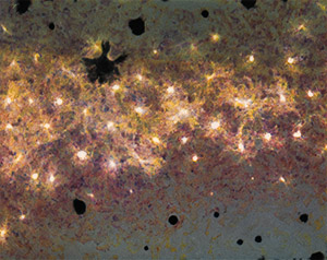
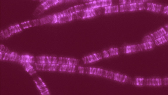
The protein family is nicknamed SEDS, because its members help control the shape, elongation, division and spore formation of bacterial cells. Now researchers have proof that SEDS proteins also play a role in constructing cell walls. This image shows the movement of a molecular machine that contains a SEDS protein as it constructs hoops of bacterial cell wall material.

Any molecule involved in building or maintaining cell walls is of immediate interest as a possible target for antibiotic drugs. That’s because animals, including humans, don’t have cell walls—we have cell membranes instead. So disabling cell walls, which bacteria need to survive, is a good way to kill bacteria without harming patients.

This strategy has worked for the first antibiotic drug, penicillin (and its many derivatives), for some 75 years. Now, many strains of bacteria have evolved to resist penicillins—and other antibiotics—making the drugs less effective.

According to the Centers for Disease Control and Prevention, drug-resistant strains of bacteria Exit icon infect at least 2 million people, killing more than 20,000 of them in the U.S. every year. Identifying potential new drug targets, like SEDS proteins, is part of a multi-faceted approach to combating drug-resistant bacteria.

Get Your Cell Biology Questions Ready for Cell Day

0 comments

What do you get when you mix a room full of scientists with a classroom full of students who have questions about cells? Cell Day 2016! During this free web chat, middle and high school students will have the opportunity to ask our scientists at NIGMS about cell biology, biochemistry, research careers and more. Join us on Thursday, November 3 anytime from 10 a.m. to 3 p.m. EDT. Registration (no longer available).

The ECM: A Dynamic System for Moving Our Cells

2 comments

In part I of this series, we mentioned that the extracellular matrix (ECM) makes our tissues stiff or squishy, solid or see-through. Here, we reveal how the ECM helps body cells move around, a process vital for wounds to heal and a fetus to grow.

Sealing and Healing Wounds

MMPs are essential for closing wounds. Credit: Stock image.

When we get injured, the first thing our body does is to form a blood clot to stop the bleeding. Skin cells then start migrating into the wound to close the cut. The ECM is essential for this step, creating a physical support structure—like a road or train track—over which skin cells travel to seal the injured spot.

The ECM is made up of a host of proteins produced before and after injury. Some other proteins called matrix metalloproteinases (MMPs) also crowd into wounds. Because humans have so many different MMPs—a full 24 of them!—it’s been difficult for scientists to figure out what roles, if any, the proteins play in healing scrapes and cuts.

Continue reading “The ECM: A Dynamic System for Moving Our Cells”

The Extracellular Matrix, a Multitasking Marvel

1 comment

In part II of this series, we reveal how the ECM helps body cells move around, a process vital for wounds to heal and a fetus to grow. Here we introduce the extracellular matrix (ECM) and discuss how it makes our tissues stiff or squishy, solid or see-through.

When we think about how our bodies are made and what they do, we usually focus on organs, tissues and cells. These structures have well-known roles. But around, within and between them is a less understood material that also plays an essential part in making us what we are.

This gelatinous filler material is known as the extracellular matrix (ECM). Once thought to be the biological equivalent of bubble wrap, we now know that the ECM is a dynamic, physiologically active component of all our tissues. It guides cell shape, orientation and function.

The ECM is found in all of our body parts. In some tissues, it’s a thin layer separating cells, like mortar between bricks. In other tissues, it’s the major constituent.

The ECM is most prevalent in connective tissue, the material that forms our skeletons, cushions our internal organs and winds between blood vessels and around nerves. In connective tissue, the ECM is more abundant than the cells suspended within it.

The extracellular matrix meets the needs of each body part. In teeth and bones, it’s rock-hard. In corneas, it’s a transparent gel that acts like a camera lens. In tendons, it forms strong fibers that bind muscle to bone. Credit: Stock image.

What makes the ECM truly unique is its variability: Its texture, composition and functions vary by body part. That’s because the ECM’s deceptively simple recipe of water, fibrous proteins and carbohydrates has virtually endless variations.

In general, the fibrous proteins give the ECM its texture and help cells adhere properly. Carbohydrates in the ECM absorb water and swell to form a gel that acts as an excellent shock absorber. Continue reading “The Extracellular Matrix, a Multitasking Marvel”

The Science of Size: Rebecca Heald Explores Size Control in Amphibians

0 comments
Rebecca Heald
Credit: Mark Hanson.
Rebecca Heald
Grew up in: Greenville, Pennsylvania
Studied at: Hamilton College, Rice University, Harvard Medical School
Job site: University of California, Berkeley
Favorite hobby: Cycling

A 50-pound frog isn’t some freak of nature or a creepy Halloween prank. It’s a thought experiment conceived by Rebecca Heald, a cell biologist at the University of California, Berkeley Exit icon, who is studying the factors that control size in animals.

Heald’s “50-pound frog project” speaks to the power of evolution and to scientists’ ability to modify the physical characteristics of an organism by altering its genome. The project also incorporates many of Heald’s fascinating discoveries studying amphibian eggs and embryos.

In amphibians, unlike in mammals, there are striking correlations among the size of the animals’ genomes (an organism’s complete set of genes) and several aspects of the animals’ size. For example, amphibians with large genomes tend to be bigger than those with smaller genomes. Larger genomes also correspond to larger cells and larger organelles (specialized cellular structures such as the nucleus). Heald has also demonstrated that these seemingly fixed parameters can be tweaked in the lab. Continue reading “The Science of Size: Rebecca Heald Explores Size Control in Amphibians”

Pigment Cells: Not Just Pretty Colors

2 comments

If you’ve ever visited an aquarium or snorkeled along a coral reef, you’ve witnessed the dazzling colors and patterns on tropical fish. The iridescent stripes and dots come from pigment cells, which also tint skin, hair and eyes in all kinds of animals, including humans. Typically, bright colors help attract mates, while dull ones provide camouflage. In humans, pigment helps protect skin from DNA-damaging UV light.

Researchers study cellular hues not only to decipher how they color our world, but also to understand skin cancers that originate from pigment cells. Some of these researchers work their way back, developmentally speaking, to focus on the type of cell, known as a neural crest cell, that is the precursor of pigment cells.

Present at the earliest stages of development, neural crest cells migrate throughout an embryo and transform into many different types of cells and tissues, including nerve cells, cartilage, bone and skin. The images here, from research on neural crest cells in fish and salamanders, showcase the beauty and versatility of pigment cells in nature’s palette.

Xanthophores
Pigment cells called xanthophores, shown here in the skin of the popular laboratory animal zebrafish, glow brightly under light. Credit: David Parichy, University of Washington.
Melanocytes
Dark pigment cells, called melanocytes, like these in pearl danio, a tropical minnow and relative of zebrafish, assemble in skin patterns that allow the animals to blend into their surroundings or attract mates. Credit: David Parichy, University of Washington.
Fin of pearl danio
Pigment cells can form all sorts of patterns, like these stripes on the fin of pearl danio. Credit: David Parichy, University of Washington.
Salamander skin
Pigment cells arise from neural crest cells. Here, pigment cells can be seen migrating in the skin of a salamander where they will form distinct color patterns. Credit: David Parichy, University of Washington.


A Labor Day-Themed Collection: Hard-Working Cell Structures

2 comments

Hard labor might be the very thing we try to avoid on Labor Day. But our cells and their components don’t have the luxury of taking a day off. Their non-stop work is what keeps us going and healthy.

Scientists often compare cells with small factories. Just like a factory, a cell contains specialized compartments and machines—including organelles and other structures—that each play their own roles in getting the job done. In the vignettes below, we give a shout out to some of these tireless cellular workers.

Energy Generators
Credit: Thomas Deerinck, National Center for Microscopy and Imaging Research
Mitochondria are the cell’s power plants. They convert energy from food into a molecule called ATP that fuels virtually every process in the cell. As shown here, mitochondria (brown) often have distinct, oblong shapes. Like most other organelles, mitochondria are encased in an outer membrane. But they also have an inner membrane that folds many times, increasing the area available for energy production. In addition, mitochondria store calcium ions, help make hemoglobin—the vital iron-containing protein that allows red blood cells to carry oxygen—and even take part in producing some hormones. Defects in mitochondria can lead to a host of rare but often incurable diseases that range from mild to devastating. Researchers are studying mitochondria to better understand their manifold jobs in the cell and to find treatments for mitochondrial diseases.

Continue reading “A Labor Day-Themed Collection: Hard-Working Cell Structures”

Interview With a Scientist: Janet Iwasa, Molecular Animator

0 comments

The world beneath our skin is full of movement. Hemoglobin in our blood grabs oxygen and delivers it throughout the body. Molecular motors in cells chug along tiny tubes, hauling cargo with them. Biological invaders like viruses enter our bodies, hijack our cells and reproduce wildly before bursting out to infect other cells.

To make sense of the subcutaneous world, Janet Iwasa, a molecular animator at the University of Utah, creates “visual hypotheses”—detailed animations that convey the latest thinking of how biological molecules interact.

“It’s really building the animated model that brings insights,” Iwasa told Biomedical Beat in 2014. “When you’re creating an animation, you’re really grappling with a lot of issues that don’t necessarily come up by any other means. In some cases, it might raise more questions, and make people go back and do some more experiments when they realize there might be something missing.”

Iwasa has collaborated with numerous scientists to develop animations of a range of biological processes and structures Exit icon. Recently, she’s undertaken an ambitious, multi-year project to animate HIV reproduction Exit icon.

Interview With a Slime Mold: Racing for New Knowledge

1 comment
Dictyostelium discoideum
Credit: Wikimedia Commons, Usman Bashir.
Dictyostelium discoideum
Natural habitat: Deciduous forest soil and moist leaf litter
Favorite food: Bacteria
Top speed: 8 micrometers per minute

Like the athletes in Rio, the world’s most highly advanced microbial runners recently gathered in Charlestown, Massachusetts, to find out which ones could use chemical cues to most quickly navigate a maze-like microfluidic racecourse. The winners’ prize: credit for helping scientists learn more about how immune system cells navigate through the human body on their way to fight disease.

The finalists were a group of soil-dwelling slime molds called Dictyostelium that were genetically engineered by a pair of Dutch biochemists to detect minuscule chemical changes in the environment. The racers used their enhanced sense of “smell” to avoid getting lost on their way to the finish line.

While researchers have been racing the genetically souped-up microbes at annual events for a few years—another competition is scheduled for October 26—scientists have been studying conventional Dictyostelium for decades to investigate other important basic life processes including early development, gene function, self/non-self recognition, cell-type regulation, chemical signaling and programmed cell death. Continue reading “Interview With a Slime Mold: Racing for New Knowledge”

Protein Paradox: Enrique De La Cruz Aims to Understand Actin

1 comment
Enrique M. De La Cruz
Credit: Jeff Foley, American Heart Association.
Enrique M. De La Cruz
Grew up in: Newark and Kearny, New Jersey
Job site: Yale University
Favorite food: His mom’s Spanish-style polenta (harina de maíz)
Alternative career: Managing a vinyl record shop
Favorite song: “Do Anything You Wanna Do” by Eddie & The Hot Rods

Enrique De La Cruz stood off to the side in a packed room. As he waited for his turn to speak, he stroked the beads of a necklace. Was he nervous? Quietly praying? When he took center stage, the purpose of the strand became clear.

Like a magician—and dressed all in black—De La Cruz held up the necklace with two hands so everyone, even those sitting in the back, could see it. It was made of snap-together beads. De La Cruz waved the strand. It wiggled in different directions. Then, with no sleight of hand, he popped off one of the beads. The necklace broke into two.

For the next hour, De La Cruz pulled out one prop after another: a piece of rope from his pocket, a pencil tucked behind his ear and even a fresh spear of asparagus stuffed in his backpack. At one point, De La Cruz assembled a conga line with people in the front row. Continue reading “Protein Paradox: Enrique De La Cruz Aims to Understand Actin”