
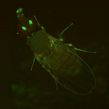
 A fruit fly expressing GFP. Credit: Jay Hirsh, University of Virginia.
During the holiday season, twinkling lights are a common sight. But no matter what time of the year, you can see colorful glows in many biology labs. Scientists have enabled many organisms to light up in the dark—from cells to fruit flies and Mexican salamanders. These glowing organisms help researchers better understand basic cell processes because their DNA has been edited to express molecules such as green fluorescent protein.
Illuminating Cell Processes
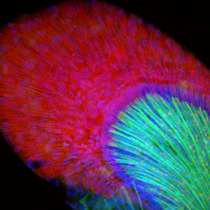 A zebrafish fin with GFP glowing (green dots) where gene sox9b is expressed. This gene plays an important role in the development of the heart, brain, and skeleton. Credit: Jessica Plavicki, University of Wisconsin, Madison.
GFP is a natural protein that scientists discovered in the North American jellyfish Aequorea victoria. The protein glows green when exposed to ultraviolet light, which is a part of sunlight that’s invisible to humans. Scientists don’t know why these jellyfish evolved their glow, but one hypothesis is that it helps them ward off predators. Researchers have transferred the gene that carries the instructions for GFP into zebrafish, fruit flies, rabbits, mice, human cells in a lab dish, and more. GFP acts as a glowing “tag” on a protein being studied, letting researchers track the protein’s activity, which would be very difficult otherwise.
Because proteins are produced only when genes are turned on by a stimulus in the cell, GFP can show whether a gene is being expressed in different parts of an organism. Researchers have used GFP to study topics such as the behavior of specific sets of cells during embryonic development, the spread of cancer cells, the progression of HIV infections, and the construction of insulin-producing cells.
Nobel Recognition
In 2008, NIGMS-supported researchers Martin Chalfie, Ph.D., Osamu Shimomura, Ph.D., and Roger Tsien, Ph.D., received the prestigious Nobel Prize in chemistry for their discovery and development of GFP. Dr. Shimomura first extracted the glowing protein from jellyfish in the 1960s. In the early 1990s, Dr. Chalfie isolated the gene that carries the instructions for GFP and transferred it into research organisms.
In subsequent years, Dr. Tsien identified the structure within GFP that produced green fluorescence and created a synthetic palette of colors that glowed longer and brighter and allowed researchers to study many different molecules at the same time.
Rainbow Glow
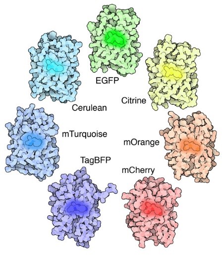 A handful of the GFP-like fluorescent proteins that researchers have developed. CC-BY-4.0.
Today, a full rainbow of proteins that are based on GFP’s structure and work similarly are available thanks to the findings of Dr. Tsien and other researchers. In addition, many fluorescent proteins besides GFP exist in nature and enable more than a hundred organisms besides Aequorea victoria to glow in a range of colors. However, these proteins often require additional chemicals, enzymes, or other substances to glow, whereas GFP does not, so GFP and proteins based on it are simpler to use. GFP and its derivatives have the additional advantage of binding to a protein at a single location so that they don’t distort it and potentially affect its function. NIGMS-funded researchers are currently using GFP and other fluorescent proteins to study a wide range of topics, from cell development to regeneration and the effects of burns on nerves and muscles.