
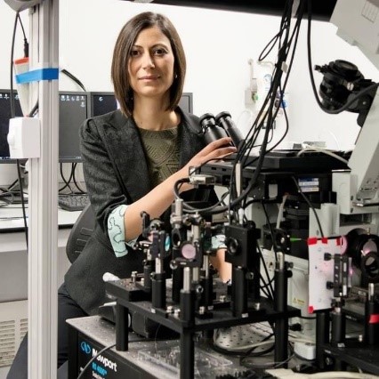
“It would be a dream come true if I could look at a cell within a tissue and have a Google Maps view to zoom in until I saw individual molecules,” says Melike Lakadamyali, Ph.D., an associate professor of physiology at the University of Pennsylvania’s Perelman School of Medicine in Philadelphia. Her lab is helping make part of that dream a reality by developing super-resolution microscopy tools that visualize cells at a near-molecular level.
Blending Physics and Biology
Science and math fascinated Dr. Lakadamyali since childhood, and she felt especially drawn to physics because she enjoyed using logic to solve problems. After graduating high school in her native country of Cyprus, she chose to study physics at the University of Texas, Austin. She never gave much thought to applying physics methods to biological
questions—a field known as biophysics—until her third year as an undergraduate, when she gained her first research experience in the lab of Josef Käs, Ph.D.
Dr. Käs assigned Dr. Lakadamyali to a project measuring properties of neuronal growth cones, which sense the environment around neurons and guide their extension. “My first day in the lab was kind of a disaster,” Dr. Lakadamyali says. She recalls breaking the tip of a valuable piece of equipment by accidentally crashing it into some glass. “But I didn’t give up, and eventually I learned how to use the machine,” she adds. Her work revealed something unexpected: The growth cones sensed mechanical pressure and would retract and start growing in a different direction when touched. The feeling of discovery that came with this new knowledge excited her, and her experience in Dr. Käs’s lab led her to pursue a career in biophysics.
After completing her bachelor’s degree, Dr. Lakadamyali entered a physics Ph.D. program at Harvard University, Cambridge, Massachusetts, and worked in the lab of Xiaowei Zhuang, Ph.D. She used fluorescence light microscopy to observe and record in real time how the influenza virus enters cells. “There were quite a lot of challenges in getting this to work,” says Dr. Lakadamyali. “The first time I saw a movie of virus particles storming cells that I recorded on the microscope, I was practically jumping up and down.”
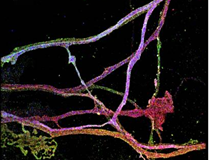
After earning her Ph.D., Dr. Lakadamyali took a postdoctoral position at Harvard in the lab of Jeff Lichtman, M.D., Ph.D. He had recently developed “brainbow” mice, whose neurons express fluorescent proteins that make the cells easier to visualize. Dr. Lakadamyali used microscopy techniques developed in Dr. Zhuang’s lab to map connections between the brainbow mice’s neurons.
Establishing a Lab
In 2010, after 4 years as a postdoc, Dr. Lakadamyali started her own lab at the Institute of Photonic Sciences, Barcelona. The institute was only about 10 years old, and the opportunity to guide its expansion into biophysics excited her. Initially, she faced challenges. “Being young and the first female group leader, I felt that I had to work much harder than my male colleagues to be taken seriously. I was constantly being confused for a graduate student,” Dr. Lakadamyali says.
Her lab ultimately flourished at the institute, but after several years, she began to miss the multidisciplinary environment of a large university where she could collaborate with biologists and other researchers outside of physics. So when two professors from the Perelman School of Medicine approached her at a conference and asked if she was interested in applying for a job, she jumped at the chance.
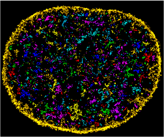
Dr. Lakadamyali officially moved to the University of Pennsylvania in 2017. There, she develops super-resolution microscopy tools and uses them to investigate the spatial organization of nucleic acids—such as DNA and RNA—and proteins inside cells. She’s particularly interested in how DNA is folded in the nucleus and how molecular motors transport organelles to where they’re needed. The precise organization of molecules in cells is vital for their proper function and, ultimately, for our health. In addition to visualizing the normal arrangement of cells’ molecules, Dr. Lakadamyali is researching how their organization is disrupted in diseases, which could support the development of diagnostic tests and treatments.
Dr. Lakadamyali largely credits her success as a scientist to the excellent mentorship she received, and she places a strong emphasis on mentoring students in her lab. “One thing that gives me constant joy is the training of the next generation of scientists,” she says. Perhaps one day, they’ll be able to use Google Maps–style microscopy in their research, thanks to her work.
Dr. Lakadamyali’s lab is supported by NIGMS grants R01GM133842 and RM1GM136511.