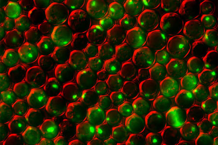
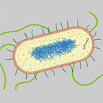
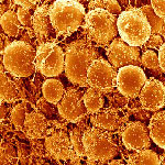
Category: Cells

Cells Merrily ‘Row’ Without Sensor Proteins

0 comments
Messenger proteins (left). When these proteins aren’t activated, the cell doesn’t move (right). Credit: Devreotes Lab, Johns Hopkins University School of Medicine.
Messenger proteins help the cell make large projections (left). When these proteins aren’t activated, the cell doesn’t move (right). Credit: Devreotes Lab, Johns Hopkins University School of Medicine. View larger image

A new study from Peter Devreotes Exit icon, Pablo Iglesias Exit icon and other scientists at Johns Hopkins University sheds light on the way in which cells get around the body to promote embryo development, wound healing and even cancer metastasis. Here’s how they describe cell movement and their findings:

Think of the cell as a rowboat. Sensor proteins on the outside pass on directional signals to messenger proteins that serve as the cell’s coxswain. The coxswain then commands other members of the molecular crew to stay in sync, propelling the cell forward. If there are no sensor signals, the coxswain can still coordinate the cell’s movement, just not in any specific direction—it’s like a boat without a rudder.

Scientists previously thought that the messenger proteins needed the sensor ones to produce both directional and random movements. Because defects in the messenger proteins have been linked to many types of cancer, the new work suggests these molecules could serve as a drug target for immobilizing tumor cells.

Learn more:

Johns Hopkins University School of Medicine News Release Exit icon

NIH Director Blogs About Value of Model Organisms in Drug Discovery Research

0 comments
(Left) Untreated yeast cells, (Right) Treated yeast cells. Credit: Daniel Tardiff, Whitehead Institute.
Treating yeast cells with the NAB compound reverses the toxic effects of elevated levels of alpha synuclein protein. Credit: Daniel Tardiff, Whitehead Institute. View larger image

These eye-catching images and the NIGMS-funded research that yielded them were recently featured by NIH Director Francis Collins on his blog. Scientists led by a team at the Whitehead Institute for Biomedical Research engineered yeast to produce too much of a protein, alpha synuclein. In Parkinson’s disease, elevated levels or mutated forms of this protein wreak havoc on the cell. Using the model system, the researchers tested tens of thousands of compounds to identify any that reversed the toxic effects. One did. The compound, abbreviated NAB, worked similarly in an animal model and in rat neurons grown in a lab dish. Collins described the approach as “an innovative strategy for drug hunting that will likely be extended to other conditions.”

Healing Wounds, Growing Hair

1 comment
Wound healing in process. Credit: Yaron Fuchs and Samara Brown in the lab of Hermann Steller, Rockefeller University.

Credit: Yaron Fuchs and Samara Brown in the lab of Hermann Steller, Rockefeller University.

Whether injured by a scrape, minor burn or knife wound, skin goes through the same steps to heal itself. Regrowing hair over new skin is one of the final steps. All the hair you can see on your body is non-living, made up of “dead” cells and protein. It sprouts from living cells in the skin called hair follicle stem cells, shown here in red and orange. For more pictures of hair follicle stem cells—and many other stunning scientific images and videos—go to the NIGMS Image and Video Gallery.

Learn more:

Rockefeller University News Release Exit icon
Steller Lab Exit icon

Flu Finds a Way In

0 comments
Influenza virus proteins in the act of self-replication. Credit: Wilson, Carragher and Potter labs, Scripps Research Institute.

Flu viruses evolve rapidly, often staying one step ahead of efforts to vaccinate against infections or treat them with antiviral drugs. Work led by Jesse Bloom of the Fred Hutchinson Cancer Research Center has uncovered a surprising new flu mutation that allows influenza to infect cells in a novel way. Normally, a protein called hemagglutinin lets flu viruses attach to cells, and a protein called neuraminidase lets newly formed viruses escape from infected cells. Bloom’s lab has characterized a mutant flu virus where neuraminidase can enable the virus to attach to host cells even when hemagglutinin’s binding is blocked. Although the researchers generated the neuraminidase mutant studied in these experiments in their lab, the same mutation occurs naturally in strains from several recent flu outbreaks. There’s a possibility that flu viruses with such mutations may be able to escape antibodies that block the binding of hemagglutinin.

This work also was funded by NIH’s National Institute of Allergy and Infectious Diseases.

Learn more:

Bloom Lab Exit icon

Fitting a Piece of the Protein-Function Puzzle

0 comments
Comparison of the predicted binding of the substrate to the active site of HpbD (blue) with the binding sites determined experimentally by crystallography (magenta). Credit: Matt Jacobson, University of California, San Francisco; Steve Almo, Albert Einstein College of Medicine.
The image shows a comparison of the predicted binding of the substrate to the active site of HpbD (blue) with the binding sites determined experimentally by crystallography (magenta). Credit: Matt Jacobson, University of California, San Francisco; Steve Almo, Albert Einstein College of Medicine.

Sequencing the genomes of almost 7,000 organisms has identified more than 40 million proteins. But how do we figure out what all these proteins do? New results from an initiative led by John Gerlt of the University of Illinois suggest a possible method for identifying the functions of unknown enzymes, proteins that speed chemical reactions within cells. Using high-powered computing, the research team modeled how the structure of a mystery bacterial enzyme, HpbD, might fit like a puzzle piece into thousands of proteins in known metabolic pathways. Since an enzyme acts on other molecules, finding its target or substrate can shed light on its function. The new method narrowed HpbD’s candidate substrate down from more than 87,000 to only four. Follow-up lab work led to the actual substrate, tHypB, and determined the enzyme’s biological role. This combination of computational and experimental methods shows promise for uncovering the functions of many more proteins.

Learn more:

University of Illinois at Urbana-Champaign News Release
Gerlt Lab
Enzyme Function Initiative

Protein Helps Chromosomes ‘Speed Date’ During Cell Division

0 comments
A cell in two stages of division: prometaphase (top) and metaphase (bottom). Credit: Lilian Kabeche, Dartmouth.
This image shows a cell in two stages of division: prometaphase (top) and metaphase (bottom). To form identical daughter cells, chromosome pairs (blue) separate via the attachment of microtubules made up of tubulin proteins (pink) to specialized structures on centromeres (green). Credit: Lilian Kabeche, Dartmouth.

Chromosome segregation during cell division is like speed dating, according to Geisel School of Medicine at Dartmouth researcher Duane Compton. He and postdoctoral fellow Lilian Kabeche learned that protein cyclin A plays moderator, helping to properly separate chromosomes via the attachment of microtubule fibers to kinetochore structures. Here’s how Compton described the process:

“The chromosomes are testing the microtubules for compatibility—that is, looking for the right match—to make sure there are correct attachments and no errors. The old view of this process held that chromosomes and microtubules pair up individually to find the correct attachment, like conventional dating. However, our results show that the system is more like speed dating. All the chromosomes have to try many connections with microtubules in a short amount of time. Then they all make their final choices at the same time. Cyclin A acts like a timekeeper or referee to make sure no one makes bad connections prematurely.”

Such bad connections can cause chromosome segregation errors that lead to cells with an abnormal number of chromosomes, a hallmark of cancer cells. So in addition to aiding our understanding of a fundamental biological process, the new insights may point to potential ways to correct such errors.

Learn more:

Dartmouth News Release Exit icon
Compton Lab Exit icon

Cell Biology Advances and Computational Techniques Earn Nobels for NIGMS Grantees

0 comments
The winners of the 2013 Nobel Prize in physiology or medicine discovered that cells import and export materials using fluid-filled sacs called vesicles. Credit: Judith Stoffer.

Every October, a few scientists receive a call from Sweden that changes their lives. From that day forward, they will be known as Nobel laureates. This year, five of the new Nobelists have received funding from NIGMS.

In physiology or medicine, NIGMS grantees James Rothman (Yale University) and Randy Schekman (University of California, Berkeley) were honored “for their discoveries of machinery regulating vesicle traffic, a major transport system in our cells.” They share the prize with Thomas C. Südhof of Stanford University.

Rothman and Schekman started out working separately and in different systems—Schekman in yeast and Rothman in reconstituted mammalian cells—and their conclusions validated each others’. It’s yet another example of the power of investigator-initiated research and the value of model systems.

Highly accurate molecular models like this one are based on computational techniques developed by the winners of the 2013 Nobel Prize in chemistry. Credit: Rommie E. Amaro, University of California, San Diego.

The three Nobelists in chemistry are NIGMS grantees Martin Karplus (Harvard University), Michael Levitt (Stanford University) and Arieh Warshel (University of Southern California), who developed “multiscale models for complex chemical systems.” They used computational techniques to obtain, for the first time, detailed structural information about proteins and other large molecules. Because of that work, scientists around the world are now able to access, with a few keystrokes, highly accurate models of nearly 100,000 molecular structures. Studying these structures has advanced our understanding of countless diseases, pharmaceuticals and basic biological processes.

Learn more:

NIGMS Nobel Prize News Announcement
NIGMS Nobelists Fact Sheet

Cool Video: How Bee Venom Toxin Kills Cells

0 comments
Credit: Huey Huang, Rice University.

Credit: Huey Huang, Rice University.

A new video, starring the toxin in bee venom, might help scientists design new drugs to combat bacterial infections. The video, by Rice University biophysicist Huey Huang Exit icon, condenses 6.5 minutes into less than a minute to show how the toxin, called melittin, destroys an animal or bacterial cell.

What looks like a red balloon is an artificial cell filled with red dye. Melittin molecules are colored green and float on the cell’s surface like twigs on a pond. As melittin accumulates on the cell’s membrane, the membrane expands to accommodate it. In the video, the membrane stretches into a column on the left.

When melittin levels reach a critical threshold, countless pinhole leaks burst open in the membrane. The cell’s vital fluids—red dye in the video—leak out through these pores. Within minutes, the cell collapses.

Many organisms use such a pore-forming technique to kill attacking bacterial cells. This research reveals molecular-level details of the strategy, bringing pharmaceutical scientists a step closer to harnessing it in the design of new antibiotics.

Cool Image: Tiny Bacterial Motor

2 comments
Phillip Klebba, Kansas State University.

Credit: Phillip Klebba, Kansas State University.

It looks like a fluorescent pill, but this image of an E. coli cell actually shows a new potential target in the fight against infectious diseases. The green highlights a protein called TonB, which is produced by many gram-negative bacteria, including those that cause typhoid fever, meningitis and dysentery. TonB lets bacteria take up iron from the host’s body, which they need to survive. New research from Phillip Klebba of Kansas State University and his colleagues shows how TonB powers iron uptake. When TonB spins within the cell envelope (the bacteria’s “skin”) like a tiny motor, it produces energy that lets another protein pull iron into the cell. This knowledge may lead to the development of antibiotics that block the motion of TonB, potentially stopping an infection in its tracks.

Learn more:
Kansas State University News Release Exit icon
Klebba Lab Exit icon
Other Cool Images