Tag: Cellular Processes

How Cells Take Out the Trash

1 comment
Proteins entering the proteasome. Credit: Office of Biological and Environmental Research of the U.S. Department of Energy Office of Science.
When proteins enter the proteasome, they’re chopped into bits for re-use. Credit: Office of Biological and Environmental Research of the U.S. Department of Energy Office of Science.

As people around the world mark Earth Day (April 22) with activities that protect the planet, our cells are busy safeguarding their own environment.

To keep themselves neat, tidy and above all healthy, cells rely on a variety of recycling and trash removal systems. If it weren’t for these systems, cells could look like microscopic junkyards—and worse, they might not function properly. Scientists funded by the National Institutes of Health are therefore working to understand the cell’s janitorial services to find ways to combat malfunctions.

Read more about how cells take out the trash and handle recycling in this Inside Life Science article.

New Life for Toxic Antibiotics?

0 comments
Pills and a bottle
Researchers found that the antibiotic trovafloxacin cuts off a channel for communication between cells and interferes with a cell-death process. Credit: Stock image.

Many compounds that show promise as new antibiotics for treating bacterial infections never make it to the clinic because they turn out to be toxic to humans as well as to bacteria. A research team led by Kodi Ravichandran of the University of Virginia recently gained insights into why one such antibiotic, trovafloxacin, harms human cells. They found that the compound cuts off a channel for communication between cells, which in turn interferes with how dying cells are broken down and recycled by the body. Roughly 200 billion cells in the human body die and are replaced every day as part of a routine cleanup process, and interference in this process by trovafloxacin may have contributed to the serious liver damage seen in some patients in clinical trials of the drug. Understanding how trovafloxacin causes toxicity in people may help researchers re-engineer this and related compounds to make them safe and effective for use in fighting bacterial infections.

Learn more:
Ravichandran Lab

What Students Want to Know About Cells

0 comments
Cell Day 2014

During a live online chat dubbed “Cell Day,” scientists at NIGMS recently fielded questions about the cell and careers in research from middle and high school students across the country. Here’s a sampling of the questions and answers, some of which have been edited for clarity or length.

What color are cells?
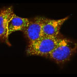
While cells with lots of iron, like red blood cells, may be red, usually cells are colorless.

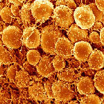
How many different types of cells can be found inside the human body?
There are about 200 cell types and a few trillion total cells in the human body. That does not include bacteria, fungi and mites that live on the body.

Is it possible to have too many or not enough cells?
The answer depends on cell type. For example, within the immune system, there are many examples of diseases that are caused by too many or not enough cells. When too many immune cells accumulate, patients get very large spleens and lymph nodes. When too few immune cells develop, patients have difficulty fighting infections.

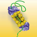
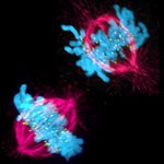
How fast does it take for a cell to produce two daughter cells?
Some cells, for example bacterial ones, can produce daughter cells very fast when nutrients are available. The doubling time for E. coli bacteria is 20 minutes. Other cells in the human body take hours or days or even years to divide.

Do skin cells stretch or multiply when you gain weight?
The size of cells is tightly regulated and maintained so they do not stretch much. As the surface area of the body increases with weight gain, the number of skin cells increases.

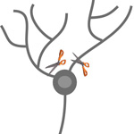
Why do cells self-destruct?
The term for cellular self-destruction is “apoptosis” or “programmed cell death.” Apoptosis is very important for normal development of humans and other animals as it ensures that we do not have too many cells and that “unhealthy” cells can be eliminated without causing harm to the surrounding cells. For instance, did you know that human embryos have webbing between their fingers and toes (just like ducks!)? Apoptosis eliminates the cells that form the web so that you are born with toes and fingers.

In what field is there a need for new scientists?
I would say that there is a need for scientists who can work at the interface between the biological and biomedical sciences and the data sciences. Knowing sophisticated mathematics and having computer skills to address questions like ‘what does this biomedical data tell us about particular diseases’ is still a challenge.

What is a scientist’s daily work day like? Is all of your time spent in a lab testing or like in an office throwing ideas around?
There are lots of different kinds of jobs a scientist can have. Many work in labs where they get to do experiments AND throw ideas around. Working in a lab is a lot of fun—you learn things about the world that no one has known before (how cool is that?). Other important jobs that scientists can do include writing about science as a journalist, helping other scientists patent new technologies they invent as a patent agent or lawyer, or working on important scientific policy issues for the government or other organizations.

Capitalizing on Cellular Conversations

0 comments
Fat cells
Fat cells such as these listen for incoming signals like FGF21, which tells them to burn more fat. Credit: David Gregory and Debbie Marshall. All rights reserved by Wellcome Images.

Living things are chatty creatures. Even when they’re not making actual sounds, organisms constantly communicate using chemical signals that course through their systems. In multicellular organisms like people, brain cells might call, “I’m in trouble!” signaling others to help mount a protective response. Single-celled organisms like bacteria may broadcast, “We have to stick together to survive!” so they can coordinate certain activities that they can’t carry out solo. In addition to sending out signals, cells have to receive information. To help them do this, they use molecular “ears” called receptors on their surfaces. When a chemical messenger attaches to a receptor, it tells the cell what’s going on and causes a response.

Scientists are following the dialogue, learning how cell signals affect health and disease. They’re also starting to take part in the cellular conversations, inserting their own comments with the goal of developing therapies that set a diseased system right.

Continue reading this new Inside Life Science article

Dendrites Show Ability to Regenerate After Injury

0 comments
Dendrites
Cutting off the dendrites from nerve cells in fruit flies revealed that they can regenerate. Credit: Melissa Rolls, Penn State University.

When a bone breaks, it might slice axons—the part of nerve cells that sends information to other cells—and potentially cause loss of mobility or feeling. Prior research had shown that a damaged nerve cell could repair such an injury through the regrowth of axons. Scientists at Penn State University wondered if dendrites—the part of nerve cells that receive information from other nerve cells—could also regenerate. To find out, Melissa Rolls and her team cut off the dendrites from nerve cells in fruit flies. Instead of dying, as was expected, the cells regrew dendrites. The research also revealed that dendrite regeneration happens independently of axon regeneration, leading investigators to believe there are two separate regeneration pathways: one for axons and one for dendrites. Learning more about this new dendrite regrowth pathway might one day lead to new approaches for healing injured nerve cells, including those damaged after a stroke.

Learn more:
Penn State News Release Exit icon
Rolls Lab

Animal Cells ‘Reach Out and Touch’ to Communicate

0 comments
Cytonemes in the fruit fly tracheal system.
Threadlike cytonemes (at right) convey signals between cells in the developing fruit fly tracheal system. Credit: Sougata Roy, University of California, San Francisco. View larger image

Scientists have long known that multicellular organisms use biological molecules produced by one cell and sensed by another to transmit messages that, for instance, guide proper development of organs and tissues. But it’s been a puzzle as to how molecules dumped out into the fluid-filled spaces between cells can precisely home in on their targets.

Using living tissue from fruit flies, a team led by Thomas Kornberg of the University of California, San Francisco, has shown that typical cells in animals can talk to each other via long, thin cell extensions called cytonemes (Latin for “cell threads”) that may span the length of 50 or 100 cells. The point of contact between a cytoneme and its target cell acts as a communications bridge between the two cells.

Until now, only nerve cells (neurons) were known to communicate this way. “This is an exciting finding,” says NIGMS’ Tanya Hoodbhoy. “Neurons are not the only ‘reach out and touch someone’ cells.”

This work also was funded by NIH’s National Heart, Lung, and Blood Institute.

Learn more:

UCSF News Release Exit icon

Mapping Approach Yields Insulin Secretion Pathway Insights

0 comments
TMEM24 protein (green) and insulin (red) in pancreatic beta cells (yellow).  Credit: Balch Lab, the Scripps Research Institute.
The interactions of TMEM24 protein (green) and insulin (red) in pancreatic beta cells are shown in yellow. Credit: Balch Lab, the Scripps Research Institute.
View larger image

The identities of the proteins that drive insulin production and release from pancreatic beta cells have largely been a mystery. In new work from the lab of William Balch of the Scripps Research Institute, researchers isolated and then identified all the insulin-bound proteins from mouse beta cells. The results provided a roadmap of the protein interactions that lead to insulin production, storage and secretion. The researchers used the roadmap to identify a protein called TMEM24, which was abundant in beta cells and binds readily to insulin. Balch and his team uncovered that TMEM24, whose involvement in insulin secretion was previously unknown, effectively regulates slower insulin release and could have a key role in maintaining control of glucose levels in the blood. The scientists hope that this roadmap of insulin-interacting proteins will lead to the development of new, targeted approaches to treating type 2 diabetes and a similar insulin-related condition called metabolic syndrome.

Learn more:

The Scripps Research Institute News Release Exit icon
Balch Lab Exit icon

Cells Merrily ‘Row’ Without Sensor Proteins

0 comments
Messenger proteins (left). When these proteins aren’t activated, the cell doesn’t move (right). Credit: Devreotes Lab, Johns Hopkins University School of Medicine.
Messenger proteins help the cell make large projections (left). When these proteins aren’t activated, the cell doesn’t move (right). Credit: Devreotes Lab, Johns Hopkins University School of Medicine. View larger image

A new study from Peter Devreotes Exit icon, Pablo Iglesias Exit icon and other scientists at Johns Hopkins University sheds light on the way in which cells get around the body to promote embryo development, wound healing and even cancer metastasis. Here’s how they describe cell movement and their findings:

Think of the cell as a rowboat. Sensor proteins on the outside pass on directional signals to messenger proteins that serve as the cell’s coxswain. The coxswain then commands other members of the molecular crew to stay in sync, propelling the cell forward. If there are no sensor signals, the coxswain can still coordinate the cell’s movement, just not in any specific direction—it’s like a boat without a rudder.

Scientists previously thought that the messenger proteins needed the sensor ones to produce both directional and random movements. Because defects in the messenger proteins have been linked to many types of cancer, the new work suggests these molecules could serve as a drug target for immobilizing tumor cells.

Learn more:

Johns Hopkins University School of Medicine News Release Exit icon

Protein Helps Chromosomes ‘Speed Date’ During Cell Division

0 comments
A cell in two stages of division: prometaphase (top) and metaphase (bottom). Credit: Lilian Kabeche, Dartmouth.
This image shows a cell in two stages of division: prometaphase (top) and metaphase (bottom). To form identical daughter cells, chromosome pairs (blue) separate via the attachment of microtubules made up of tubulin proteins (pink) to specialized structures on centromeres (green). Credit: Lilian Kabeche, Dartmouth.

Chromosome segregation during cell division is like speed dating, according to Geisel School of Medicine at Dartmouth researcher Duane Compton. He and postdoctoral fellow Lilian Kabeche learned that protein cyclin A plays moderator, helping to properly separate chromosomes via the attachment of microtubule fibers to kinetochore structures. Here’s how Compton described the process:

“The chromosomes are testing the microtubules for compatibility—that is, looking for the right match—to make sure there are correct attachments and no errors. The old view of this process held that chromosomes and microtubules pair up individually to find the correct attachment, like conventional dating. However, our results show that the system is more like speed dating. All the chromosomes have to try many connections with microtubules in a short amount of time. Then they all make their final choices at the same time. Cyclin A acts like a timekeeper or referee to make sure no one makes bad connections prematurely.”

Such bad connections can cause chromosome segregation errors that lead to cells with an abnormal number of chromosomes, a hallmark of cancer cells. So in addition to aiding our understanding of a fundamental biological process, the new insights may point to potential ways to correct such errors.

Learn more:

Dartmouth News Release Exit icon
Compton Lab Exit icon

Cool Video: How Bee Venom Toxin Kills Cells

0 comments
Credit: Huey Huang, Rice University.

Credit: Huey Huang, Rice University.

A new video, starring the toxin in bee venom, might help scientists design new drugs to combat bacterial infections. The video, by Rice University biophysicist Huey Huang Exit icon, condenses 6.5 minutes into less than a minute to show how the toxin, called melittin, destroys an animal or bacterial cell.

What looks like a red balloon is an artificial cell filled with red dye. Melittin molecules are colored green and float on the cell’s surface like twigs on a pond. As melittin accumulates on the cell’s membrane, the membrane expands to accommodate it. In the video, the membrane stretches into a column on the left.

When melittin levels reach a critical threshold, countless pinhole leaks burst open in the membrane. The cell’s vital fluids—red dye in the video—leak out through these pores. Within minutes, the cell collapses.

Many organisms use such a pore-forming technique to kill attacking bacterial cells. This research reveals molecular-level details of the strategy, bringing pharmaceutical scientists a step closer to harnessing it in the design of new antibiotics.