Tag: Cellular Processes

Dendrites Show Ability to Regenerate After Injury

0 comments
Dendrites
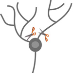
Cutting off the dendrites from nerve cells in fruit flies revealed that they can regenerate. Credit: Melissa Rolls, Penn State University.

When a bone breaks, it might slice axons—the part of nerve cells that sends information to other cells—and potentially cause loss of mobility or feeling. Prior research had shown that a damaged nerve cell could repair such an injury through the regrowth of axons. Scientists at Penn State University wondered if dendrites—the part of nerve cells that receive information from other nerve cells—could also regenerate. To find out, Melissa Rolls and her team cut off the dendrites from nerve cells in fruit flies. Instead of dying, as was expected, the cells regrew dendrites. The research also revealed that dendrite regeneration happens independently of axon regeneration, leading investigators to believe there are two separate regeneration pathways: one for axons and one for dendrites. Learning more about this new dendrite regrowth pathway might one day lead to new approaches for healing injured nerve cells, including those damaged after a stroke.

Learn more:
Penn State News Release Exit icon
Rolls Lab

Animal Cells ‘Reach Out and Touch’ to Communicate

0 comments
Cytonemes in the fruit fly tracheal system.
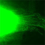
Threadlike cytonemes (at right) convey signals between cells in the developing fruit fly tracheal system. Credit: Sougata Roy, University of California, San Francisco. View larger image

Scientists have long known that multicellular organisms use biological molecules produced by one cell and sensed by another to transmit messages that, for instance, guide proper development of organs and tissues. But it’s been a puzzle as to how molecules dumped out into the fluid-filled spaces between cells can precisely home in on their targets.

Using living tissue from fruit flies, a team led by Thomas Kornberg of the University of California, San Francisco, has shown that typical cells in animals can talk to each other via long, thin cell extensions called cytonemes (Latin for “cell threads”) that may span the length of 50 or 100 cells. The point of contact between a cytoneme and its target cell acts as a communications bridge between the two cells.

Until now, only nerve cells (neurons) were known to communicate this way. “This is an exciting finding,” says NIGMS’ Tanya Hoodbhoy. “Neurons are not the only ‘reach out and touch someone’ cells.”

This work also was funded by NIH’s National Heart, Lung, and Blood Institute.

Learn more:

UCSF News Release Exit icon

Mapping Approach Yields Insulin Secretion Pathway Insights

0 comments
TMEM24 protein (green) and insulin (red) in pancreatic beta cells (yellow).  Credit: Balch Lab, the Scripps Research Institute.
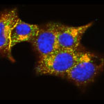
The interactions of TMEM24 protein (green) and insulin (red) in pancreatic beta cells are shown in yellow. Credit: Balch Lab, the Scripps Research Institute.
View larger image

The identities of the proteins that drive insulin production and release from pancreatic beta cells have largely been a mystery. In new work from the lab of William Balch of the Scripps Research Institute, researchers isolated and then identified all the insulin-bound proteins from mouse beta cells. The results provided a roadmap of the protein interactions that lead to insulin production, storage and secretion. The researchers used the roadmap to identify a protein called TMEM24, which was abundant in beta cells and binds readily to insulin. Balch and his team uncovered that TMEM24, whose involvement in insulin secretion was previously unknown, effectively regulates slower insulin release and could have a key role in maintaining control of glucose levels in the blood. The scientists hope that this roadmap of insulin-interacting proteins will lead to the development of new, targeted approaches to treating type 2 diabetes and a similar insulin-related condition called metabolic syndrome.

Learn more:

The Scripps Research Institute News Release Exit icon
Balch Lab Exit icon

Cells Merrily ‘Row’ Without Sensor Proteins

0 comments
Messenger proteins (left). When these proteins aren’t activated, the cell doesn’t move (right). Credit: Devreotes Lab, Johns Hopkins University School of Medicine.
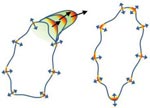
Messenger proteins help the cell make large projections (left). When these proteins aren’t activated, the cell doesn’t move (right). Credit: Devreotes Lab, Johns Hopkins University School of Medicine. View larger image

A new study from Peter Devreotes Exit icon, Pablo Iglesias Exit icon and other scientists at Johns Hopkins University sheds light on the way in which cells get around the body to promote embryo development, wound healing and even cancer metastasis. Here’s how they describe cell movement and their findings:

Think of the cell as a rowboat. Sensor proteins on the outside pass on directional signals to messenger proteins that serve as the cell’s coxswain. The coxswain then commands other members of the molecular crew to stay in sync, propelling the cell forward. If there are no sensor signals, the coxswain can still coordinate the cell’s movement, just not in any specific direction—it’s like a boat without a rudder.

Scientists previously thought that the messenger proteins needed the sensor ones to produce both directional and random movements. Because defects in the messenger proteins have been linked to many types of cancer, the new work suggests these molecules could serve as a drug target for immobilizing tumor cells.

Learn more:

Johns Hopkins University School of Medicine News Release Exit icon

Protein Helps Chromosomes ‘Speed Date’ During Cell Division

0 comments
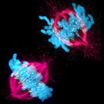
A cell in two stages of division: prometaphase (top) and metaphase (bottom). Credit: Lilian Kabeche, Dartmouth.
This image shows a cell in two stages of division: prometaphase (top) and metaphase (bottom). To form identical daughter cells, chromosome pairs (blue) separate via the attachment of microtubules made up of tubulin proteins (pink) to specialized structures on centromeres (green). Credit: Lilian Kabeche, Dartmouth.

Chromosome segregation during cell division is like speed dating, according to Geisel School of Medicine at Dartmouth researcher Duane Compton. He and postdoctoral fellow Lilian Kabeche learned that protein cyclin A plays moderator, helping to properly separate chromosomes via the attachment of microtubule fibers to kinetochore structures. Here’s how Compton described the process:

“The chromosomes are testing the microtubules for compatibility—that is, looking for the right match—to make sure there are correct attachments and no errors. The old view of this process held that chromosomes and microtubules pair up individually to find the correct attachment, like conventional dating. However, our results show that the system is more like speed dating. All the chromosomes have to try many connections with microtubules in a short amount of time. Then they all make their final choices at the same time. Cyclin A acts like a timekeeper or referee to make sure no one makes bad connections prematurely.”

Such bad connections can cause chromosome segregation errors that lead to cells with an abnormal number of chromosomes, a hallmark of cancer cells. So in addition to aiding our understanding of a fundamental biological process, the new insights may point to potential ways to correct such errors.

Learn more:

Dartmouth News Release Exit icon
Compton Lab Exit icon

Cool Video: How Bee Venom Toxin Kills Cells

0 comments
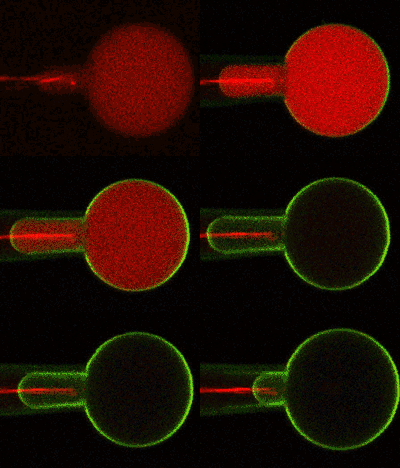
Credit: Huey Huang, Rice University.

Credit: Huey Huang, Rice University.

A new video, starring the toxin in bee venom, might help scientists design new drugs to combat bacterial infections. The video, by Rice University biophysicist Huey Huang Exit icon, condenses 6.5 minutes into less than a minute to show how the toxin, called melittin, destroys an animal or bacterial cell.

What looks like a red balloon is an artificial cell filled with red dye. Melittin molecules are colored green and float on the cell’s surface like twigs on a pond. As melittin accumulates on the cell’s membrane, the membrane expands to accommodate it. In the video, the membrane stretches into a column on the left.

When melittin levels reach a critical threshold, countless pinhole leaks burst open in the membrane. The cell’s vital fluids—red dye in the video—leak out through these pores. Within minutes, the cell collapses.

Many organisms use such a pore-forming technique to kill attacking bacterial cells. This research reveals molecular-level details of the strategy, bringing pharmaceutical scientists a step closer to harnessing it in the design of new antibiotics.