Tag: Computational Biology

PECASE Honoree Sohini Ramachandran Studies the Genetic Foundations of Traits in Diverse Populations

0 comments
Headshot of Sohini Ramachandran. Sohini Ramachandran, Brown University.
Credit: Danish Saroee/Swedish Collegium for Advanced Study.

Recent advances in computing enable researchers to explore the life sciences in ways that would have been impossible a few decades ago. One new tool is the ability to sequence genomes, revealing people’s full DNA blueprints. The collection of more and more genetic data allows researchers to compare the DNA of many people and observe variations, including those shared by people with a common ancestry.

Sohini Ramachandran Link to external web site, Ph.D., is director of the Center for Computational Molecular Biology and associate professor of biology and computer science at Brown University in Providence, Rhode Island. She is also a recent recipient of the Presidential Early Career Award for Scientists and Engineers (PECASE). Dr. Ramachandran researches the causes and consequences of human genetic variations using computer models. Starting with genomic data from living people, her lab applies statistical methods, mathematical modeling, and computer simulations to discover how human populations moved and changed genetically over time.

Continue reading “PECASE Honoree Sohini Ramachandran Studies the Genetic Foundations of Traits in Diverse Populations”

Interview With a Scientist – Rommie Amaro: Computational and Theoretical Model Builder

0 comments

Many researchers who search for anti-cancer drugs have labs filled with chemicals and tissue samples. Not Rommie Amaro. Her work uses computers to analyze the shape and behavior of a protein called p53. Defective versions of p53 are associated with more human cancers than any other malfunctioning protein.

Continue reading “Interview With a Scientist – Rommie Amaro: Computational and Theoretical Model Builder”

How Cells Manage Chance

3 comments

We asked the heads of our scientific divisions to tell us about some of the big questions in fundamental biomedical science that researchers are investigating with NIGMS support. This article is the second in an occasional series that explores these questions and explains how pursuing the answers could advance understanding of important biological processes.

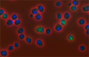
Sample slide, variability of mRNA in yeast cells
The number of copies of mRNA molecules (bright green) observed here in yeast cells (dark blue) fluctuates randomly. Credit: David Ball, Virginia Tech.

For some health conditions, the cause is clear: A single altered gene is responsible. But for many others, the path to disease is more complex. Scientists are working to understand how factors like genetics, lifestyle and environmental exposures all contribute to disease. Another important, but less well-known, area of investigation is the role of chance at the molecular level.

One team working in this field is led by John Tyson at Virginia Tech. The group focuses on how chance events affect the cell division cycle, in which a cell duplicates its contents and splits into two. This cycle is the basis for normal growth, reproduction and the replenishment of skin, blood and other cells throughout the body. Errors in the cycle are associated with a number of conditions, including birth defects and cancer. Continue reading “How Cells Manage Chance”

The Simple Rules Bacteria Follow to Survive

1 comment
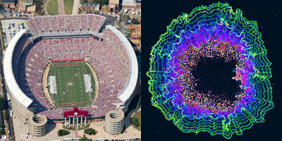
Left: Football stadium. Right: Colored contoured lines showing the periodic stops in the growth of a bacterial colony
Football image credit: Stock image. The colored contoured lines show the periodic stops in the growth of a bacterial colony. Credit: Süel Lab, UCSD.

What do these images of football fans and bacterial cells have in common? By following simple rules, each individual allows the group to accomplish tasks none of them could do alone—a stadium wave that ripples through the crowd or a cell colony that rebounds after antibiotic treatment.

These collective behaviors are just a few examples of what scientists call emergent phenomena. While the reasons for the emergence of such behavior in groups of birds, fish, ants and other creatures is well understood, they’ve been less clear in bacteria. Two independent research teams have now identified some of the rules bacterial cells follow to enable the colony to persist. Continue reading “The Simple Rules Bacteria Follow to Survive”

Unprecedented Views of HIV

0 comments

Visualizations can give scientists unprecedented views of complex biological processes. Here’s a look at two new ones that shed light on how HIV enters host cells.

Animation of HIV’s Entry Into Host Cells

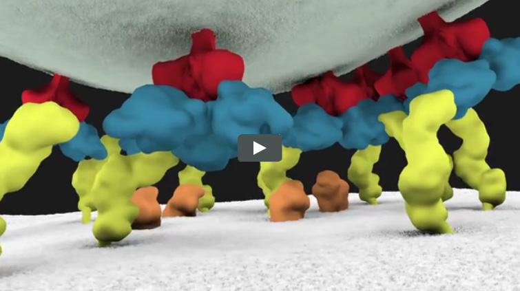
Screen shot of the video
This video animation of HIV’s entry into a human immune cell is the first one released in Janet Iwasa’s current project to visualize the virus’ life cycle. As they’re completed, the animations will be posted at http://scienceofhiv.org Exit icon.

We previously introduced you to Janet Iwasa, a molecular animator who’s visualized complex biological processes such as cells ingesting materials and proteins being transported across a cell membrane. She has now released several animations from her current project of visualizing HIV’s life cycle Exit icon. The one featured here shows the virus’ entry into a human immune cell.

“Janet’s animations add great value by helping us consider how complex interactions between viruses and their host cells actually occur in time and space,” says Wes Sundquist, who directs the Center for the Structural Biology of Cellular Host Elements in Egress, Trafficking, and Assembly of HIV Exit icon at the University of Utah. “By showing us how different steps in viral replication must be linked together, the animations suggest hypotheses that hadn’t yet occurred to us.”

Continue reading “Unprecedented Views of HIV”

Intercepting Amyloid-Forming Proteins

0 comments
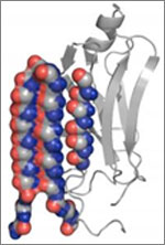
Structure of a protein involved in disease-associated amyloid fibrils.
A molecule targets the intermediary structure of a protein involved in disease-associated amyloid fibrils. Credit: University of Washington.

Alzheimer’s disease, type 2 diabetes and many other illnesses are linked to the buildup of proteins whose structures have changed into shapes that enable the formation of cell-entangling threads called amyloid fibrils. About 10 years ago, researchers led by Valerie Daggett of the University of Washington used computer simulations to suggest that such proteins, on their way to creating fibrils, form an intermediary structure called an alpha sheet that’s even more toxic to cells than fibrils. Now Daggett’s team has experimentally investigated this possibility. The scientists made alpha sheet molecules expected to bind to amyloid-forming proteins in the computationally predicted intermediate state. When they tested the molecules on two amyloid disease-related proteins, they observed a substantial reduction in fibril formation. The work is still very preliminary, but it highlights a potential new avenue for treating a range of amyloid-related diseases.

This work also was funded by NIH’s National Institute of Allergy and Infectious Diseases.

Learn more:
University of Washington News Release Exit icon
Daggett Lab Exit icon
Monster Mash: Protein Folding Gone Wrong Article from Inside Life Science

Meet Rhiju Das

1 comment
Rhiju Das
Credit: Rhiju Das
Rhiju Das
Fields: Biophysics and biochemistry
Works at: Stanford University
Born and raised in: The greater Midwest (Texas, Indiana and Oklahoma)
Studied at: Harvard University, Stanford University
When he’s not in the lab he’s: Enjoying the California outdoors with his wife and 3-year-old daughter
If he could recommend one book about science to a lay reader, it would be: “The Eighth Day of Creation,” about the revolution in molecular biology in the 1940s and 50s.

At the turn of the 21st century, Rhiju Das saw a beautiful picture that changed his life. Then a student of particle physics with a focus on cosmology, he attended a lecture unveiling an image of the ribosome—the cellular machinery that assembles proteins in every living creature. Ribosomes are enormous, complicated machines made up of many proteins and nucleic acids similar to DNA. Deciphering the structure of a ribosome—the 3-D image Das saw—was such an impressive feat that the scientists who accomplished it won the 2009 Nobel Prize in chemistry.

Das, who had been looking for a way to apply his physics background to a research question he could study in a lab, had found his calling.

“It was an epiphany—it was just flabbergasting to me that a hundred thousand atoms could find their way into such a well-defined structure at atomic resolution. It was like miraculously a bunch of nuts and bolts had self-assembled into a Ferrari,” recounted Das. “That inspired me to drop everything and learn everything I could about nucleic acid structure.”

Das focuses on the nucleic acid known as RNA, which, in addition to forming part of the ribosome, plays many roles in the body. As is the case for most proteins, RNA folds into a 3-D shape that enables it to work properly.

Das is now the head of a lab at Stanford University that unravels how the structure and folding of RNA drives its function. He has taken a unique approach to uncovering the rules behind nucleic acid folding: harnessing the wisdom of the crowd.

Together with his collaborator, Adrien Treuille of Carnegie Mellon University, Das created an online, multiplayer video game called EteRNA Exit icon. More than a mere game, it does far more than entertain. With its tagline “Played by Humans, Scored by Nature,” it’s upending how scientists approach RNA structure discovery and design.

Das’ Findings

Treuille and Das launched EteRNA after working on another computer game called Foldit, which lets participants play with complex protein folding questions. Like Foldit, EteRNA asks players to assemble, twist and revise structures—this time of RNA—onscreen.

But EteRNA takes things a step further. Unlike Foldit, where the rewards are only game points, the winners of each round of EteRNA actually get to have their RNA designs synthesized in a wet lab at Stanford. Das and his colleagues then post the results—which designs resulted in a successful, functional RNAs and which didn’t—back online for the players to learn from.

In a paper published in the Proceedings of the National Academy of Sciences Exit icon, Das and his colleagues showed how effective this approach could be. The collective effort of the EteRNA participants—which now number over 100,000—was better and faster than several established computer programs at solving RNA design problems, and even came up with successful new structural rules never before proposed by scientists or computers.

“What was surprising to me was their speed,” said Das. “I had just assumed that it would take a year or so before players were really able to analyze experimental data, make conclusions and come up with robust rules. But it was one of the really shocking moments of my life when, about 2 months in, we plotted the performance of players against computers and they were out-designing the computers.”

“As far as I can tell, none of the top players are academic scientists,” he added. “But if you talk to them, the first thing they’ll tell you is not how many points they have in the game but how many times they’ve had a design synthesized. They’re just excited about seeing whether or not their hypotheses were correct or falsified. So I think the top players truly are scientists—just not academic ones. They get a huge kick out of the scientific method, and they’re good at it.”

To capture lessons learned through the crowd-sourcing approach, Das and his colleagues incorporated successful rules and features into a new algorithm for RNA structure discovery, called EteRNABot, which has performed better than older computer algorithms.

“We thought that maybe the players would react badly [to EteRNABot], that they would think they were going to be automated out of existence,” said Das. “But, as it turned out, it was exciting for them to have their old ideas put into an algorithm so they could move on to the next problems.”

You can try EteRNA for yourself at http://eternagame.org Exit icon. Das and Treuille are always looking for new players and soliciting feedback.

Fitting a Piece of the Protein-Function Puzzle

0 comments
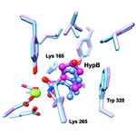
Comparison of the predicted binding of the substrate to the active site of HpbD (blue) with the binding sites determined experimentally by crystallography (magenta). Credit: Matt Jacobson, University of California, San Francisco; Steve Almo, Albert Einstein College of Medicine.
The image shows a comparison of the predicted binding of the substrate to the active site of HpbD (blue) with the binding sites determined experimentally by crystallography (magenta). Credit: Matt Jacobson, University of California, San Francisco; Steve Almo, Albert Einstein College of Medicine.

Sequencing the genomes of almost 7,000 organisms has identified more than 40 million proteins. But how do we figure out what all these proteins do? New results from an initiative led by John Gerlt of the University of Illinois suggest a possible method for identifying the functions of unknown enzymes, proteins that speed chemical reactions within cells. Using high-powered computing, the research team modeled how the structure of a mystery bacterial enzyme, HpbD, might fit like a puzzle piece into thousands of proteins in known metabolic pathways. Since an enzyme acts on other molecules, finding its target or substrate can shed light on its function. The new method narrowed HpbD’s candidate substrate down from more than 87,000 to only four. Follow-up lab work led to the actual substrate, tHypB, and determined the enzyme’s biological role. This combination of computational and experimental methods shows promise for uncovering the functions of many more proteins.

Learn more:

University of Illinois at Urbana-Champaign News Release
Gerlt Lab
Enzyme Function Initiative