Tag: Diseases

Nature’s Medicine Cabinet

2 comments

More than 70 percent of new drugs approved within the past 30 years originated from trees, sea creatures and other organisms that produce substances they need to survive. Since ancient times, people have been searching the Earth for natural products to use—from poison dart frog venom for hunting to herbs for healing wounds. Today, scientists are modifying them in the laboratory for our medicinal use. Here’s a peek at some of the products in nature’s medicine cabinet.

Vampire bat

A protein called draculin found in the saliva of vampire bats is in the last phases of clinical testing as a clot-buster for stroke patients. Vampire bats are able to drink blood from their victims because draculin keeps blood from clotting. The first phases of clinical trials have shown that the protein’s anti-coagulative properties could give doctors more time to treat stroke patients and lower the risk of bleeding in the brain.

Continue reading “Nature’s Medicine Cabinet”

Digging Deeply Into Data for the Causes of Disease

1 comment

Hunting for the cause of a disease can be like tracing a river back to its many sources. Myriad factors, large and small, may contribute to a condition. One approach to the search focuses on the massive amounts of genomic and other biological data that scientists are gathering in the course of their studies. To examine this data and look for meaningful patterns and other clues, scientists turn to bioinformatics, a field focused on the development of analytical methods and software tools.

Here are a few examples of how National Institutes of Health-funded scientists are using bioinformatics to dig deeply into data and learn more about the development of diseases, including Huntington’s, preeclampsia and asthma.

Huntington’s Disease

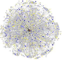
Network of proteins that interact with huntingtin

Researchers have mapped a network of 2,141 proteins that all interact either directly or through one other protein with huntingtin (red), the protein associated with Huntington’s disease. Credit: Cendrine Tourette, Buck Institute for Research on Aging, J Biol Chem 2014 Mar 7;289(10):6709-26 Exit icon.

The cause of Huntington’s disease, a degenerative neurological disorder with no known cure, may appear simple. It begins with a change in a single gene that alters the shape and functioning of the huntingtin protein. But this protein, whether in its normal or altered form, does not act alone. It interacts with other proteins, which in turn interact with others.

A research team led by Robert Hughes of the Buck Institute for Research on Aging set out to understand how this ripple effect contributes to the breakdown in normal cellular function associated with Huntington’s disease. The scientists used experimental and computational approaches to map a network of 2,141 proteins that interact with the huntingtin protein either directly or through one other protein. They found that many of these proteins were involved in cell movement and intercellular communication. Understanding how the huntingtin protein leads to mistakes in these cellular processes could help scientists pursue new approaches to developing treatments. Continue reading “Digging Deeply Into Data for the Causes of Disease”

Correcting a Cellular Routing Error Could Treat Rare Kidney Disease

0 comments
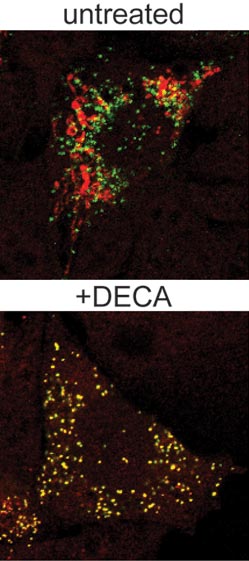
AGT protein and peroxisomes in untreated and treated cells.
The altered AGT protein (red) and peroxisomes (green) appear in different places in untreated cells (top), but they appear together (shown in yellow) in cells treated with DECA (bottom). Credit: Carla Koehler/Reproduced with permission from Proceedings of the National Academy of Sciences USA. View larger image.

Our cells have organized systems to route newly created proteins to the places where they’re needed to do their jobs. For some people born with a rare and potentially fatal genetic kidney disorder called PH1, a genetically altered form of a particular protein mistakenly ends up in mitochondria instead of in another organelle, the peroxisome. This cellular routing error of the AGT protein results in the harmful buildup of oxalate, which leads to kidney failure and other problems at an early age.

In new work led by UCLA biochemist Carla Koehler Exit icon, researchers used a robotic screening system to identify a compound that interferes with the delivery of proteins to mitochondria. Koehler’s team Exit icon showed that adding a small amount of the compound, known as DECA, to cells grown in the laboratory prevented the altered form of the AGT protein from going to the mitochondria and sent it to the peroxisome. The compound also reduced oxalate levels in a cell model of PH1.

The team’s findings suggest that DECA, which is already approved by the Food and Drug Administration for treating certain bacterial infections, could be a promising candidate for treating children affected by PH1. And, Koehler notes, the screening strategy that she and her team used to identify DECA as a potential therapy may help researchers identify other new therapies for the disorder.

This work was funded in part by NIH under grant R01GM061721.

Mighty Mitochondria

0 comments
Mitochondria from the heart muscle cell of a rat.
Mitochondria (red) from the heart muscle cell of a rat. Nearly all our cells have these structures. Credit: Thomas Deerinck, National Center for Microscopy and Imaging Research Exit icon.

Meet mitochondria: cellular compartments, or organelles, that are best known as the powerhouses that convert energy from the food we eat into energy that runs a range of biological processes.

As you can see in this close-up of mitochondria from a rat’s heart muscle cell, the organelles have an inner membrane that folds in many places (and that appears here as striations). This folding vastly increases the surface area for energy production. Nearly all our cells have mitochondria, but cells with higher energy demands have more. For instance, a skin cell has just a few hundred, while the cell pictured here has about 5,000.

Scientists are discovering there’s more to mitochondria than meets the eye, especially when it comes to understanding and treating disease.

Read more about mitochondria in this Inside Life Science article.

Restoring the Function of an Immune Receptor Involved in Crohn’s Disease

4 comments
Gut bacteria
Receptor proteins bind to bacterial cell wall fragments, initiating an immune response to remove bad gut bacteria. Credit: S. Melanie Lee, Caltech; Zbigniew Mikulski and Klaus Ley, La Jolla Institute for Allergy and Immunology.

Our bodies depend on a set of immune receptors to remove harmful bacteria and control the growth of helpful bacteria in our guts. Genetic changes that alter the function of the receptors can have an adverse effect and result in chronic inflammatory diseases like Crohn’s disease. Catherine Leimkuhler Grimes and Vishnu Mohanan of the University of Delaware researched a Crohn’s-associated immune receptor, NOD2, to figure out how it can lose the ability to respond properly to bacteria. In the process, they identified the involvement of a protective protein called HSP70. Increasing HSP70 levels in kidney, colon and white blood cells appeared to restore NOD2 function. This work represents a first step toward developing drugs to treat Crohn’s disease.

This work was funded in part by an Institutional Development Award (IDeA) Network of Biomedical Research Excellence (INBRE) grant.

Raking the Family Tree for Disease-Causing Variations

1 comment
Silhouettes of people with nucleic acid sequences and a stethoscope.
A new software tool analyzes disease-causing genetic variations within a family. Credit: NIH’s National Human Genome Research Institute.

Changes in your DNA sequence occur randomly and rarely. But when they do happen, they can increase your risk of developing common, complex diseases, such as cancer. One way to identify disease-causing variations is to study the genomes of family members, since the changes typically are passed down to subsequent generations.

To rake through a family tree for genetic variations with the highest probabilities of causing a disease, researchers combined several commonly-used statistical methods into a new software tool called pVAAST. The scientific team, which included Mark Yandell and Lynn Jorde of the University of Utah and Chad Huff of the University of Texas MD Anderson Cancer Center, used the tool to identify the genetic causes of a chronic intestinal inflammation disease and of developmental defects affecting the heart, face and limbs.

The results confirmed previously identified genetic variations for the developmental diseases and pinpointed a previously unknown variation for the intestinal inflammation. Together, the findings confirm the ability of the tool to detect disease-causing genetic changes within a family. Another research team has already used the software tool to discover rare genetic changes associated with family cases of breast cancer. These studies are likely just the beginning for studying genetic patterns of diseases than run in a family.

This work also was funded by NIH’s National Institute of Diabetes and Digestive and Kidney Diseases; National Cancer Institute; National Human Genome Research Institute; National Heart, Lung, and Blood Institute; and National Institute of Mental Health.

Learn more:
University of Utah News Release (no longer available)
Yandell Exit icon, Jorde and Huff Exit icon Labs

New Compound Improves Insulin Levels in Preliminary Studies

1 comment
compound
A new compound (chemical structure shown here) blocks the activity of an enzyme involved in glucose regulation.

The discovery of a compound that slows the natural degradation of insulin in mice opens up a new area of investigation in the search for drugs to treat diabetes. The research team, which included David Liu Exit icon and Alan Saghatelian Exit icon of Harvard University, Markus Seeliger of Stony Brook University School of Medicine, and Wei-Jen Tang Exit icon of the University of Chicago focused on insulin-degrading enzyme, or IDE. Using a method called DNA-templated synthesis, the scientists made 14,000 small molecules and found one that bound to the enzyme, suggesting it might modulate the enzyme’s activity. Work in test tubes and in animal models confirmed this—and showed that blocking IDE activity improved insulin levels and glucose tolerance. The researchers also learned that the enzyme is misnamed: In addition to insulin, it degrades two other hormones involved in glucose regulation.

NIGMS’ Peter Preusch says, “This is a very interesting fusion of chemical methods and biology that has uncovered new basic science findings about insulin processing with potential clinical impact.”

This work also was funded by NIH’s National Cancer Institute and the Office of the Director.

Learn more:
Harvard University News Article Exit icon
Chemistry of Health Booklet

Meet Elizabeth Grice

0 comments
Elizabeth Grice
Elizabeth Grice
First job: Detasseling corn
Favorite food: Chocolate
Pets: An adopted shelter cat, Dolce
Favorite city: Athens, Greece
Hidden talent: Baking creative desserts
Credit: Bill Branson, NIH

Imagine a landscape with peaks and valleys, folds and niches, cool, dry zones and hot, wet ones. Every inch is swarming with diverse communities, but there are no cities, no buildings, no fields and no forests.

You’ve probably thought little about the inhabitants, but you see their environment every day. It’s your largest organ—your skin.

Elizabeth Grice, an assistant professor at the University of Pennsylvania, studies the skin microbiome to learn how and why bacteria colonize particular places on the body. Already, she’s found that the bacterial communities on healthy skin are different from those on diseased skin.

She hopes her work will point to ways of treating certain skin diseases, especially chronic wounds. “I like to think that I am making discoveries that will impact the way medicine is practiced,” she says.

Grice’s Findings

To investigate what role bacteria play in diabetic wounds, Grice and her colleagues took skin swabs from both diabetic and healthy mice, and then compared the two. They found that diabetic mice had about 40 times more bacteria on their skin, but it was concentrated into few species. A more diverse array of bacteria colonized the skin of healthy mice.

The researchers then gave each mouse a small wound and spent 28 days swabbing the sites to collect bacteria and observing how the skin healed. They found that wounds on diabetic mice started to increase in size at the same time as wounds on healthy mice began to heal. In about 2 weeks, most healthy mice looked as good as new. But most of the wounds on diabetic mice had barely healed even after a month.

Interestingly, bacterial communities in the wounds became more diverse in both groups of mice as they healed—although the wounds on diabetic mice still had less diversity than the ones on healthy mice.

“Bacterial diversity is probably a good thing, especially in wounds,” says Grice. “Often, potentially infectious bacteria are found on normal skin and are kept in check by the diversity of bacteria surrounding them.”

Grice and her colleagues also found distinctly different patterns of gene activity between the two groups of mice. As a result, the diabetic mice put out a longer-lasting immune response, including inflamed skin. Scientists believe prolonged inflammation might slow the healing process.

Grice’s team suspects that one of the main types of bacteria found on diabetic wounds, Staphylococcus, makes one of the inflammation-causing genes more active.

Now that they know more about the bacteria that thrive on diabetic wounds, Grice and her colleagues are a step closer to looking at whether they could reorganize these colonies to help the wounds heal.

Content adapted from the NIGMS Findings magazine article Body Bacteria.

Basic Research Fuels Medical Advances

0 comments
Genetic defect that causes myotonic dystrophy type 2 and used that information to design drug candidates to counteract the disease. Credit: Ilyas Yildirim, Northwestern University.
Scientists revealed a detailed image of the genetic change that causes myotonic dystrophy type 2 and used that information to design drug candidates to counteract the disease. Credit: Ilyas Yildirim, Northwestern University. View larger image

This image may look complicated, but it tells a fairly straightforward tale about basic research: Learning more about basic life processes can pave the way for medical and other advances.

In this example, researchers led by Matthew Disney of the Scripps Research Institute’s Florida campus focused on better understanding the structural underpinnings of myotonic dystrophy type 2, a relatively rare, inherited form of adult-onset muscular dystrophy. While this work is still in the preliminary stages, it may hold potential for someday treating the disorder.

Some 300,000 NIH-funded scientists are working on projects aimed at improving disease diagnosis, treatment and prevention, often through increasing understanding of basic life processes.

Read the complete Inside Life Science article.

Chemist Phil Baran Joins “Genius” Ranks as MacArthur Fellow

1 comment
Cake decorated with a two-dimensional structure of the molecule, stephacidin B
When Baran’s research team succeeds in synthesizing an important natural product, the group sometimes celebrates with a cake decorated with a two-dimensional structure of the molecule. This molecule, stephacidin B, was isolated from a fungus and has anticancer properties. See images of other Baran lab cakes.

As a newly appointed MacArthur Fellow, Phil Baran is now officially a genius. The MacArthur award recognizes “exceptionally creative” individuals who have made significant contributions to their field and are expected to continue doing so. Baran, a synthetic organic chemist at Scripps Research Institute in La Jolla, Calif., was recognized today for “inventing efficient, scalable, and environmentally sound methods” for building, from scratch, molecules produced in nature. Many of these natural products have medicinal properties. Baran has already concocted a host of natural products, including those with the ability to kill bacteria or cancer cells. In addition to emphasizing the important pharmaceutical applications of his work, Baran embraces its creative aspects: “The area of organic chemistry is such a beautiful one because one can be both an artist and an explorer at the same time,” he said in the MacArthur video interview Exit icon.

Learn more:

NIGMS “Meet a Chemist” Profile of Baran
NIH Director’s Blog Post on Baran’s Recent Work