Tag: Viruses

Interview with a Scientist: Wes Sundquist, How the Host Immune System Fights HIV

1 comment

For more than 30 years, NIGMS has supported the structural characterization of human immunodeficiency virus (HIV) enzymes and viral proteins. This support has been instrumental in the development of crucial drugs for antiretroviral therapy such as protease inhibitors. NIGMS continues to support further characterization of viral proteins as well as cellular and viral complexes. These complexes represent the fundamental interactions between the virus and its host target cell and, as such, represent potential new targets for therapeutic development.

In this second in a series of three video interviews with NIGMS-funded researchers probing the structure of HIV, Wes Sundquist, professor of biochemistry at the University of Utah, discusses his lab’s studies of how HIV uses factors in host cells to replicate itself. In particular, Sundquist focuses on the ESCORT pathway that enables HIV to leave infected cells and spread infection elsewhere.

Continue reading “Interview with a Scientist: Wes Sundquist, How the Host Immune System Fights HIV”

Interview With a Scientist: Irwin Chaiken, Rendering HIV Inert

0 comments

For more than 30 years, NIGMS has supported the structural characterization of human immunodeficiency virus (HIV) enzymes and viral proteins. This support has been instrumental in the development of crucial drugs for antiretroviral therapy such as protease inhibitors. NIGMS continues to support further characterization of viral proteins as well as cellular and viral complexes. These complexes represent the fundamental interactions between the virus and its host target cell and, as such, represent potential new targets for therapeutic development.

Continue reading “Interview With a Scientist: Irwin Chaiken, Rendering HIV Inert”

Viruses: Manufacturing Tycoons?

0 comments
Pseudomonas chlororaphis

A computer image shows a bacterial cell invaded by a virus. The virus uses the cell to copy itself many times. It has built a protein compartment (red, rough circle surrounding the center) to house its DNA. Viral heads (blue, smaller pentagonal shapes spread through out) and tails (pink, rod shaped near the edges) are essential parts of a finished viral particle. The small, light blue particles are the bacterium’s own protein-making ribosomes. Credit: Vorrapon Chaikeeratisak, Kanika Khanna, Axel Brilot and Katrina Nguyen.

As inventors and factory owners learned during the Industrial Revolution, the best way to manufacture a lot of products is with an assembly line that follows a set of precisely organized steps employing many copies of identical and interchangeable parts. Some viruses are among life’s original mass producers: They use sophisticated organization principles to turn bacterial cells into virus particle factories.

Scientists at the University of California, San Diego, and the University of California, San Francisco, used cutting-edge techniques to watch a bacteria-infecting virus (bacteriophage) set up its particle-making factory inside a host cell.

The image above shows this happening inside a Pseudomonas chlororaphis, a soil-borne bacterium that protects plants against fungal pathogens. The virus builds a compartment (red, rough circle surrounding the center) that helps organize an assembly line for making copies of itself. The compartment looks like a cell nucleus, which bacteria do not have, and it functions like a nucleus by keeping activities that directly involve DNA separate from other cellular functions. Continue reading “Viruses: Manufacturing Tycoons?”

Researchers Score Goal with New Atomic-Scale Model of Salmonella-Infecting Virus

1 comment

An atomic-scale model of a virus that infects the Salmonella bacterium. Credit: C. Hryc and the Chiu Lab, Baylor College of Medicine.

This sphere could be a prototype design for the 2018 World Cup official match soccer ball, but you won’t see it dribbled around any soccer fields. The image is actually an atomic-scale model of a virus that infects the Salmonella bacterium. Like a soccer ball, both are approximately spherical shapes created by a combination of hexagonal (six-sided) and pentagonal (five-sided) units. Wah Chiu, a biochemist at Baylor College of Medicine, and his colleagues used new computational methods to construct the model from more than 20,000 cryo-electron microscopy (cryo-EM) images. Cryo-EM is a sophisticated technique that uses electron beams for visualizing frozen samples of proteins and other biological specimens.

The researchers’ model, published in a recent issue of PNAS, shows the virus’ protein shell, or capsid, that encloses the virus’ genetic material. Each color shows capsid proteins having the same interactions with their neighbors. The fine resolution allowed researchers to identify the protein interactions essential to building a stable shell. They developed a new approach to checking the accuracy and reliability of the virus model and reporting what parts are the most certain. The approach could be used to evaluate other complex biological structures, potentially leading to better quality models and new avenues for drug design and development.

This research is funded in part by NIH under grants R01GM079429, P01GM063210, P41GM103832, PN2EY016525, T15LM007093.

Online Virus Tracking Tool Nextstrain Wins Inaugural Open Science Prize

0 comments
Nextstrain’s analysis of the genomes from Zika virus obtained in 25 countries over the past few years. 
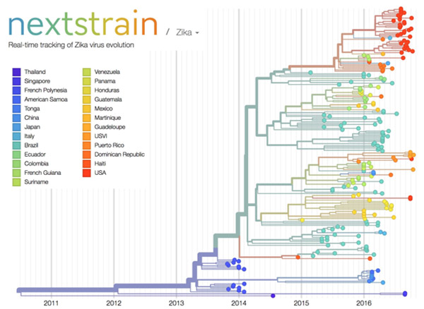
Credit: Trevor Bedford and Richard Neher, nextstrain.org.

Over the past decade, scientists and clinicians have eagerly deposited their burgeoning biomedical data into publicly accessible databases. However, a lack of computational tools for sharing and synthesizing the data has prevented this wealth of information from being fully utilized.

In an attempt to unleash the power of open-access data, the National Institutes of Health, in collaboration with the Howard Hughes Medical Institute and Britain’s Wellcome Trust, launched the Open Science Prize. Last week, after a multi-stage public voting process, the inaugural award was announced. The winner of the grand prize—and $230,000—is a prototype computational tool called nextstrain Exit icon that tracks the spread of emerging viruses such as Ebola and Zika. This tool could be especially valuable in revealing the transmission patterns and geographic spread of new outbreaks before vaccines are available, such as during the 2013-2016 Ebola epidemic and the current Zika epidemic.

An international team of scientists—led by NIGMS grantee Trevor Bedford of the Fred Hutchinson Cancer Research Center, Seattle, and Richard Neher Exit icon of Biozentrum at the University of Basel, Switzerland—developed nextstrain as an open-access system capable of sharing and analyzing viral genomes. The system mines viral genome sequence data that researchers have made publicly available online. nextstrain then rapidly determines the evolutionary relationships among all the viruses in its database and displays the results of its analyses on an interactive public website.

The image here shows nextstrain’s analysis of the genomes from Zika virus obtained in 25 countries over the past few years. Plotting the relatedness of these viral strains on a timeline provides investigators a sense of how the virus has spread and evolved, and which strains are genetically similar. Researchers can upload genome sequences of newly discovered viral strains—in this case Zika—and find out in short order how their new strain relates to previously discovered strains, which could potentially impact treatment decisions.

Nearly 100 interdisciplinary teams comprising 450 innovators from 45 nations competed for the Open Science Prize. More than 3,500 people from six continents voted online for the winner. Other finalists for the prize focused on brain maps, gene discovery, air-quality monitoring, neuroimaging and drug discovery.

nextstrain was funded in part by NIH under grant U54GM111274.

Our Complicated Relationship With Viruses

9 comments
Illustration of Influenza Virus H1N1. Swine Flu.
Nearly 10 percent of the human genome is derived from the genes of viruses. Credit: Stock image.

When viruses infect us, they can embed small chunks of their genetic material in our DNA. Although infrequent, the incorporation of this material into the human genome has been occurring for millions of years. As a result of this ongoing process, viral genetic material comprises nearly 10 percent of the modern human genome. Over time, the vast majority of viral invaders populating our genome have mutated to the point that they no longer lead to active infections. But they are not entirely dormant.

Sometimes, these stowaway sequences of viral genes, called “endogenous retroviruses” (ERVs), can contribute to the onset of diseases such as cancer. They can also make their hosts susceptible to infections from other viruses. However, scientists have identified numerous cases of viral hitchhikers bestowing crucial benefits to their human hosts—from protection against disease to shaping important aspects of human evolution, such as the ability to digest starch.

Protecting Against Disease

Geneticists Cedric Feschotte, Edward Chuong and Nels Elde Exit icon at the University of Utah have discovered that ERVs lodged in the human genome can jump start the immune system.

For a virus to successfully make copies of itself inside a host cell, it needs molecular tools similar to the ones its host normally uses to translate genes into proteins. As a result, viruses have tools meticulously shaped by evolution to commandeer the protein-producing machinery of human cells.

Continue reading “Our Complicated Relationship With Viruses”

Viral Views: New Insights on Infection Strategies

0 comments

The following images show a few ways in which cutting-edge research tools are giving us clearer views of viruses—and possible ways to disarm them. The examples, which highlight work involving HIV and the coronavirus, were funded in part by our Biomedical Technology Research Resources program.

Uncloaking HIV’s Camouflage

HIV capsid with (right, red) and without (left) a camouflaging human protein.
HIV capsid with (right, red) and without (left) a camouflaging human protein. Credit: Juan R. Perilla, Klaus Schulten and the Theoretical and Computational Biophysics Group, University of Illinois at Urbana-Champaign.

To sneak past our immune defenses and infect human cells, HIV uses a time-honored strategy—disguise. The virus’ genome is enclosed in a protein shell called a capsid (on left) that’s easily recognized and destroyed by the human immune system. To evade this fate, the chrysalis-shaped capsid cloaks itself with a human protein known as cyclophilin A (in red, on right). Camouflaged as human, the virus gains safe passage into and through a human cell to deposit its genetic material in the nucleus and start taking control of cellular machinery.

Biomedical and technical experts teamed up to generate these HIV models at near-atomic resolution. First, structural biologists at the Pittsburgh Center for HIV Protein Interactions Exit icon used a technique called cryo-electron microscopy (cryo-EM) to get information on the shape of an HIV capsid as well as the capsid-forming proteins’ connections to each other and to cyclophilin A. Then experts at the Resource for Macromolecular Modeling and Bioinformatics fed the cryo-EM data into their visualization and simulation programs to computationally model the physical interactions among every single atom of the capsid and the cyclophilin A protein. The work revealed a previously unknown site where cyclophilin A binds to the capsid, offering new insights on the biology of HIV infection. Continue reading “Viral Views: New Insights on Infection Strategies”

Finding Adventure: Blake Wiedenheft’s Path to Gene Editing

1 comment
Blake Wiedenheft
Blake Wiedenheft
Grew up in: Fort Peck, Montana
Fields: Microbiology, biochemistry, structural biology
Job site: Montana State University
Secret talent: Being a generalist; enjoying many different subjects and activities
When not in the lab, he’s: Running, biking, skiing or playing scrabble with his grandmother

Scientific discoveries are often stories of adventure. This is the realization that set Blake Wiedenheft on a path toward one of the hottest areas in biology.

His story begins in Montana, where he grew up and now lives. Always exploring different interests, Wiedenheft decided in his final semester at Montana State University (MSU) in Bozeman to volunteer for Mark Young, a scientist who studies plant viruses. Even though he majored in biology, Wiedenheft had spent little time in a lab and hadn’t even considered research as a career option. Continue reading “Finding Adventure: Blake Wiedenheft’s Path to Gene Editing”

Unusual DNA Form May Help Virus Withstand Extreme Conditions

0 comments
A, B and Z DNA.
DNA comes in three forms: A, B and Z. Credit: A-DNA, B-DNA and Z-DNA by Zephyris (Richard Wheeler) under CC BY-SA 3.0.

DNA researcher Rosalind Franklin Exit icon first described an unusual form of DNA called the A-form in the early 1950s (Franklin, who died in 1958, would have turned 95 next month). New research on a heat- and acid-loving virus has revealed surprising information about this DNA form, which is one of three known forms of DNA: A, B and Z.

“Many people have felt that this A-form of DNA is only found in the laboratory under very non-biological conditions, when DNA is dehydrated or dry,” says Edward Egelman Exit icon in a University of Virginia news release Exit icon about the recent study. But considered with earlier studies on bacteria by other researchers, the new findings suggest that the A-form “appears to be a general mechanism in biology for protecting DNA.” Continue reading “Unusual DNA Form May Help Virus Withstand Extreme Conditions”

Meet Nels Elde and His Team’s Amazing, Expandable Viruses

0 comments
Nels Elde, Ph.D.
Credit: Kristan Jacobsen
Nels Elde, Ph.D.
Fields: Evolutionary genetics, virology, microbiology, cell biology
Works at: University of Utah, Salt Lake City
When not in the lab, he’s: Gardening, supervising pets, procuring firewood
Hobbies: Canoeing, skiing, participating in facial hair competitions

“I really look at my job as an adventure,” says Nels Elde. “The ability to follow your nose through different fields is what motivates me.”

Elde has used that approach to weave evolutionary genetics, bacteriology, virology, genomics and cell biology into his work. While a graduate student at the University of Chicago and postdoctoral researcher at the Fred Hutchinson Cancer Research Center in Seattle, he became interested in how interactions between pathogens (like viruses and bacteria) and their hosts (like humans) drive the evolution of both parties. He now works in Salt Lake City, where, as an avid outdoorsman, he draws inspiration from the wild landscape.

Outside the lab, Elde keeps diverse interests and colorful company. His best friend wrote a song about his choice of career as a cell biologist. (You can hear this song at the end of the 5-minute video Exit icon in which Elde explains his work.) Continue reading “Meet Nels Elde and His Team’s Amazing, Expandable Viruses”