Year: 2018

Molecular Fireworks: How Microtubules Form Inside Cells

1 comment
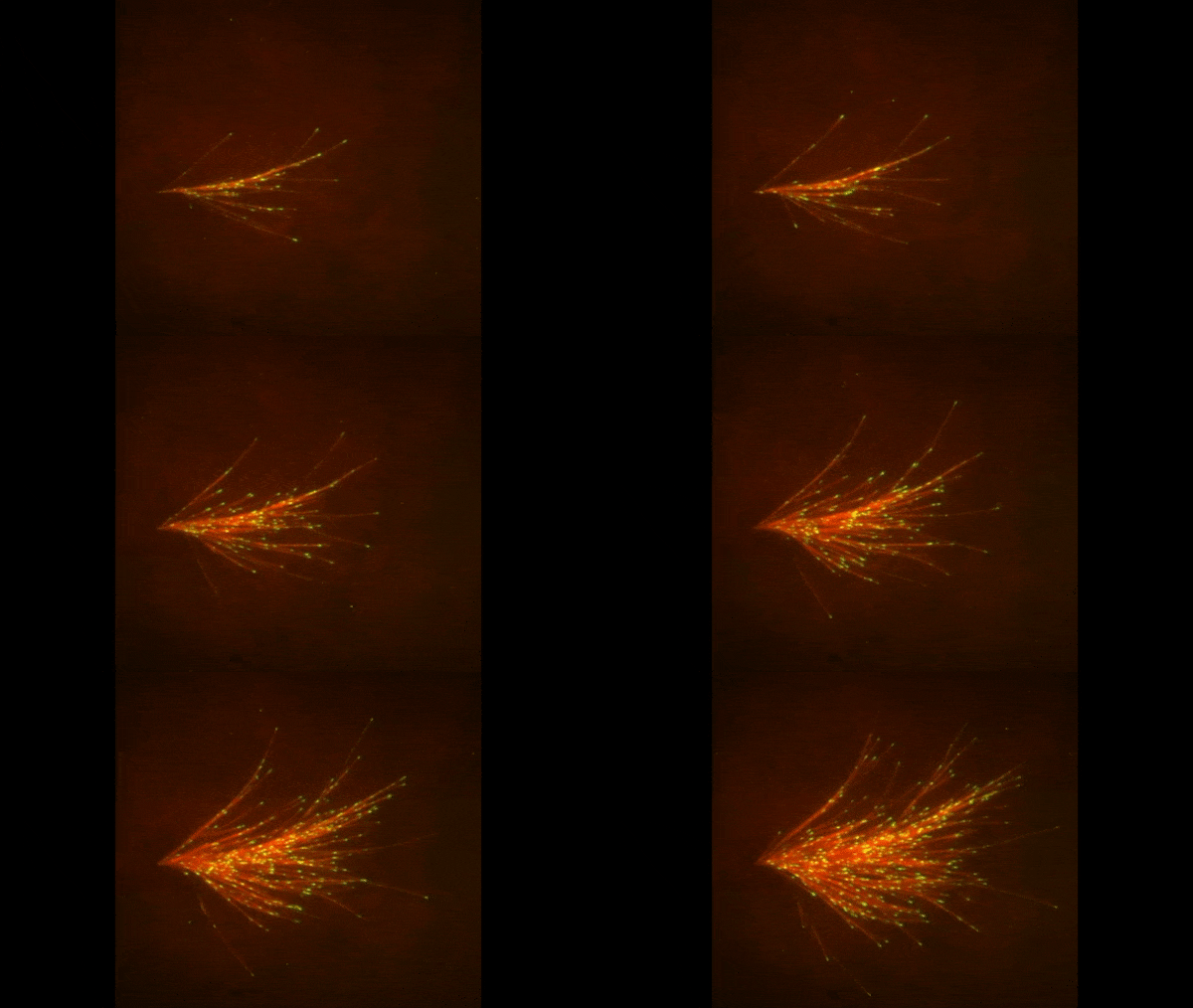
A video depicting red strands of various lengths exploding outward from a focal point at the left. The strands are tipped in neon green.
       Microtubules sprout from one another. Credit: Petry lab, Princeton University.

The red spray pictured here may look like fireworks erupting across the night sky on July 4th, but it’s actually a rare glimpse of tiny protein strands called microtubules sprouting and growing from one another in a lab. Microtubules are the largest of the molecules that form a cell’s skeleton. When a cell divides, microtubules help ensure that each daughter cell has a complete set of genetic information from the parent. They also help organize the cell’s interior and even act as miniature highways for certain proteins to travel along.

As their name suggests, microtubules are hollow tubes made of building blocks called tubulins. Scientists know that a protein called XMAP215 adds tubulin proteins to the ends of microtubules to make them grow, but until recently, the way that a new microtubule starts forming remained a mystery.

Sabine Petry Link to external web site and her colleagues at Princeton University developed a new imaging method for watching microtubules as they develop and found an important clue to the mystery. They adapted a technique called total internal reflection fluorescence (TIRF) microscopy, which lit up only a tiny sliver of a sample from frog egg (Xenopus) tissue. This allowed the scientists to focus clearly on a few of the thousands of microtubules in a normal cell. They could then see what happened when they added certain proteins to the sample.

Continue reading “Molecular Fireworks: How Microtubules Form Inside Cells”

Interview With a Scientist: Andrew Goodman, Separating Causation and Correlation in the Microbiome

0 comments

You’ve likely heard some variation of the statistic that there are at least as many microbial cells in our body as human cells. You may have also heard that the microscopic bugs that live in our guts, on our skins, and every crevice they can find, collectively referred to as the human microbiome, are implicated in human health. But do these bacteria, fungi, archaea, protists, and viruses cause disease, or are the specific populations of microbes inside us a result of our state of health? That’s the question that drives the research in the lab of Andrew Goodman Link to external web site, associate professor of microbial pathogenesis at Yale University.

Continue reading “Interview With a Scientist: Andrew Goodman, Separating Causation and Correlation in the Microbiome”

Best Documentary: Cells Record Their Own Lives Using CRISPR

1 comment

Suppose you were a police detective investigating a robbery. You’d appreciate having a stack of photographs of the crime in progress, but you’d be even happier if you had a detailed movie of the robbery. Then, you could see what happened and when. Research on cells is somewhat like this. Until recently, scientists could take snapshots of cells in action, but they had trouble recording what cells were doing over time. They were getting an incomplete picture of the events occurring in cells.

Researchers have started solving this problem by combining some old knowledge—that DNA is spectacularly good at storing information—with a popular new research tool called CRISPR. CRISPR (clustered regularly interspaced short palindromic repeats) is an immune system feature in bacteria that helps them to remember and destroy viruses that infect them. CRISPR can change DNA sequences in precise ways; and it’s programmable, meaning that researchers can tell CRISPR where to make a change on a DNA strand, and even what kind of change to make. By linking cellular events to CRISPR, researchers can make something like a movie that captures many pieces of information in the form of DNA changes that researchers can read back later. These pieces of information include timing, duration, and intensity of events, such as the turning on of a specific protein pathway or the exposure of the cell to pathogens (i.e. germs). Here, we look at some of the ways NIGMS-funded research teams and others are using CRISPR to capture these kinds of data within DNA sequences.

Left: Rectangle containing magnetic tape illustrated as a black strip wound on two spools. Closeup of the magnetic tape beneath as a blue strip with orange lines to indicate stored audio signals. Text reads: data in magnetic tape. Center: Four, white capsule-shaped bacteria, with three rows of connected shapes (black diamonds, blue and orange rectangles) beneath to illustrate stored biological signals in bacteria. Text reads: data in CRISPR tape in cells. Right: Numerous capsule-shaped bacteria in different colors, each containing a black strip wound on two spools

An audio recorder stores audio signals into a magnetic tape medium (left). Similarly, a microscopic data recorder stores biological signals into a CRISPR tape in bacteria (middle). An enormous amount of information can be stored across multiple bacterial cells (right). Credit: Wang Lab/Columbia University Medical Center.

Round and Round: mSCRIBE Creates a Continuous Recording Loop

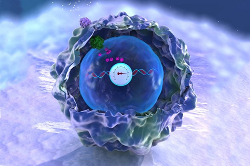
A dark blue-green cell with textured surface containing a round, blue meter with a white dial. The dial reads a magenta ribbon of DNA and records over time the number of cellular events that occur. The cellular events are depicted by purple, green, and smaller magenta clusters moving through the cell.
MIT bioengineers, led by Timothy Lu, have devised a memory storage system illustrated here as a DNA-embedded meter that records the activity of a signaling pathway in a human cell. Credit: Timothy Lu lab, MIT.

CRISPR uses an enzyme called Cas9 like a surgical knife, to slice both strands of a cell’s DNA at precise points. A cut like this sends the cell scrambling to repair the damage. Often, the repair effort results in changes, or errors, in the repaired strand that pile up at a known rate. Timothy Lu Link to external web site and his colleagues at the Massachusetts Institute of Technology (MIT) decided to turn this cut-repair-error system into a way to record certain events inside a cell. They call their method mSCRIBE (mammalian synthetic cellular recorder integrating biological events).

Continue reading “Best Documentary: Cells Record Their Own Lives Using CRISPR”

Teens Explore Science and Health through Game Design

0 comments

Educators often struggle to teach teens about sexual and reproductive health. Hexacago Health Academy (HHA) Link to external web site, an education program from the University of Chicago, leverages the fun activity of gameplay to impart these lessons to young people from Chicago’s South Side community. Funded by the Student Education Partnership Award (SEPA)Link to external web site, part of the National Institute of General Medical Sciences (NIGMS), in 2015, HHA assists teachers in their goal of helping teen students gain awareness and control over their health and also learn about careers in STEM Link to external web site and health fields.

Woman in a black buisness suit with arms crossed standing against a wall and smiling
Melissa Gilliam, founder of Ci3. Credit: Anna Knott, Chicago Magazine.

Genesis of HHA

HHA was cofounded by Melissa Gilliam Link to external web site, a University of Chicago professor of Obstetrics/Gynecology and Pediatrics and founder of the Center for Interdisciplinary Inquiry & Innovation in Sexual and Reproductive Health (Ci3) Link to external web site. During a 2013 summer program with high school students, Gilliam and Patrick Jagoda Link to external web site, associate professor of English and Cinema & Media Studies, and cofounder of Ci3’s Game Changer Chicago Design Lab, introduced the students to their STEM-based alternate reality game called The Source, in which a young woman crowdsources player help to solve a mystery that her father has created for her.

From their experience with The Source, Gilliam and Jagoda quickly learned that students not only wanted to play games but to design them too. What followed was the Game Changer Lab’s creation of the Hexacago game board, as well as the launch of HHA, a SEPA-funded project that the lab oversees.

Hexacago Game Board

At the core of HHA is the Hexacago game board, which displays the city of Chicago, along with Lake Michigan, a train line running through the city, and neighborhoods gridded into a hexagonal pattern.

HHA students not only play games designed from the Hexacago board template, but also design their own games from it that are intended to inspire behavior change in health-related situations and improve academic performance.

High school students seated at a table with a glossy, laminate test model of the Hexacago game and game pieces on top of it
Credit: Ci3 at the University of Chicago.

In this way, HHA is much more than just game design and play. “Students have no idea that what they’re doing is learning. In their minds, they’re really focused on designing games,” says Gilliam. “That’s the idea behind Hexacago Health Academy: helping people acquire deep knowledge of science and health issues by putting on the hat of a game designer.” Moreover, through the process of gameplay and design, students practice all the rich skills that result from teamwork, including collaborative learning, leadership, and communication.

Continue reading “Teens Explore Science and Health through Game Design”

Interview with a Scientist: Michael Summers, Using Nuclear Magnetic Resonance to Study HIV

0 comments

For more than 30 years, NIGMS has supported the structural characterization of human immunodeficiency virus (HIV) enzymes and viral proteins. This support has been instrumental in the development of crucial drugs for antiretroviral therapy such as protease inhibitors. NIGMS continues to support further characterization of viral proteins as well as cellular and viral complexes. These complexes represent the fundamental interactions between the virus and its host target cell and, as such, represent potential new targets for therapeutic development.

In this third in a series of three video interviews with NIGMS-funded researchers probing the structure of HIV, Michael Summers,Link to external web site professor of biochemistry at the University of Maryland, Baltimore County, discusses his use of nuclear magnetic resonance (NMR) technology to study HIV. Of recent interest to Summers has been using NMR to investigate how HIV’s RNA enables the virus to reproduce. His goals for this line of research are to develop treatments against HIV as well as learning how to best engineer viruses to deliver helpful therapies to individuals with a variety of diseases.

Continue reading “Interview with a Scientist: Michael Summers, Using Nuclear Magnetic Resonance to Study HIV”

Interview with a Scientist: Wes Sundquist, How the Host Immune System Fights HIV

1 comment

For more than 30 years, NIGMS has supported the structural characterization of human immunodeficiency virus (HIV) enzymes and viral proteins. This support has been instrumental in the development of crucial drugs for antiretroviral therapy such as protease inhibitors. NIGMS continues to support further characterization of viral proteins as well as cellular and viral complexes. These complexes represent the fundamental interactions between the virus and its host target cell and, as such, represent potential new targets for therapeutic development.

In this second in a series of three video interviews with NIGMS-funded researchers probing the structure of HIV, Wes Sundquist, professor of biochemistry at the University of Utah, discusses his lab’s studies of how HIV uses factors in host cells to replicate itself. In particular, Sundquist focuses on the ESCORT pathway that enables HIV to leave infected cells and spread infection elsewhere.

Continue reading “Interview with a Scientist: Wes Sundquist, How the Host Immune System Fights HIV”

Interview With a Scientist: Irwin Chaiken, Rendering HIV Inert

0 comments

For more than 30 years, NIGMS has supported the structural characterization of human immunodeficiency virus (HIV) enzymes and viral proteins. This support has been instrumental in the development of crucial drugs for antiretroviral therapy such as protease inhibitors. NIGMS continues to support further characterization of viral proteins as well as cellular and viral complexes. These complexes represent the fundamental interactions between the virus and its host target cell and, as such, represent potential new targets for therapeutic development.

Continue reading “Interview With a Scientist: Irwin Chaiken, Rendering HIV Inert”

CLAMP Helps Lung Cells Pull Together

0 comments
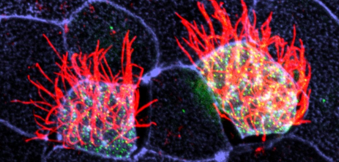
ALT TEXTCells covered with cilia (red strands) on the surface of frog embryos. Credit: The Mitchell Lab.
The outermost cells that line blood vessels, lungs, and other organs also act like guards, alert and ready to thwart pathogens, toxins, and other invaders that can do us harm. Called epithelial cells, they don’t just lie passively in place. Instead, they communicate with each other and organize their internal structures in a single direction, like a precisely drilled platoon of soldiers lining up together and facing the same way.

Lining up this way is crucial during early development, when tissues and organs are forming and settling into their final positions in the developing body. In fact, failure to line up in the correct way is linked to numerous birth defects. In the lungs, for instance, epithelial cells’ ability to synchronize with one another is important since these cells have special responsibilities such as carrying mucus up and out of lung tissue. When these cells can’t coordinate their functions, disease results.

Some lung epithelial cells are covered in many tiny, hair-like structures called cilia. All the cilia on lung epithelial cells must move uniformly in a tightly choreographed way to be effective in their mucus-clearing job. This is a unique example of a process called planar cell polarity (PCP) that occurs in cells throughout the body. Researchers are seeking to identify the signals cells use to implement PCP. Continue reading “CLAMP Helps Lung Cells Pull Together”

Interview with a Scientist: Jeramiah Smith on the Genomic Antics of an Ancient Vertebrate

0 comments

The first known descriptions of cancer come from ancient Egypt more than 3,500 years ago. Early physicians attributed the disease to several factors, including an imbalance in the body’s humoral fluids, trauma, and parasites. Only in the past 50 years or so have we figured out that mutations in critical genes are often the trigger. The sea lamprey, a slimy, snake-like blood sucker, is proving to be an ideal tool for understanding these mutations.

The sea lamprey, often called the jawless fish, is an ancient vertebrate whose ancestor diverged from the other vertebrate lineages (fish, reptiles, birds and mammals) more than 500 million years ago. Jeramiah Smith,Link to external web site associate professor of biology at the University of Kentucky, has discovered that lamprey have two separate genomes: a complete genome specific to their reproductive cells, consisting of 99 chromosomes (humans have 23 pairs) and another genome in which about 20 percent of genes have been deleted after development. Using the lamprey model, Smith and his colleagues have learned that many of these deleted genes—such as those that initiate growth pathways—are similar to human oncogenes (i.e., cancer-causing genes).

Continue reading “Interview with a Scientist: Jeramiah Smith on the Genomic Antics of an Ancient Vertebrate”

Pericytes: Capillary Guardians in the Brain

0 comments
ALT TEXT
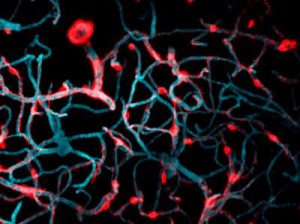
The long arms of pericytes cells (red) stretch along capillaries (blue) in a mouse brain. Credit: Andy Shih.

Nerve cells, or neurons, in our brains do amazing work, from telling our hearts to beat to storing our memories. But neurons cannot operate alone. Many kinds of cells support and regulate neurons and—like neurons—they can come under attack due to injuries or disorders, such as stroke or Alzheimer’s disease. Learning what jobs these cells do and how they respond to disease may show researchers new ways to treat central nervous system disorders. One type of support cell, the pericyte, plays some key roles in brain health. These cells are readily adaptable, even in adult brains, and can support a variety of functions.

Pericytes help with blood flow to nerve cells in the brain. They lie wrapped all along the huge networks of capillaries—the tiniest blood vessels—that both feed neurons and form the blood-brain barrier, which filters out certain substances from blood to protect the brain. Pericytes have a body that appears as a bump protruding from a capillary surface. Pericytes also have long thin arms that stretch along each capillary like a snake on a tree branch. These arms, called processes, reach almost to where the next pericyte process begins, without overlapping. This creates a pericyte chain that covers nearly the entire capillary network.

Pericytes are critical for blood vessel stability and blood-brain barrier function. They’re also known to die off as a result of trauma and disease. Andy ShihLink to external web site, Andree-Ann Berthiaume, and colleagues at the Medical University of South Carolina in Charleston, set up an imaging technique in mouse brains that allowed them to see what pericytes do under normal conditions as well as how these cells respond when some are damaged.

Continue reading “Pericytes: Capillary Guardians in the Brain”