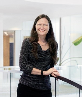
When she started college, Anne Carpenter, Ph.D., never guessed she’d one day create software for analyzing images of cells that would help identify potential medicines and that thousands of researchers would use. She wasn’t planning to become a computational biologist, or even to focus on science at all, but she’s now an institute scientist and the senior director of the Imaging Platform at the Broad Institute of Massachusetts Institute of Technology (MIT) and Harvard in Cambridge.
Starting Out in Science
Before beginning her undergraduate studies at Purdue University in West Lafayette, Indiana, Dr. Carpenter’s strongest interests were reading and writing. Then, her subjects expanded. “In college, I liked science as much as anything else, and I realized that was unusual, as a lot of other people really struggled with it. I decided to pursue science because I enjoyed it and the field had good job prospects,” she says. Dr. Carpenter majored in biology because she felt it had the “juiciest questions” as well as a direct impact on human health.
After completing her bachelor’s degree, Dr. Carpenter pursued a Ph.D. in cell biology at the University of Illinois Urbana-Champaign. She studied how chromatin responds to signals from estrogen receptors, and her research plan required capturing thousands of microscopy images—a task that would have taken months to do manually. Instead, Dr. Carpenter decided to try automating the microscope with computer programming, and after about a month of tinkering, she was successful, saving herself weeks of tedious, manual image collection.
Dr. Carpenter then had a different challenge: thousands of images to analyze. Again, she turned to computational tools, using automated image analysis to pull precise data from large collections of microscopy images. Dr. Carpenter knew she wanted to pursue this newfound passion after finishing her Ph.D., which led her to a postdoctoral researcher position at the Whitehead Institute in Cambridge, Massachusetts, where she focused on computational image analysis.
Applying Computer Science to Cells
Dr. Carpenter didn’t know it at the time, but her work at the Whitehead Institute would lead to the development of CellProfiler—open-source software for gathering quantitative data from cell images that has now been cited in more than 15,000 scientific papers. She started working on the tool to overcome a problem she encountered while using existing software to analyze images of fruit fly cells. Because that software was designed for human cells, it didn’t perform well with her fruit fly samples.
To solve the problem, Dr. Carpenter decided to develop more versatile software, enlisting the help of Thouis “Ray” Jones, Sc.D., who at the time was a computer science graduate student at MIT. “As I talked about the software we were developing, more and more people at the Whitehead Institute started coming to me and asking for help on their microscopy analysis projects. It became clear that the existing tools couldn’t accomplish many tasks that biologists were interested in, so I gradually spent more time writing software,” says Dr. Carpenter.
From Cell Images to Medicines
From the time she was an undergraduate, Dr. Carpenter had planned on working at a biomedical company, not an academic institution. However, she felt that her software would be valuable for the research community and realized that if she started a career in industry, the tool would likely never be fully developed. So instead of following her original plan, Dr. Carpenter founded a lab with Dr. Jones at the Broad Institute in 2007 that focused solely on computational work. They continued refining CellProfiler, and it’s now a ubiquitous tool in biology research. “All the major pharmaceutical companies use my software. It’s astonishing to me that something I created has that kind of impact,” says Dr. Carpenter.
Researchers in pharmaceutical companies and academia can use CellProfiler to analyze cell images in a variety of ways. For example, comparing cells affected by a rare genetic disease with healthy cells, CellProfiler may detect that the mitochondria in the disease-affected cells tend to be distributed differently than those in unaffected cells. Then, the scientists can treat the disease-affected cells with various potential medicines and determine if any of the treatments cause the distribution of the mitochondria to look more like that of a healthy cell. Using CellProfiler, researchers have identified at least six potential medicines that have entered clinical trials.
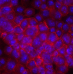
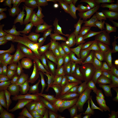
Dr. Carpenter went on to develop another tool, Cell Painting, in collaboration with Stuart Schreiber, Ph.D., that researchers can use to stain components of cells before analyzing images of them with CellProfiler. (Most cells are transparent, so scientists typically stain cellular structures of interest before analysis to make them easier for people or software to observe.) Cell Painting uses six fluorescent dyes to reveal eight cellular components—ranging from the nucleus to the plasma membrane—that are often affected by medicines and disease-causing genetic variants. Using these tools in tandem, or in combination, with other tools, enables researchers to examine many important features at the same time.
Moving Forward in Interdisciplinary Science
Dr. Carpenter continues to mine the rich information gleaned from images of cells to better understand human diseases and to identify potential medicines as a co-leader, with Shantanu Singh, Ph.D., of the Carpenter-Singh lab. The lab has compiled the largest public database of Cell Painting images in the world and plans to spend the next several years working with it and other huge libraries of cell images to support their current work. In addition, they hope to explore other areas such as creating a database linking genes with potential medicines that may affect their function, as well as predicting whether potential medicines might have negative side effects.
About half the members of the Broad Institute’s Imaging Platform team come from biology backgrounds, while the other half have focused on computational science. Dr. Carpenter looks for scientists who are curious about areas outside of their expertise and can communicate well with people who work in other fields. “Some of the most challenging biology problems remain because they require expertise from multiple disciplines. No human being can have the level of expertise in multiple fields that would be needed to solve those problems. That’s why team science, where you bring together experts from different domains, is so important,” she says.
Dr. Carpenter’s work is supported by the NIGMS Maximizing Investigators’ Research Award program through grant R35GM122547. She also receives funding through NIGMS grant P41GM135019.
This post is a great supplement to Pathways: The Imaging Issue.
Both this post and Pathways describe tools used for imaging microscopic biological structures such as cells.
Learn more in our Educator’s Corner.