Happy Valentine’s Day! In place of red roses, we hope you’ll accept a bouquet of beautiful scientific images featuring rich, red hues. Be sure to click all the way through to see the festive protein flowing through your blood!
For more scientific photos, illustrations, and videos in all the colors of the rainbow, visit our image and video gallery.
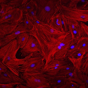
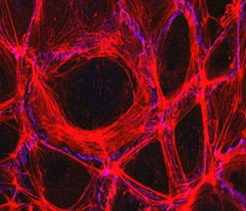
This image shows heart fibroblast cells from mice. A protein called actin in shown in red, and the cell nuclei are stained blue. Fibroblasts contribute to the formation of connective tissue, which supports and connects other tissues or organs in the body.
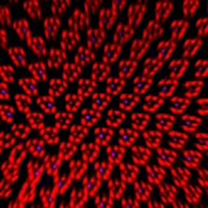
A close-up of a fruit fly's retina reveals a layer of cells at the back of the eye. Every red spot is a light-sensitive structure called a rhabdomere, and each blue dot is a light-sensing molecule known as rhodopsin-4.
This vibrant web is the endothelium—the thin layer of cells that lines our arteries and veins. The endothelium controls the movement of materials into and out of the bloodstream. Holding endothelial cells tightly together are specialized proteins that function like strong ropes (red) and others that act like cement (blue).
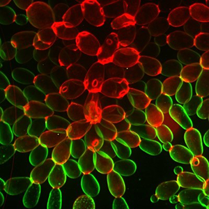
Researchers created this multicellular yeast, called snowflake yeast, from unicellular yeast through many generations of directed evolution. Stained cell membranes (green) and cell walls (red) reveal the connections between cells. Younger cells take up more cell membrane stain, while older cells take up more cell wall stain, leading to the color differences shown here.
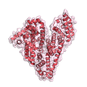
This heart is actually the molecular structure of serum albumin, the most abundant protein in the blood plasma of mammals, including humans. Serum albumin helps maintain normal water levels in our tissues and carries almost half of all calcium ions in our blood. This protein also transports some hormones, nutrients, and metals throughout the bloodstream.