Category: Genes

The Simple Rules Bacteria Follow to Survive

1 comment
Left: Football stadium. Right: Colored contoured lines showing the periodic stops in the growth of a bacterial colony
Football image credit: Stock image. The colored contoured lines show the periodic stops in the growth of a bacterial colony. Credit: Süel Lab, UCSD.

What do these images of football fans and bacterial cells have in common? By following simple rules, each individual allows the group to accomplish tasks none of them could do alone—a stadium wave that ripples through the crowd or a cell colony that rebounds after antibiotic treatment.

These collective behaviors are just a few examples of what scientists call emergent phenomena. While the reasons for the emergence of such behavior in groups of birds, fish, ants and other creatures is well understood, they’ve been less clear in bacteria. Two independent research teams have now identified some of the rules bacterial cells follow to enable the colony to persist. Continue reading “The Simple Rules Bacteria Follow to Survive”

Field Focus: Progress in RNA Interference Research

3 comments
Scientists first noticed what would later prove to be RNA interference when puzzling over an unexpected loss of color in petunia petals. Subsequent studies in roundworms revealed that double-stranded RNA can inactivate specific genes. Credit: Alisa Z. Machalek.

In less than two decades, RNA interference (RNAi)—a natural process cells use to inactivate, or silence, specific genes—has progressed from a fundamental finding to a powerful research tool and a potential therapeutic approach. To check in on this fast-moving field, I spoke to geneticists Craig Mello of the University of Massachusetts Medical School and Michael Bender of NIGMS. Mello shared the 2006 Nobel Prize in physiology or medicine with Andrew Fire Exit icon of Stanford University School of Medicine for the discovery of RNAi. Bender manages NIGMS grants in areas that include RNAi research.

How have researchers built on the initial discovery of RNAi?

A scientific floodgate opened after the 1998 discovery that it was possible to switch off specific genes by feeding microscopic worms called C. elegans double-stranded RNA that had the same sequence of genetic building blocks as a target gene. (Double-stranded RNA is a type of RNA molecule often found in, or produced by, viruses.) Scientists investigating gene function quickly began to test RNAi as a gene-silencing technique in other organisms and found that they could use it to manipulate gene activity in many different model systems. Additional studies led the way toward getting the technique to work in cells from mammals, which scientists first demonstrated in 2001. Soon, researchers were exploring the potential of RNAi to treat human disease.

Continue reading “Field Focus: Progress in RNA Interference Research”

Meet Sarkis Mazmanian and the Bacteria That Keep Us Healthy

1 comment
Sarkis K. Mazmanian
Credit: New York Academy of Sciences
Sarkis K. Mazmanian, Ph.D.
Born in: The country of Lebanon, moved to Los Angeles when he was 1
Fields: Microbiology, immunology, neuroscience
Works at: California Institute of Technology
Awards won: Many, including the MacArthur Foundation “Genius” grant
Most proud of: The success of his trainees! “There’s nothing that comes close to the gratification and joy I feel when a student or research fellow goes on to be an independent scientist.”
When not in the lab or mentoring students, he’s: Spending time with his family, including his 1-year-old-son or going for an occasional run

As a child, Sarkis Mazmanian frequently took things apart to figure out how they worked. At the age of 12, he dismantled his family’s entire television set—to the dismay of his parents and the unsuccessful TV repairman.

“I wasn’t aware of this at the time, but maybe that was some sort of a foreshadowing that I would enjoy science,” Mazmanian says. “Scientists take biological systems apart to understand how they work.”

Mazmanian never thought he’d become a microbiologist, let alone a leading expert in the field. He began studying microbiology at the University of California, Los Angeles (UCLA), because it was the major that allowed him to do the most hands-on research. But as soon as he entered the field, he fell in love with the complexities of microbial organisms and the efficiency of their functions. Continue reading “Meet Sarkis Mazmanian and the Bacteria That Keep Us Healthy”

How a Cell Knows Friend From Foe

1 comment

We asked the heads of our scientific divisions to tell us about some of the big questions in fundamental biomedical science that researchers are investigating with NIGMS support. This article is the first in an occasional series that will explore these questions and explain how pursuing the answers could advance understanding of important biological processes.

Video screen shot showing different strains of amoeba cells in red and green.
This video shows different strains of amoeba cells in red and green. As cells move toward one another, they use two sets of proteins to recognize others from the same strain. When close relatives meet, their proteins match and the cells join together to form a multicellular structure. When cells from different strains meet, their proteins don’t match, so they can’t aggregate. Credit: Shigenori Hirose, Baylor College of Medicine.

Cells are faced with many decisions: When’s the best time to produce a new protein? To grow and split into two? To treat another cell as an invader? Scientists are working to understand how cells make these and many other decisions, and how these decisions contribute to health and disease.

An active area of research on cell decisions focuses on allorecognition, the ability of an organism to distinguish its own cells from those of another. Immune cells use a system called the major histocompatibility complex (MHC) to identify which cells belong to the body and which are foreign. The particular set of MHC proteins on the outer surface of a cell helps immune cells decide whether it does not belong and should be attacked.

But the system isn’t perfect. Invading pathogens can go undetected, and the body can mistake its own cells for intruders. Continue reading “How a Cell Knows Friend From Foe”

Meet Nels Elde and His Team’s Amazing, Expandable Viruses

0 comments
Nels Elde, Ph.D.
Credit: Kristan Jacobsen
Nels Elde, Ph.D.
Fields: Evolutionary genetics, virology, microbiology, cell biology
Works at: University of Utah, Salt Lake City
When not in the lab, he’s: Gardening, supervising pets, procuring firewood
Hobbies: Canoeing, skiing, participating in facial hair competitions

“I really look at my job as an adventure,” says Nels Elde. “The ability to follow your nose through different fields is what motivates me.”

Elde has used that approach to weave evolutionary genetics, bacteriology, virology, genomics and cell biology into his work. While a graduate student at the University of Chicago and postdoctoral researcher at the Fred Hutchinson Cancer Research Center in Seattle, he became interested in how interactions between pathogens (like viruses and bacteria) and their hosts (like humans) drive the evolution of both parties. He now works in Salt Lake City, where, as an avid outdoorsman, he draws inspiration from the wild landscape.

Outside the lab, Elde keeps diverse interests and colorful company. His best friend wrote a song about his choice of career as a cell biologist. (You can hear this song at the end of the 5-minute video Exit icon in which Elde explains his work.) Continue reading “Meet Nels Elde and His Team’s Amazing, Expandable Viruses”

Field Focus: Our Microbial Menagerie

0 comments

Trillions of microorganisms inhabit us—inside and out. Scientists are surveying these microbial metropolises to learn more about their role in health. Microbiologists Darren Sledjeski of NIGMS and Andrew Goodman Exit icon of Yale University share a few details of what researchers have learned so far.

Vitruvian man filled with bacteria.
Researchers are surveying the microbes that inhabit us to learn more about their role in health. Credit: Andrew Goodman, Yale University.
  1. The majority of the microbes that inhabit us are bacteria. The rest of the microbial menagerie is fungi and viruses, including ones that infect the bacteria! Collectively, our resident microorganisms are referred to as the human microbiota, and their genomes are called the human microbiome.
  2. Our bodies harbor more bacterial cells than human ones. Even so, the microbiota accounts for less than 3 percent of a person’s body mass. That’s because our cells are up to 10,000 times bigger in volume than bacterial cells.
  3. Your collection of bacteria has more genes than you do. Scientists estimate that the genomes of gut bacteria contain 100-fold or more genes than our own genomes. For this reason, the human microbiome is sometimes called our second genome.
  4. Most of our microbes are harmless, and some are helpful. For example, harmless microbes on the skin keep infectious microbes from occupying that space. Microbes in the colon break down lactose and other complex carbohydrates that our bodies can’t naturally digest.
  5. Different microbes occupy different parts of the body. Some skin bacteria prefer the oily nooks near the nose, while others like the dry terrain of the forearm. Bacteria don’t all fare well in the same environment and have adapted to live in certain niches. The NIGMS Findings Magazine article Body Bacteria: Exploring the Skin’s Microbial Metropolis shows what types of bacteria colonize where.
Screenshot from the iBiology video.
Are we more microbial than human? Richard Losick, a microbiologist at Harvard University, explores that question in this video lecture produced by iBiology Exit icon.
  1. Each person’s microbiota is unique. The demographics of microbiota differ among individuals. Diet is one reason. Also, while a type of microbe might be part of one person’s normal microbial flora, it might not be part of another’s, and could potentially make that person sick.
  2. Host-microbial interactions are universal. Microbial communities may vary from person to person, but everyone’s got them, including other creatures. For this reason, researchers can use model organisms to tease apart the complexities of host-microbial interactions and develop broad principles for understanding them. The mouse is the most widely used animal model for microbiome studies.
  3. The role of microbiota in our health isn’t entirely clear. While it’s now well accepted that the microbial communities that inhabit us are actively involved in a range of conditions—from asthma to obesity—research studies have not yet pinpointed why or how. In other words, the results may suggest that the presence of a bacterial community is associated with a disease, but they don’t show cause and effect.
  4. Most of our microbes have not been grown in the lab. Microbes require a certain mix of nutrients and other microbes to survive, making it challenging to replicate their natural environments in a petri dish. New culturing techniques are enabling scientists to study previously uncultivated microbes.
  5. The impact of probiotic and prebiotic products isn’t clear. Fundamental knowledge gaps remain regarding how these products may work and what effects they might have on host-microbial interactions. A new NIH effort to stimulate research in this area is under way.
  6. There’s even more we don’t know! Additional areas of research include studying the functions of microbial genes and the effects of gut microbes on medicines. The more we learn from these and other studies, the more we’ll understand how our normal microbiota interacts with us and how to apply that knowledge to promote our health.

Digging Deeply Into Data for the Causes of Disease

1 comment

Hunting for the cause of a disease can be like tracing a river back to its many sources. Myriad factors, large and small, may contribute to a condition. One approach to the search focuses on the massive amounts of genomic and other biological data that scientists are gathering in the course of their studies. To examine this data and look for meaningful patterns and other clues, scientists turn to bioinformatics, a field focused on the development of analytical methods and software tools.

Here are a few examples of how National Institutes of Health-funded scientists are using bioinformatics to dig deeply into data and learn more about the development of diseases, including Huntington’s, preeclampsia and asthma.

Huntington’s Disease

Network of proteins that interact with huntingtin

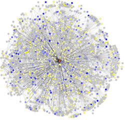
Researchers have mapped a network of 2,141 proteins that all interact either directly or through one other protein with huntingtin (red), the protein associated with Huntington’s disease. Credit: Cendrine Tourette, Buck Institute for Research on Aging, J Biol Chem 2014 Mar 7;289(10):6709-26 Exit icon.

The cause of Huntington’s disease, a degenerative neurological disorder with no known cure, may appear simple. It begins with a change in a single gene that alters the shape and functioning of the huntingtin protein. But this protein, whether in its normal or altered form, does not act alone. It interacts with other proteins, which in turn interact with others.

A research team led by Robert Hughes of the Buck Institute for Research on Aging set out to understand how this ripple effect contributes to the breakdown in normal cellular function associated with Huntington’s disease. The scientists used experimental and computational approaches to map a network of 2,141 proteins that interact with the huntingtin protein either directly or through one other protein. They found that many of these proteins were involved in cell movement and intercellular communication. Understanding how the huntingtin protein leads to mistakes in these cellular processes could help scientists pursue new approaches to developing treatments. Continue reading “Digging Deeply Into Data for the Causes of Disease”

5 Reasons Biologists Love Math

4 comments

Biologists use math in a variety of ways, from designing experiments to mapping complex biological systems. Credit: Stock image.

On Saturday (at 9:26:53 to be exact), math lovers and others around the world will celebrate Pi—that really long number that represents the ratio of the circumference of a circle to its diameter. I asked our scientific experts why math is important to biomedical research. Here are a few reasons.

  1. Math allows biologists to describe how molecules move in and out of cells, how bacteria shuttle through blood vessels, how drugs get broken down in the body and many other physiological processes.
  2. Studying the geometry, topology and other physical characteristics of DNA, proteins and cellular structures has shed light on their functions and on approaches for enhancing or disrupting those functions.
  3. Math helps scientists design their experiments, including clinical trials, so they result in meaningful data, a.k.a statistical significance.
  4. Scientists use math to piece together all the different parts of a cell, an organ or an entire organism to better understand how the parts interact and how perturbations in these complex systems may contribute to disease.
  5. Sometimes it’s impossible or too difficult to answer a research question through traditional lab experiments, so biologists rely on math to develop models that represent the system they’re studying, whether it’s a metastasizing cancer cell or an emerging infectious disease. These approaches allow scientists to indicate the likelihood of certain outcomes as well as refine the research questions.

Want more? Here’s a video with 10 reasons biologists should know some math.

Scientists Shine Light on What Triggers REM Sleep

1 comment
Illustration of a brain.
While studying how the brain controls REM sleep, researchers focused on areas abbreviated LDT and PPT in the mouse brainstem. This illustration shows where these two areas are located in the human brain. Credit: Wikimedia Commons. View larger image

Has the “spring forward” time change left you feeling drowsy? While researchers can’t give you back your lost ZZZs, they are unraveling a long-standing mystery about sleep. Their work will advance the scientific understanding of the process and could improve ways to foster natural sleep patterns in people with sleep disorders.

Working at Massachusetts General Hospital and MIT, Christa Van Dort, Matthew Wilson, and Emery Brown focused on the stage of sleep known as REM. Our most vivid dreams occur during this period, as do rapid eye movements, for which the state is named. Many scientists also believe REM is crucial for learning and memory.

REM occurs several times throughout the night, interspersed with other sleep states collectively called non-REM sleep. Although REM is clearly necessary—it occurs in all land mammals and birds—researchers don’t really know why. They also don’t understand how the brain turns REM on and off.

Continue reading “Scientists Shine Light on What Triggers REM Sleep”

Remotely and Noninvasively Controlling Genes and Cells in Living Animals

0 comments
Remote control car key.
Researchers are developing a system to remotely control genes or cells in living animals with radio wave technology similar to that used to operate remote control car keys. Credit: Stock image.

One of the items on biomedical researchers’ “to-do” list is devising noninvasive ways to control the activity of specific genes or cells in order to study what those genes or cells do and, ultimately, to treat a range of human diseases and disorders.

A team of scientists recently reported progress on a new, noninvasive system that could remotely and rapidly control biological targets in living animals. The system can be activated remotely using either low-frequency radio waves or a magnetic field. Similar radio wave technology operates automatic garage-door openers and remote control car keys and is used in medicine to control electronic pacemakers noninvasively. Magnetic fields are used to activate sensors in burglar alarm systems and to turn your laptop to hibernate mode when the cover is closed. Continue reading “Remotely and Noninvasively Controlling Genes and Cells in Living Animals”