Tag: Cellular Processes

Pass the Salt: Sodium’s Role in Nerve Signaling and Stress on Blood Vessels

5 comments

Most of the mouthwatering dishes in a Thanksgiving feast share a vital ingredient: salt! Though the words “salt” and “sodium” are often used interchangeably, table salt is actually a compound combining the elements sodium and chloride. Table salt is the most common form that sodium takes on Earth. Many other sodium compounds are also useful to us. For instance, you might use baking soda, also known as sodium bicarbonate, in preparing Thanksgiving treats. Sodium compounds are also used in soaps and cosmetics and in producing paper, glass, metals, medicines, and more.

A graphic showing sodium’s abbreviation, atomic number, and atomic weight connected by lines to illustrations of a saltshaker, a streetlight, and a human arm flexing its muscle. The best-known sodium compound is table salt (sodium chloride). Sodium also gives traditional streetlights their yellow glow and is essential for muscle and nerve function. Credit: Compound Interest. CC BY-NC-ND 4.0. Click to enlarge
Continue reading “Pass the Salt: Sodium’s Role in Nerve Signaling and Stress on Blood Vessels”

Quiz: How Does Your Knowledge of Life’s Building Blocks Stack Up?

4 comments

Cells are the smallest units of life, providing structure and function for all living things, from microorganisms—like bacteria, algae, and yeast—to humans. They come in a wide range of sizes and shapes, and they’re complex machines with many smaller components that work together.

Some NIGMS-funded researchers use imaging techniques to peer inside cells, examine their structures, and study how they divide, grow, communicate, and carry out basic functions. Others use biochemical and genetic tests to study how cells interact with their environments, including those that may be toxic. Understanding cells’ biological processes helps to keep us healthy and identify new methods for treating disease.

Take our quiz to test how well you know cells. Afterward, check out our other blog posts on cell biology.

Continue reading “Quiz: How Does Your Knowledge of Life’s Building Blocks Stack Up?”

Freezing a Moment in Time: Snapshots of Cryo-EM Research

0 comments

To get a look at cell components that are too small to see with a normal light microscope, scientists often use cryo-electron microscopy (cryo-EM). As the prefix cryo- means “cold” or “freezing,” cryo-EM involves rapidly freezing a cell, virus, molecular complex, or other structure to prevent water molecules from forming crystals. This preserves the sample in its natural state and keeps it still so that it can be imaged with an electron microscope, which uses beams of electrons instead of light. Some electrons are scattered by the sample, while others pass through it and through magnetic lenses to land on a detector and form an image.

Typically, samples contain many copies of the object a scientist wants to study, frozen in a range of orientations. Researchers take images of these various positions and combine them into a detailed 3D model of the structure. Electron microscopes allow us to see much smaller structures than light microscopes do because the wavelengths of electrons are much shorter than the wavelength of light. NIGMS-funded researchers are using cryo-EM to investigate a range of scientific questions.

Caught in Translation

One cluster that is yellow, purple, and orange and another that is beige, purple, and green. 
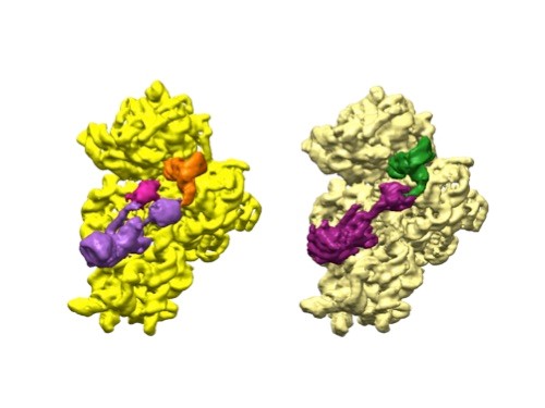
3D reconstructions of two stages in the assembly of the bacterial ribosome created from time-resolved cryo-EM images. Credit: Joachim Frank, Columbia University.

Joachim Frank, Ph.D., a professor of biochemistry and molecular biophysics and of biological sciences at Columbia University in New York, New York, along with two other researchers, won the 2017 Nobel Prize in Chemistry for developing cryo.

Dr. Frank’s lab focuses on the process of translation, where structures called ribosomes turn genetic instructions into proteins, which are needed for many chemical reactions that support life. Recently, Dr. Frank has adopted and further developed a technique called time-resolved cryo-EM. This method captures images of short-lived states in translation that disappear too quickly (after less than a second) for standard cryo-EM to capture. The ability to fully visualize translation could help researchers identify errors in the process that lead to disease and also to develop treatments.

Continue reading “Freezing a Moment in Time: Snapshots of Cryo-EM Research”
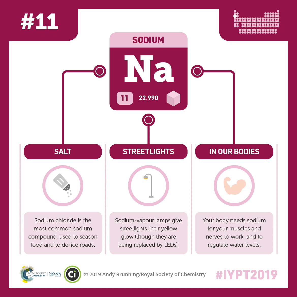
Explore Our STEM Education Resources for the New School Year

0 comments

If you’re looking for ways to engage students in science this school year, NIGMS offers a range of free resources that can help. All of our STEM materials are online and print-friendly, making them easy to use for remote teaching.

Pathways , developed in collaboration with Scholastic, is aligned with STEM and ELA education standards for grades 6 through 12. Materials include:

  • Student magazines with corresponding teaching guides
  • Related lessons with interactives
  • Videos
  • Vocabulary lists
Cover of Pathways student magazine showing a microscopy image of a fruit fly’s head with bright blue eyes and the featured questions: What is this? And what does it have to do with how you sleep? 
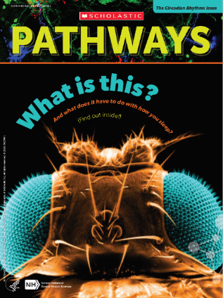
Cover of Pathways student magazine, third issue.

Available lessons examine basic science careers, regeneration, and circadian rhythms.

Continue reading “Explore Our STEM Education Resources for the New School Year”

Phosphorus: Glowing, Flammable, and Essential to Our Cells

3 comments

Of the 118 known elements, scientists believe that 25 are essential for human biology. Four of these (hydrogen, oxygen, nitrogen, and carbon) make up a whopping 96 percent of our bodies. The other 21 elements, though needed in smaller quantities, perform fascinating and vital functions. Phosphorus is one such element. It has diverse uses outside of biology. For example, it can fuel festive Fourth of July fireworks! Inside our bodies, it’s crucial for a wide range of cell functions.

A graphic showing phosphorus’s abbreviation, atomic number, and atomic weight connected by lines to illustrations of DNA helixes, a match, and a glowing white pyramid. Phosphorus plays a vital role in life as part of DNA’s backbone. Red phosphorus helps ignite matches, and white phosphorus glows in the presence of oxygen. Credit: Compound Interest.
CC BY-NC-ND 4.0 Link to external web site. Click to enlarge
Continue reading “Phosphorus: Glowing, Flammable, and Essential to Our Cells”

The Maternal Magic of Mitochondria

5 comments
An oblong purple shape with ripples throughout against a light blue background. 
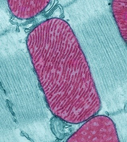
Mitochondria (purple) in a rodent heart muscle cell. Credit: Thomas Deerinck, National Center for Microscopy and Imaging Research.

Mitochondria (mitochondrion in singular) are indispensable. Every cell of our bodies, apart from mature red blood cells, contains the capsule-shaped organelles that generate more than 90 percent of our energy, which is why they’re often called “the powerhouse of the cell.” They produce this energy by forming adenosine triphosphate (ATP), our cells’ most common energy source. But mitochondria also support cells in other ways. For example, they help cells maintain the correct concentration of calcium ions, which are involved in blood clotting and muscle contraction. Mitochondria are also the only structure in our cells with their own unique DNA, which with rare exceptions, is inherited only from mothers. That’s why, in honor of Mother’s Day, we’re exploring this special cellular connection to moms.

Continue reading “The Maternal Magic of Mitochondria”

Twisting and Turning: Unraveling What Causes Asymmetry

0 comments

Note to our Biomedical Beat readers: Echoing the sentiments NIH Director Francis Collins made on his blog, NIGMS is making every effort during the COVID-19 pandemic to keep supporting the best and most powerful science. In that spirit, we’ll continue to bring you stories across a wide range of NIGMS topics. We hope these posts offer a respite from the coronavirus news when needed.

Asymmetry in our bodies plays an important role in how they work, affecting everything from function of internal systems to the placement and shape of organs. Take a look at your hands. They are mirror images of each other, but they’re not identical. No matter how you rotate them or flip them around, they will never be the same. This is an example of chirality, which is a particular type of asymmetry. Something is chiral if it can’t overlap on its mirror image.

An image of a pair of hands, palms facing up. An arrow points to another image of the left hand on top of the right, both palms still facing up, illustrating that they can’t be superimposed. 
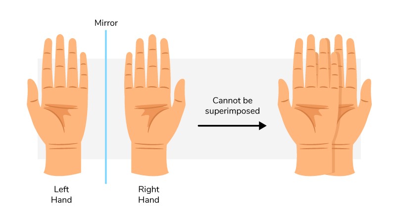
Our hands are chiral: They’re mirror images but aren’t identical.

Scientists are exploring the role of chirality and other types of asymmetry in early embryonic development. Understanding this relationship during normal development is important for figuring out how it sometimes goes wrong, leading to birth defects and other medical problems.

Continue reading “Twisting and Turning: Unraveling What Causes Asymmetry”

How Errors in Divvying Up Chromosomes Lead to Defects in Cells

0 comments

Note to our Biomedical Beat readers: Echoing the sentiments NIH Director Francis Collins made on his blog, NIGMS is making every effort during the COVID-19 pandemic to keep supporting the best and most powerful science. In that spirit, we’ll continue to bring you stories across a wide range of NIGMS topics. We hope these posts offer a respite from the coronavirus news when needed.

Mitosis is fundamental among all organisms for reproduction, growth, and cell replacement. When a cell divides, it’s vital that the two new daughter cells maintain the same genes as the parent.

In one step of mitosis, chromosomes are segregated into two groups, which will go into the two new daughter cells. But if the chromosomes don’t divide properly, one daughter cell may have too many and the other too few. Having the wrong number of chromosomes, a condition called aneuploidy, can trigger cells to grow out of control.

Illustration of two sets of chromosomes being pulled apart. One pair separates evenly and is labeled normal, but the other doesn’t and is labeled aneuploidy.
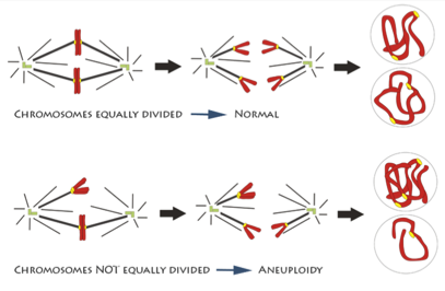
An illustration of chromosomes being segregated equally and unequally during mitosis. Credit: Deluca Lab, Colorado State University.

How chromosome segregation errors disrupt cell division is an important area of research. Although it’s been studied for decades, new aspects are still being uncovered and much remains unknown. NIGMS-funded scientists are studying different aspects of mitosis and chromosome segregation. Understanding the details can provide vital insight into an essential biological process and may also be the key to developing better drugs for cancer and other diseases.

Continue reading “How Errors in Divvying Up Chromosomes Lead to Defects in Cells”

Explore Our Virtual Learning STEM Resources

1 comment

If you’re looking for engaging ways to teach science from home, NIGMS offers a range of resources that can help.

Cover of the graphic novel Occupied by Microbes!, showing four teens racing downhill on skateboards. 
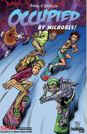
A SEPA-funded resource about microbes. Credit: University of Nebraska, Lincoln.

Our Science Education and Partnership Award (SEPA) webpage features free, easy-to-access STEM and informal science education projects for pre-K through grade 12. Aligned with state and national standards for STEM teaching and learning, the program has tools such as:

  • Apps
  • Interactives
  • Online books
  • Curricula and lesson plans
  • Short movies

Students can learn about sleep, cells, growth, microbes, a healthy lifestyle, genetics, and many other subjects.

Continue reading “Explore Our Virtual Learning STEM Resources”

PECASE Honoree James Olzmann Investigates the Secrets of Lipid Droplets

0 comments

Note to our Biomedical Beat readers: Echoing the sentiments NIH Director Francis Collins made on his blog, NIGMS is making every effort during the COVID-19 pandemic to keep supporting the best and most powerful science. In that spirit, we’ll continue to bring you stories across a wide range of NIGMS topics. We hope these posts offer a respite from the coronavirus news when needed.

A large, blue oval surrounded by much smaller yellow circles. 
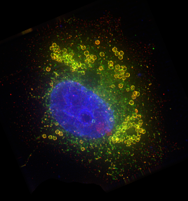
A cell nucleus (blue) surrounded by lipid droplets (yellow). Credit: James Olzmann.

Within our cells, lipids are often stored in droplets, membrane-bound packages of lipids produced by the endoplasmic reticulum. For many years, scientists thought lipid droplets were simple globs of fat and rarely studied them. But over the past few decades, research has revealed that they’re full-fledged organelles, or specialized structures that perform important cellular functions. The field of lipid droplet research has been growing ever since.

Continue reading “PECASE Honoree James Olzmann Investigates the Secrets of Lipid Droplets”