Tag: DNA

Computational Biologist Melissa Wilson on Sex Chromosomes, Gila Monsters, and Career Advice

1 comment
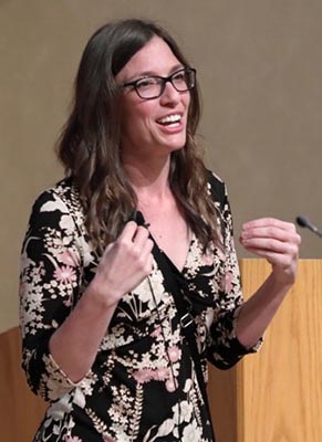
Melissa Wilson wearing a floral dress and speaking beside a podium during her lecture. Dr. Melissa Wilson.
Credit: Chia-Chi Charlie Chang.

The X and Y chromosomes, also known as sex chromosomes, differ greatly from each other. But in two regions, they are practically identical, said Melissa Wilson Link to external web site, assistant professor of genomics, evolution, and bioinformatics at Arizona State University.

“We’re interested in studying how the process of evolution shaped the X and the Y chromosome in gene content and expression and how that subsequently affects literally everything else that comes with being a human,” she said at the April 10 NIGMS Director’s Early-Career Investigator (ECI) Lecture at NIH.

Continue reading “Computational Biologist Melissa Wilson on Sex Chromosomes, Gila Monsters, and Career Advice”

The Skull’s Petrous Bone and the Rise of Ancient Human DNA: Q & A with Genetic Archaeologist David Reich

1 comment
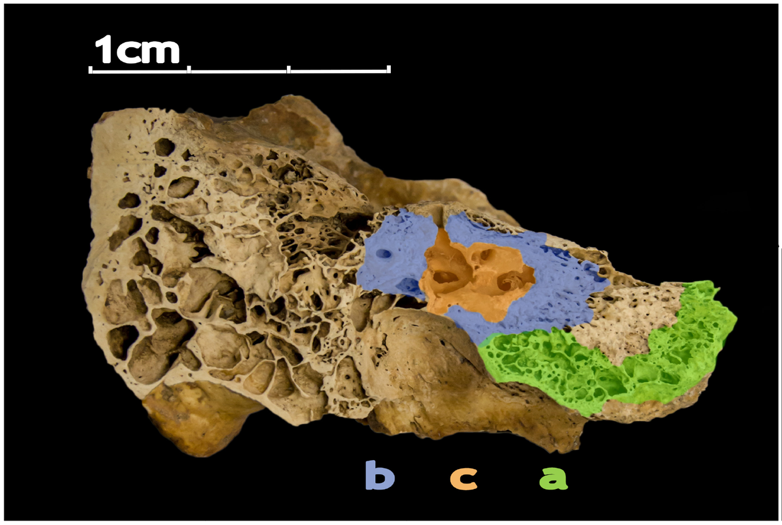
A macro image of the petrous bone. 3 sections are color coded A (green), B (blue), and C (red)The human petrous bone in the skull protects the inner ear structures. Though it is one of the hardest, densest bones in the body, some portions (such as the area in orange, protecting the cochlea) are denser than others. Possibly because the petrous bone is so dense, DNA within the petrous bone is better preserved than in other bones. In some cases, scientists have extracted more than 100 times more DNA from the petrous bone than other bones, including teeth. Credit: Pinhasi et al., 2015, PLOS ONE.

For the past few decades, new evidence about ancient humans—in the form of skeletal remains, tools, and other artifacts—has trickled in, inching us closer to an understanding of how our species evolved and spread out across the planet. In just the past few years, however, knowledge of our deep past expanded significantly thanks to a series of technological breakthroughs in sequencing of ancient human genomes. This technology can be used to find genetic links among populations of human ancestors dating back hundreds of thousands of years.

In addition to advances in genomic technology, another factor is driving the explosion of new discoveries—an inch-long section of the human skull. Found near our ears, this pyramid-shaped portion of the temporal bone is nicknamed the petrous bone. The bone is very hard, possibly because it needs to protect fragile structures such as the cochlea, which translates sound into brain signals, and the semicircular canals, which help us maintain our balance. Perhaps because the petrous bone is so dense, it also is the bone in the body that best preserves DNA after a person dies. As a result, archaeologists are scrambling to study samples taken from this pyramid-shaped structure to unlock the mysteries of our species’ formative years.

Here’s a sampling of headlines declaring new findings about ancient peoples from around the globe that were based on genetic information obtained from the petrous bone (NIGMS-funded research indicated in black):

“How the introduction of farming changed the human genome” November 2015

“Fourth strand’ of European ancestry originated with hunter-gatherers isolated by Ice Age” November 2015

“Scientists sequence first ancient Irish human genomes” December 2015

“Genetic studies provide insight into ancient Britain’s diversity” January 2016

“The world’s first farmers were surprisingly diverse” June 2016

“Study reveals Asian ancestry of Pacific islanders” October 2016

“Ancient DNA solves mystery of the Canaanites, reveals the biblical people’s fate” July 2017

“Ancient DNA data fills in thousands of years of human prehistory in Africa” September 2017

“European Hunter-Gatherers Interbred With Farmers From the Near East” November 2017

“Surprise as DNA reveals new group of Native Americans: the ancient Beringians” January 2018

“Ancient DNA reveals genetic replacement despite language continuity in the South Pacific” February 2018

“Stone Age Moroccan Genomes Reveal Sub-Saharan African, Near Eastern Ancestry” March 2018

“Some early modern populations in Britain may have had dark skin” March 2018

Continue reading “The Skull’s Petrous Bone and the Rise of Ancient Human DNA: Q & A with Genetic Archaeologist David Reich”

Genomic Gymnastics of a Single-Celled Ciliate and How It Relates to Humans

0 comments
Laura Landweber
Credit: Denise Applewhite.
Laura Landweber
Grew up in: Princeton, New Jersey
Job site: Columbia University, New York City
Favorite food: Dark chocolate and dark leafy greens
Favorite music: 1940’s style big band jazz
Favorite hobby: Swing dancing
If I weren’t a scientist I would be a: Chocolatier (see “Experiments in Chocolate” sidebar at bottom of story)

One day last fall, molecular biologist Laura Landweber surveyed the Princeton University lab where she’d worked for 22 years. She and her team members had spent many hours that day laboriously affixing yellow Post-it notes to the laboratory equipment—microscopes, centrifuges, computers—they would bring with them to Columbia University, where Landweber had just been appointed full professor. Each Post-it specified the machinery’s location in the new lab. Items that would be left behind—glassware, chemical solutions, furniture, office supplies—were left unlabeled.

As Landweber viewed the lab, decorated with a field of sunny squares, her thoughts turned to another sorting process—the one used by her primary research subject, a microscopic organism, to sift through excess DNA following mating. Rather than using Post-it notes, the creature, a type of single-celled organism called a ciliate, uses small pieces of RNA to tag which bits of genetic material to keep and which to toss.

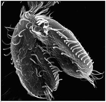
Landweber is particularly fond of Oxytricha trifallax, a ciliate with relatives that live in soil, ponds and oceans all over the world. The kidney-shaped cell is covered with hair-like projections called cilia that help it move around and devour bacteria and algae. Oxytricha is not only bizarre in appearance, it’s also genetically creative.

Unlike humans, whose cells are programmed to die rather than pass on genomic errors, Oxytricha cells appear to delight in genomic chaos. During sexual reproduction, the ciliate shatters the DNA in one of its two nuclei into hundreds of thousands of pieces, descrambles the DNA letters, throws most away, then recombines the rest to create a new genome.

Landweber has set out to understand how—and possibly why—Oxytricha performs these unusual genomic acrobatics. Ultimately, she hopes that learning how Oxytricha rearranges its genome can illuminate some of the events that go awry during cancer, a disease in which the genome often suffers significant reorganization and damage.

Oxytricha’s Unique Features

Oxytricha carries two separate nuclei—a macronucleus and a micronucleus. The macronucleus, by far the larger of the two, functions like a typical genome, the source of gene transcription for proteins. The tiny micronucleus only sees action occasionally, when Oxytricha reproduces sexually.

Oxytricha trifallax cells in the process of mating
Two Oxytricha trifallax cells in the process of mating. Credit, Robert Hammersmith.

What really makes Oxytricha stand out is what it does with its DNA during the rare occasions that it has sex. When food is readily available, Oxytricha procreates without a partner, like a plant grown from a cutting. But when food is scarce, or the cell is stressed, it seeks a mate. When two Oxytricha cells mate, the micronuclear genomes in each cell swap DNA, then replicate. One copy of the new hybrid micronucleus remains intact, while the other breaks its DNA into hundreds of thousands of pieces, some of which are tagged, recombined, then copied another thousand-fold to form a new macronucleus. Continue reading “Genomic Gymnastics of a Single-Celled Ciliate and How It Relates to Humans”
Have Nucleus, Will Travel (in Three Dimensions)

0 comments
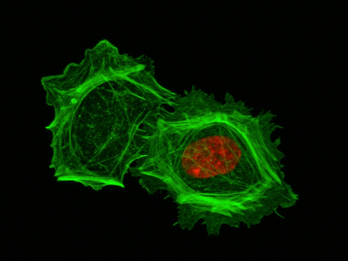
A closeup of two human cells with the cells dyed green and the necleaus dyed red.These two human cells are nearly identical, except that the cell on the left had its nucleus (dyed red) removed. The structures dyed green are protein strands that give cells their shape and coherence. Credit: David Graham, UNC-Chapel Hill.
Both of the cells above can scoot across a microscope slide equally well. But when it comes to moving in 3D, the one without the red blob in the center (the nucleus) stalls out. That’s sort of like an Olympic speed skater who wouldn’t be able to perform even a single leap in a figure skating competition.

Scientists have known for some time that the nucleus is involved in moving cells across a flat surface—it slides to one side of the cell and “pushes” from behind. However, scientists have also shown that cells with their nuclei removed can migrate along a flat surface just as well as their brethren with intact nuclei. But they had no idea that, without a nucleus, a cell could no longer move in three dimensions.

This discovery was made by UNC-Chapel Hill biologists Keith Burridge and James Bear and their colleagues. These NIGMS-funded researchers also observed that cells whose nuclei had been disconnected from the cytoskeleton could not move through 3D matrices. The cytoskeleton is the microscopic network of actin protein filaments and tubules in the cytoplasm of many cells that provides the cell’s shape and coherence. It has also has been thought to play a major role in cell movement.

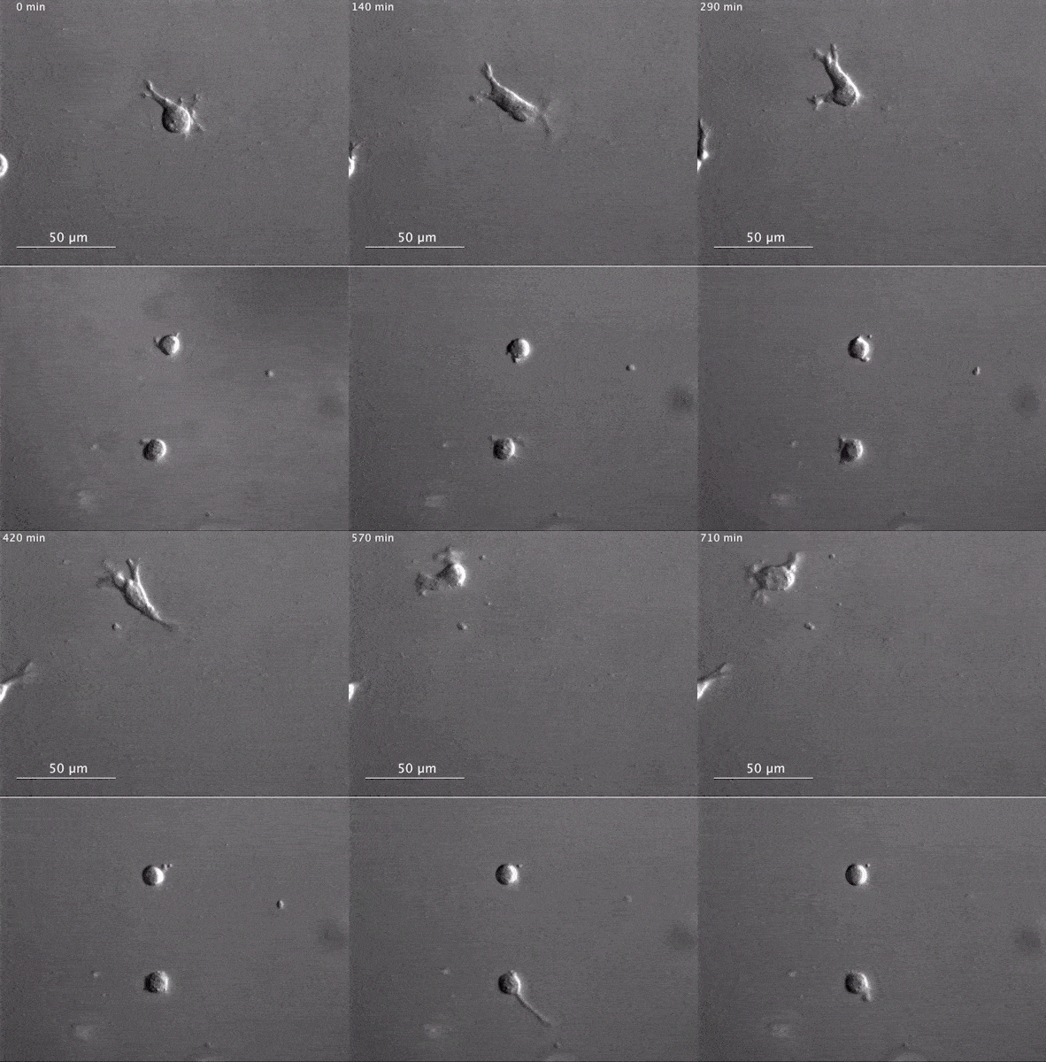
Two views of cells one on top of the other. The top animation shows a cell moving across the frame while the cells in the bottom box are static.The gray, stringy background of these videos is a 3D jello-like matrix. The cell in the top half of this video has a nucleus and can migrate through the matrix. Both cells in the bottom half have been enucleated (a fancy term for having its nucleus removed) and cannot travel through the matrix. Credit: Graham et al., Journal of Cell Biology, 2018.
The researchers speculate that the reason cells without nuclei (or those whose nuclei have been disconnected from the cytoskeleton) don’t navigate in 3D has to do with complex mechanical interactions between the cytoskeleton and the nucleoskeleton. The nucleoskeleton is a molecular scaffold within the nucleus supporting many functions such as DNA replication and transcription, chromatin remodeling, and mRNA synthesis. The interface between the cytoskeleton and nucleoskeleton consists of interlocking proteins that together provide the physical traction that cells need to push their way through 3D environments. Disrupting this interface is the equivalent of breaking the clutch in a car: the motor revs, but the wheels don’t spin, and the car goes nowhere.

A better understanding of the physical connections between the nucleus and the cytoskeleton and how they influence cell migration may provide additional insight into the role of the nucleus in diseases, such as cancer, in which the DNA-containing organelle is damaged or corrupted.

This research was funded in part by NIGMS grants 5R01GM029860-35, 5P01GM103723-05, and 5R01GM111557-04.

“Selfish” Gene Enhances Own Transmission at Expense of Organism’s Fertility

2 comments
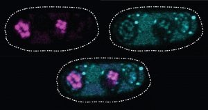
These glowing images of yeast (Schizosaccharomyces kambucha) reproductive cells show an example of a selfish gene at work.
These glowing images of yeast (Schizosaccharomyces kambucha) reproductive cells show an example of a selfish gene at work. Here, the selfish gene boosts its chances of being passed to the next generation by producing both a toxin (stained cyan) and an antitoxin (stained magenta). Cells with a copy of the selfish gene are protected by the antitoxin, left and bottom ovals. Those without the selfish gene are destroyed by the toxin. Scientists suspect that selfish genes could be operating throughout many organisms’ genomes, possibly having a major impact on how genetic material is inherited over generations. Credit: Image courtesy of María Angélica Bravo Núñez and Nicole Nuckolls.

There’s an old saying that rules are meant to be broken. In the 1860s, Gregor Mendel found that each copy of a gene in an organism has an equal chance of being passed to the next generation. According to this simple rule, each version of a gene gets passed to offspring with the same frequency. The natural selection process can then operate efficiently, favoring the genes that produce an advantage for an organism’s survival or reproductive success and, over successive generations, eliminating genes from the gene pool that bring a disadvantage.

Of course, the way organisms inherit genes is not as straightforward as Mendel’s work predicted. In natural systems, inheritance often fails to follow the rules. One culprit scientists are identifying again and again are what are called “selfish genes”: one or more genes that have evolved a method of skewing inheritance in their favor.

Scientists refer to these selfish genes—which are often complexes of multiple genes working together—as “selfish” because they enhance their own transmission to the next generation, sometimes by killing off any of the organism’s reproductive cells that lack copies of those genes. Using a variety of techniques, the genes are effective at passing themselves on to future generations. However, their methods set up a conflict within the organism by damaging its fertility; overall, fewer reproductive cells or offspring survive to produce a new generation.

Selfish genes are a challenge for scientists to identify, but researchers say that knowing more about these genes could help explain a range of genetic mysteries, from causes of infertility to details on how species evolve. The methods these genes use could also be harnessed to help control the reproduction of certain populations such as mosquitos that spread disease.

One recently described selfish gene system is found in the yeast cells pictured above. Sarah Zanders and her colleagues at the Stowers Institute for Medical Research in Kansas City, Missouri, and the Fred Hutchinson Cancer Research Center in Seattle, Washington, study selfish gene systems in yeast to understand how common they are and how they affect a species’ fertility and evolution. “Usually we think about infertility stemming from the good guys failing. For example, a gene that normally promotes fertility could be mutated so that it can no longer do its job,” says Zanders. “But selfish genes are another potential source of infertility. Learning general principles about selfish genes in simple models will guide future searches for selfish genes that could be contributing to human infertility.”

Recently, the scientists discovered a single selfish gene, wtf4, that encodes both a toxin and an antitoxin protein. When yeast produce their reproductive cells, called spores, the wtf4 toxin protein is released into the immediate vicinity, but the antitoxin remains inside spores that contain a copy of wtf4. The toxin destroys all the spores that don’t have the antitoxin protein. Although the yeast has fewer spores—and therefore reduced fertility—each spore carries wtf4, ensuring that the gene will be passed to the next generation of yeast.

Continue reading ““Selfish” Gene Enhances Own Transmission at Expense of Organism’s Fertility”

Viruses: Manufacturing Tycoons?

0 comments
Pseudomonas chlororaphis

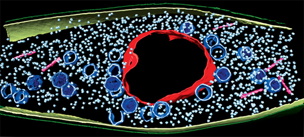
A computer image shows a bacterial cell invaded by a virus. The virus uses the cell to copy itself many times. It has built a protein compartment (red, rough circle surrounding the center) to house its DNA. Viral heads (blue, smaller pentagonal shapes spread through out) and tails (pink, rod shaped near the edges) are essential parts of a finished viral particle. The small, light blue particles are the bacterium’s own protein-making ribosomes. Credit: Vorrapon Chaikeeratisak, Kanika Khanna, Axel Brilot and Katrina Nguyen.

As inventors and factory owners learned during the Industrial Revolution, the best way to manufacture a lot of products is with an assembly line that follows a set of precisely organized steps employing many copies of identical and interchangeable parts. Some viruses are among life’s original mass producers: They use sophisticated organization principles to turn bacterial cells into virus particle factories.

Scientists at the University of California, San Diego, and the University of California, San Francisco, used cutting-edge techniques to watch a bacteria-infecting virus (bacteriophage) set up its particle-making factory inside a host cell.

The image above shows this happening inside a Pseudomonas chlororaphis, a soil-borne bacterium that protects plants against fungal pathogens. The virus builds a compartment (red, rough circle surrounding the center) that helps organize an assembly line for making copies of itself. The compartment looks like a cell nucleus, which bacteria do not have, and it functions like a nucleus by keeping activities that directly involve DNA separate from other cellular functions. Continue reading “Viruses: Manufacturing Tycoons?”

Six Things to Know About DNA and DNA Repair

3 comments

Deoxyribonucleic acid, better known as DNA, was first identified on a discarded surgical bandage almost 150 years ago. Increasingly sophisticated tools and techniques have allowed scientists to learn more about this chemical compound that includes all the instructions necessary for building a living organism. From among the dozens of fascinating things known about DNA, here are six items touching on the make up of DNA’s double helix, the vast amounts of DNA packed into every human’s cells, common DNA errors and a few ways DNA can repair itself.

1. DNA is in every living thing.

Nucleotide
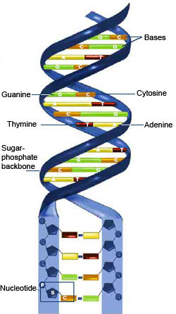
DNA consists of two long, twisted chains made of nucleotides. Each nucleotide contains one base, one phosphate molecule and the sugar molecule deoxyribose. The bases in DNA nucleotides are adenine, cytosine, guanine and thymine. Credit: NIGMS.

The chemical instructions for building a person—and every other creature on Earth—are contained in DNA. DNA is shaped like a corkscrew-twisted ladder, called a double helix. The two ladder rails are referred to as backbones, made of alternating groups of sugar and phosphate. The ladder’s rungs are made from four different building blocks called bases, arranged in pairs: adenine (A) paired with thymine (T), and cytosine (C) paired with guanine (G). Humans have about 3 billion base pairs in each cell. The order of the base pairs determines the exact instructions encoded in that part of the DNA molecule. Also, the sequence of DNA base pairs in one person is about 99.9 percent identical to that of everyone else.

2. Humans have a lot of DNA.

Humans begin as a single fertilized cell containing (with some rare exceptions) the full complement of DNA—the genome—arranged into 46 discrete chromosomes (23 pairs, with mom and dad each contributing half of each pair) in the cell’s nucleus. There are 6 feet of DNA coiled up tightly in that first cell. All the information in the DNA is replicated each time the cell divides. The amount of DNA packed into all of an adult’s cells is on the order of 100 trillion feet (about 19 billion miles)—so that if the DNA chain was stretched out, it would be long enough to reach back and forth between the Earth and the Sun more than 200 times. Continue reading “Six Things to Know About DNA and DNA Repair”

Birthdays, Nobel Prizes and Basic Research

1 comment
James D. Watson
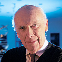
James D. Watson. Credit: Wikimedia Commons, Cold Spring Harbor Laboratory.

April 6 is the birthday of two Nobel Prize winners in physiology or medicine—James Watson and Edmond H. Fischer. They have also both been NIGMS-supported researchers.

Double helix model
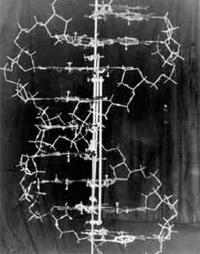
In 1953, Watson and Crick created their historic model of the shape of DNA: the double helix. Credit: Cold Spring Harbor Laboratory archives.

James D. Watson, born on this day in 1928, was honored with the Nobel Prize in 1962. He shared it with Francis H. Compton Crick and Maurice Wilkins “for their discoveries concerning the molecular structure of nucleic acids and its significance for information transfer in living material.” This laid the groundwork for future discoveries. In the early 1950s, Wilkins and another scientist, Rosalind Franklin, worked to determine DNA’s structure. In 1953, Watson and Crick discovered its shape as a double helix. This twisted ladder structure enabled other researchers to unlock the secret of how genetic information is stored, transferred and copied. Franklin is widely recognized as having played a significant role in revealing the physical structure of DNA; due to her death at age 37 in 1958, Franklin did not earn a share of the prize. Read more about DNA.

Continue reading “Birthdays, Nobel Prizes and Basic Research”

Field Focus: High-Quality Genome Sequences Inform the Study of Human Evolution

1 comment

Leafing through my favorite biology textbook from a handful of years ago, I was struck by the relative brevity of the chapter on human evolution. While other fields of biological research have enjoyed a steady gallop of productivity over the last few decades due in part to advances in computing power, imaging technology and experimental methods, the study of human evolution can be seen as having lagged behind until recently due to an almost complete dependence on fossil evidence.

Fortunately, contemporary biology textbook chapters on human evolution are being primed for a serious upgrade thanks to the recent availability of high-quality genome sequences from diverse modern human populations as well as from ancient humans and other non-human hominids, including the Neanderthals and Denisovans (but, for purposes of this story, not the Great Apes).

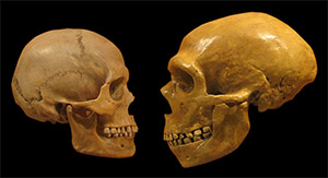
Modern human skull (left) and Neanderthal skull (right), shown to scale. There are not enough Denisovan bone fragments to reconstruct its skull. Credit: Wikimedia Commons, hairymuseummatt.

What are the new resources for studying human evolution?

The cost of DNA sequencing has dropped precipitously in the last decade. As a result, more complete human genome sequences become available for analysis with each passing year.

For example, the 1000 Genomes Project Exit icon includes more than 1,000 full human genome sequences of individuals from European, Asian, American and Sub-Saharan African populations. Earlier this year, the Simons Genome Diversity Project Exit icon further increased the number of available human genomes by adding 300 individuals representing 142 populations around the globe.

Continue reading “Field Focus: High-Quality Genome Sequences Inform the Study of Human Evolution”

Our Complicated Relationship With Viruses

9 comments
Illustration of Influenza Virus H1N1. Swine Flu.
Nearly 10 percent of the human genome is derived from the genes of viruses. Credit: Stock image.

When viruses infect us, they can embed small chunks of their genetic material in our DNA. Although infrequent, the incorporation of this material into the human genome has been occurring for millions of years. As a result of this ongoing process, viral genetic material comprises nearly 10 percent of the modern human genome. Over time, the vast majority of viral invaders populating our genome have mutated to the point that they no longer lead to active infections. But they are not entirely dormant.

Sometimes, these stowaway sequences of viral genes, called “endogenous retroviruses” (ERVs), can contribute to the onset of diseases such as cancer. They can also make their hosts susceptible to infections from other viruses. However, scientists have identified numerous cases of viral hitchhikers bestowing crucial benefits to their human hosts—from protection against disease to shaping important aspects of human evolution, such as the ability to digest starch.

Protecting Against Disease

Geneticists Cedric Feschotte, Edward Chuong and Nels Elde Exit icon at the University of Utah have discovered that ERVs lodged in the human genome can jump start the immune system.

For a virus to successfully make copies of itself inside a host cell, it needs molecular tools similar to the ones its host normally uses to translate genes into proteins. As a result, viruses have tools meticulously shaped by evolution to commandeer the protein-producing machinery of human cells.

Continue reading “Our Complicated Relationship With Viruses”