Tag: DNA

An Enlightening Protein

0 comments
A fly glowing green. 
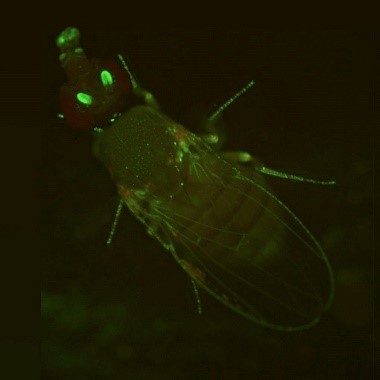
A fruit fly expressing GFP. Credit: Jay Hirsh, University of Virginia.

During the holiday season, twinkling lights are a common sight. But no matter what time of the year, you can see colorful glows in many biology labs. Scientists have enabled many organisms to light up in the dark—from cells to fruit flies and Mexican salamanders. These glowing organisms help researchers better understand basic cell processes because their DNA has been edited to express molecules such as green fluorescent protein.

Continue reading “An Enlightening Protein”

Q&A With Nobel Laureate and CRISPR Scientist Jennifer Doudna

0 comments
A headshot of Dr. Doudna. Jennifer Doudna, Ph.D. Credit: University of California, Berkeley.

The 2020 Nobel Prize in Chemistry was awarded to Jennifer Doudna, Ph.D., and Emmanuelle Charpentier, Ph.D., for the development of the gene-editing tool CRISPR. Dr. Doudna shared her thoughts on the award and answered questions about CRISPR in a live chat with NIH Director Francis S. Collins, M.D., Ph.D. Here are a few highlights from the interview.

Q: How did you find out that you won the Nobel Prize?

A: It’s a little bit of an embarrassing story. I slept through a very important phone call and finally woke up when a reporter called me. I was just coming out of a deep sleep, and the reporter was asking, “What do you think about the Nobel?” And I said, “I don’t know anything about it. Who won it?” I thought they were asking for comments on somebody else who won it. And she said, “Oh my gosh! You don’t know! You won it!”

Continue reading “Q&A With Nobel Laureate and CRISPR Scientist Jennifer Doudna”

Scientist Interview: Investigating Circadian Rhythms With Michael W. Young

1 comment

Sudden changes to our schedules, like the end of daylight saving time this Sunday or flying across time zones, often leave us feeling off kilter because they disrupt our bodies’ circadian rhythms. Circadian rhythms are physical, mental, and behavioral changes that follow a daily cycle. When these “biological clocks” are disrupted, our bodies eventually readjust. However, some people have conditions that cause their circadian rhythms to be permanently out of sync with their surroundings.

Continue reading “Scientist Interview: Investigating Circadian Rhythms With Michael W. Young”

Phosphorus: Glowing, Flammable, and Essential to Our Cells

3 comments

Of the 118 known elements, scientists believe that 25 are essential for human biology. Four of these (hydrogen, oxygen, nitrogen, and carbon) make up a whopping 96 percent of our bodies. The other 21 elements, though needed in smaller quantities, perform fascinating and vital functions. Phosphorus is one such element. It has diverse uses outside of biology. For example, it can fuel festive Fourth of July fireworks! Inside our bodies, it’s crucial for a wide range of cell functions.

A graphic showing phosphorus’s abbreviation, atomic number, and atomic weight connected by lines to illustrations of DNA helixes, a match, and a glowing white pyramid. Phosphorus plays a vital role in life as part of DNA’s backbone. Red phosphorus helps ignite matches, and white phosphorus glows in the presence of oxygen. Credit: Compound Interest.
CC BY-NC-ND 4.0 Link to external web site. Click to enlarge
Continue reading “Phosphorus: Glowing, Flammable, and Essential to Our Cells”

RNA Polymerase: A Target for New Antibiotic Drugs?

1 comment

DNA, with its double-helix shape, is the stuff of genes. But genes themselves are only “recipes” for protein molecules, which are molecules that do the real heavy lifting (or do much of the work) inside cells.

RNAP illustrated as a crab claw, clamping on a DNA double helix. 
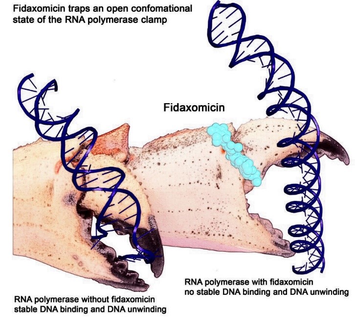
Artist interpretation of RNAP grasping and unwinding a DNA double helix. Credit: Wei Lin and Richard H. Ebright.

Here’s how it works. A molecular machine called RNA polymerase (RNAP) travels along DNA to find a place where a gene begins. RNAP uses a crab-claw-like structure to grasp and unwind the DNA double helix at that spot. RNAP then copies (“transcribes”) the gene into messenger RNA (mRNA), a molecule similar to DNA.

The mRNA molecule travels to one of the cell’s many protein-making factories (ribosomes), which use the mRNA message as instructions for making a specific protein.

Continue reading “RNA Polymerase: A Target for New Antibiotic Drugs?”

Computational Biologist Melissa Wilson on Sex Chromosomes, Gila Monsters, and Career Advice

1 comment
Melissa Wilson wearing a floral dress and speaking beside a podium during her lecture. Dr. Melissa Wilson.
Credit: Chia-Chi Charlie Chang.

The X and Y chromosomes, also known as sex chromosomes, differ greatly from each other. But in two regions, they are practically identical, said Melissa Wilson Link to external web site, assistant professor of genomics, evolution, and bioinformatics at Arizona State University.

“We’re interested in studying how the process of evolution shaped the X and the Y chromosome in gene content and expression and how that subsequently affects literally everything else that comes with being a human,” she said at the April 10 NIGMS Director’s Early-Career Investigator (ECI) Lecture at NIH.

Continue reading “Computational Biologist Melissa Wilson on Sex Chromosomes, Gila Monsters, and Career Advice”

The Skull’s Petrous Bone and the Rise of Ancient Human DNA: Q & A with Genetic Archaeologist David Reich

1 comment
A macro image of the petrous bone. 3 sections are color coded A (green), B (blue), and C (red)The human petrous bone in the skull protects the inner ear structures. Though it is one of the hardest, densest bones in the body, some portions (such as the area in orange, protecting the cochlea) are denser than others. Possibly because the petrous bone is so dense, DNA within the petrous bone is better preserved than in other bones. In some cases, scientists have extracted more than 100 times more DNA from the petrous bone than other bones, including teeth. Credit: Pinhasi et al., 2015, PLOS ONE.

For the past few decades, new evidence about ancient humans—in the form of skeletal remains, tools, and other artifacts—has trickled in, inching us closer to an understanding of how our species evolved and spread out across the planet. In just the past few years, however, knowledge of our deep past expanded significantly thanks to a series of technological breakthroughs in sequencing of ancient human genomes. This technology can be used to find genetic links among populations of human ancestors dating back hundreds of thousands of years.

In addition to advances in genomic technology, another factor is driving the explosion of new discoveries—an inch-long section of the human skull. Found near our ears, this pyramid-shaped portion of the temporal bone is nicknamed the petrous bone. The bone is very hard, possibly because it needs to protect fragile structures such as the cochlea, which translates sound into brain signals, and the semicircular canals, which help us maintain our balance. Perhaps because the petrous bone is so dense, it also is the bone in the body that best preserves DNA after a person dies. As a result, archaeologists are scrambling to study samples taken from this pyramid-shaped structure to unlock the mysteries of our species’ formative years.

Here’s a sampling of headlines declaring new findings about ancient peoples from around the globe that were based on genetic information obtained from the petrous bone (NIGMS-funded research indicated in black):

“How the introduction of farming changed the human genome” November 2015

“Fourth strand’ of European ancestry originated with hunter-gatherers isolated by Ice Age” November 2015

“Scientists sequence first ancient Irish human genomes” December 2015

“Genetic studies provide insight into ancient Britain’s diversity” January 2016

“The world’s first farmers were surprisingly diverse” June 2016

“Study reveals Asian ancestry of Pacific islanders” October 2016

“Ancient DNA solves mystery of the Canaanites, reveals the biblical people’s fate” July 2017

“Ancient DNA data fills in thousands of years of human prehistory in Africa” September 2017

“European Hunter-Gatherers Interbred With Farmers From the Near East” November 2017

“Surprise as DNA reveals new group of Native Americans: the ancient Beringians” January 2018

“Ancient DNA reveals genetic replacement despite language continuity in the South Pacific” February 2018

“Stone Age Moroccan Genomes Reveal Sub-Saharan African, Near Eastern Ancestry” March 2018

“Some early modern populations in Britain may have had dark skin” March 2018

Continue reading “The Skull’s Petrous Bone and the Rise of Ancient Human DNA: Q & A with Genetic Archaeologist David Reich”

Genomic Gymnastics of a Single-Celled Ciliate and How It Relates to Humans

0 comments
Laura Landweber
Credit: Denise Applewhite.
Laura Landweber
Grew up in: Princeton, New Jersey
Job site: Columbia University, New York City
Favorite food: Dark chocolate and dark leafy greens
Favorite music: 1940’s style big band jazz
Favorite hobby: Swing dancing
If I weren’t a scientist I would be a: Chocolatier (see “Experiments in Chocolate” sidebar at bottom of story)

One day last fall, molecular biologist Laura Landweber surveyed the Princeton University lab where she’d worked for 22 years. She and her team members had spent many hours that day laboriously affixing yellow Post-it notes to the laboratory equipment—microscopes, centrifuges, computers—they would bring with them to Columbia University, where Landweber had just been appointed full professor. Each Post-it specified the machinery’s location in the new lab. Items that would be left behind—glassware, chemical solutions, furniture, office supplies—were left unlabeled.

As Landweber viewed the lab, decorated with a field of sunny squares, her thoughts turned to another sorting process—the one used by her primary research subject, a microscopic organism, to sift through excess DNA following mating. Rather than using Post-it notes, the creature, a type of single-celled organism called a ciliate, uses small pieces of RNA to tag which bits of genetic material to keep and which to toss.

Landweber is particularly fond of Oxytricha trifallax, a ciliate with relatives that live in soil, ponds and oceans all over the world. The kidney-shaped cell is covered with hair-like projections called cilia that help it move around and devour bacteria and algae. Oxytricha is not only bizarre in appearance, it’s also genetically creative.

Unlike humans, whose cells are programmed to die rather than pass on genomic errors, Oxytricha cells appear to delight in genomic chaos. During sexual reproduction, the ciliate shatters the DNA in one of its two nuclei into hundreds of thousands of pieces, descrambles the DNA letters, throws most away, then recombines the rest to create a new genome.

Landweber has set out to understand how—and possibly why—Oxytricha performs these unusual genomic acrobatics. Ultimately, she hopes that learning how Oxytricha rearranges its genome can illuminate some of the events that go awry during cancer, a disease in which the genome often suffers significant reorganization and damage.

Oxytricha’s Unique Features

Oxytricha carries two separate nuclei—a macronucleus and a micronucleus. The macronucleus, by far the larger of the two, functions like a typical genome, the source of gene transcription for proteins. The tiny micronucleus only sees action occasionally, when Oxytricha reproduces sexually.

Oxytricha trifallax cells in the process of mating
Two Oxytricha trifallax cells in the process of mating. Credit, Robert Hammersmith.

What really makes Oxytricha stand out is what it does with its DNA during the rare occasions that it has sex. When food is readily available, Oxytricha procreates without a partner, like a plant grown from a cutting. But when food is scarce, or the cell is stressed, it seeks a mate. When two Oxytricha cells mate, the micronuclear genomes in each cell swap DNA, then replicate. One copy of the new hybrid micronucleus remains intact, while the other breaks its DNA into hundreds of thousands of pieces, some of which are tagged, recombined, then copied another thousand-fold to form a new macronucleus. Continue reading “Genomic Gymnastics of a Single-Celled Ciliate and How It Relates to Humans”

Have Nucleus, Will Travel (in Three Dimensions)

0 comments
A closeup of two human cells with the cells dyed green and the necleaus dyed red.These two human cells are nearly identical, except that the cell on the left had its nucleus (dyed red) removed. The structures dyed green are protein strands that give cells their shape and coherence. Credit: David Graham, UNC-Chapel Hill.

Both of the cells above can scoot across a microscope slide equally well. But when it comes to moving in 3D, the one without the red blob in the center (the nucleus) stalls out. That’s sort of like an Olympic speed skater who wouldn’t be able to perform even a single leap in a figure skating competition.

Scientists have known for some time that the nucleus is involved in moving cells across a flat surface—it slides to one side of the cell and “pushes” from behind. However, scientists have also shown that cells with their nuclei removed can migrate along a flat surface just as well as their brethren with intact nuclei. But they had no idea that, without a nucleus, a cell could no longer move in three dimensions.

This discovery was made by UNC-Chapel Hill biologists Keith Burridge and James Bear and their colleagues. These NIGMS-funded researchers also observed that cells whose nuclei had been disconnected from the cytoskeleton could not move through 3D matrices. The cytoskeleton is the microscopic network of actin protein filaments and tubules in the cytoplasm of many cells that provides the cell’s shape and coherence. It has also has been thought to play a major role in cell movement.

Two views of cells one on top of the other. The top animation shows a cell moving across the frame while the cells in the bottom box are static.The gray, stringy background of these videos is a 3D jello-like matrix. The cell in the top half of this video has a nucleus and can migrate through the matrix. Both cells in the bottom half have been enucleated (a fancy term for having its nucleus removed) and cannot travel through the matrix. Credit: Graham et al., Journal of Cell Biology, 2018.

The researchers speculate that the reason cells without nuclei (or those whose nuclei have been disconnected from the cytoskeleton) don’t navigate in 3D has to do with complex mechanical interactions between the cytoskeleton and the nucleoskeleton. The nucleoskeleton is a molecular scaffold within the nucleus supporting many functions such as DNA replication and transcription, chromatin remodeling, and mRNA synthesis. The interface between the cytoskeleton and nucleoskeleton consists of interlocking proteins that together provide the physical traction that cells need to push their way through 3D environments. Disrupting this interface is the equivalent of breaking the clutch in a car: the motor revs, but the wheels don’t spin, and the car goes nowhere.

A better understanding of the physical connections between the nucleus and the cytoskeleton and how they influence cell migration may provide additional insight into the role of the nucleus in diseases, such as cancer, in which the DNA-containing organelle is damaged or corrupted.

This research was funded in part by NIGMS grants 5R01GM029860-35, 5P01GM103723-05, and 5R01GM111557-04.

“Selfish” Gene Enhances Own Transmission at Expense of Organism’s Fertility

2 comments
These glowing images of yeast (Schizosaccharomyces kambucha) reproductive cells show an example of a selfish gene at work.
These glowing images of yeast (Schizosaccharomyces kambucha) reproductive cells show an example of a selfish gene at work. Here, the selfish gene boosts its chances of being passed to the next generation by producing both a toxin (stained cyan) and an antitoxin (stained magenta). Cells with a copy of the selfish gene are protected by the antitoxin, left and bottom ovals. Those without the selfish gene are destroyed by the toxin. Scientists suspect that selfish genes could be operating throughout many organisms’ genomes, possibly having a major impact on how genetic material is inherited over generations. Credit: Image courtesy of María Angélica Bravo Núñez and Nicole Nuckolls.

There’s an old saying that rules are meant to be broken. In the 1860s, Gregor Mendel found that each copy of a gene in an organism has an equal chance of being passed to the next generation. According to this simple rule, each version of a gene gets passed to offspring with the same frequency. The natural selection process can then operate efficiently, favoring the genes that produce an advantage for an organism’s survival or reproductive success and, over successive generations, eliminating genes from the gene pool that bring a disadvantage.

Of course, the way organisms inherit genes is not as straightforward as Mendel’s work predicted. In natural systems, inheritance often fails to follow the rules. One culprit scientists are identifying again and again are what are called “selfish genes”: one or more genes that have evolved a method of skewing inheritance in their favor.

Scientists refer to these selfish genes—which are often complexes of multiple genes working together—as “selfish” because they enhance their own transmission to the next generation, sometimes by killing off any of the organism’s reproductive cells that lack copies of those genes. Using a variety of techniques, the genes are effective at passing themselves on to future generations. However, their methods set up a conflict within the organism by damaging its fertility; overall, fewer reproductive cells or offspring survive to produce a new generation.

Selfish genes are a challenge for scientists to identify, but researchers say that knowing more about these genes could help explain a range of genetic mysteries, from causes of infertility to details on how species evolve. The methods these genes use could also be harnessed to help control the reproduction of certain populations such as mosquitos that spread disease.

One recently described selfish gene system is found in the yeast cells pictured above. Sarah Zanders Exit icon and her colleagues at the Stowers Institute for Medical Research in Kansas City, Missouri, and the Fred Hutchinson Cancer Research Center in Seattle, Washington, study selfish gene systems in yeast to understand how common they are and how they affect a species’ fertility and evolution. “Usually we think about infertility stemming from the good guys failing. For example, a gene that normally promotes fertility could be mutated so that it can no longer do its job,” says Zanders. “But selfish genes are another potential source of infertility. Learning general principles about selfish genes in simple models will guide future searches for selfish genes that could be contributing to human infertility.”

Recently, the scientists discovered a single selfish gene, wtf4, that encodes both a toxin and an antitoxin protein. When yeast produce their reproductive cells, called spores, the wtf4 toxin protein is released into the immediate vicinity, but the antitoxin remains inside spores that contain a copy of wtf4. The toxin destroys all the spores that don’t have the antitoxin protein. Although the yeast has fewer spores—and therefore reduced fertility—each spore carries wtf4, ensuring that the gene will be passed to the next generation of yeast. Continue reading ““Selfish” Gene Enhances Own Transmission at Expense of Organism’s Fertility”