Tag: Nobel Prize

The Chemistry Clicked: Two NIGMS-Funded Researchers Receive Nobel Prize

0 comments

Since its creation in 1962, NIGMS has supported the work of the recipients of 94 Nobel Prizes—44 in physiology or medicine and 50 in chemistry. NIGMS-funded investigators perform cutting-edge basic research that is foundational to understanding normal life processes and disease. Such important breakthroughs in chemistry and biology often fuel more focused research that, years later, leads to important medical advances or products such as medicines or biotechnology tools.

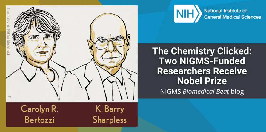
Sketches of Drs. Carolyn R. Bertozzi and K. Barry Sharpless above their printed names.
Credit: Niklas Elmehed.

The most recent NIGMS-supported Nobel laureates are Carolyn R. Bertozzi, Ph.D., the Anne T. and Robert M. Bass Professor in the School of Humanities and Sciences at Stanford University in Stanford, California, and K. Barry Sharpless, Ph.D., the W.M. Keck Professor of Chemistry at the Scripps Research Institute in La Jolla, California. They, along with Morten Meldal, Ph.D., a professor of chemistry at the University of Copenhagen in Denmark, are being recognized with the 2022 Nobel Prize in chemistry for their work on a transformative scientific approach known as “click chemistry.” The three scientists will receive their awards during a ceremony in Stockholm, Sweden, on December 10, 2022.

Continue reading “The Chemistry Clicked: Two NIGMS-Funded Researchers Receive Nobel Prize”

Q&A With Nobel Laureate and CRISPR Scientist Jennifer Doudna

0 comments
A headshot of Dr. Doudna. Jennifer Doudna, Ph.D. Credit: University of California, Berkeley.
The 2020 Nobel Prize in Chemistry was awarded to Jennifer Doudna, Ph.D., and Emmanuelle Charpentier, Ph.D., for the development of the gene-editing tool CRISPR. Dr. Doudna shared her thoughts on the award and answered questions about CRISPR in a live chat with NIH Director Francis S. Collins, M.D., Ph.D. Here are a few highlights from the interview.

Q: How did you find out that you won the Nobel Prize?

A: It’s a little bit of an embarrassing story. I slept through a very important phone call and finally woke up when a reporter called me. I was just coming out of a deep sleep, and the reporter was asking, “What do you think about the Nobel?” And I said, “I don’t know anything about it. Who won it?” I thought they were asking for comments on somebody else who won it. And she said, “Oh my gosh! You don’t know! You won it!”

Continue reading “Q&A With Nobel Laureate and CRISPR Scientist Jennifer Doudna”

Scientist Interview: Investigating Circadian Rhythms With Michael W. Young

1 comment

Sudden changes to our schedules, like the end of daylight saving time this Sunday or flying across time zones, often leave us feeling off kilter because they disrupt our bodies’ circadian rhythms. Circadian rhythms are physical, mental, and behavioral changes that follow a daily cycle. When these “biological clocks” are disrupted, our bodies eventually readjust. However, some people have conditions that cause their circadian rhythms to be permanently out of sync with their surroundings.

Continue reading “Scientist Interview: Investigating Circadian Rhythms With Michael W. Young”

Birthdays, Nobel Prizes and Basic Research

1 comment
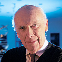
James D. Watson
James D. Watson. Credit: Wikimedia Commons, Cold Spring Harbor Laboratory.

April 6 is the birthday of two Nobel Prize winners in physiology or medicine—James Watson and Edmond H. Fischer. They have also both been NIGMS-supported researchers.

Double helix model
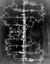
In 1953, Watson and Crick created their historic model of the shape of DNA: the double helix. Credit: Cold Spring Harbor Laboratory archives.

James D. Watson, born on this day in 1928, was honored with the Nobel Prize in 1962. He shared it with Francis H. Compton Crick and Maurice Wilkins “for their discoveries concerning the molecular structure of nucleic acids and its significance for information transfer in living material.” This laid the groundwork for future discoveries. In the early 1950s, Wilkins and another scientist, Rosalind Franklin, worked to determine DNA’s structure. In 1953, Watson and Crick discovered its shape as a double helix. This twisted ladder structure enabled other researchers to unlock the secret of how genetic information is stored, transferred and copied. Franklin is widely recognized as having played a significant role in revealing the physical structure of DNA; due to her death at age 37 in 1958, Franklin did not earn a share of the prize. Read more about DNA.

Continue reading “Birthdays, Nobel Prizes and Basic Research”

Cool Images: A Holiday-Themed Collection

1 comment

Here are some images from our gallery that remind us of the winter holidays—and showcase important findings and innovations in biomedical research.

Ribbons and Wreaths

Wreath

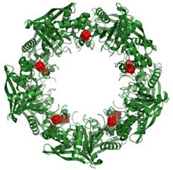
This wreath represents the molecular structure of a protein, Cas4, which is part of a system, known as CRISPR, that bacteria use to protect themselves against viral invaders. The green ribbons show the protein’s structure, and the red balls show the location of iron and sulfur molecules important for the protein’s function. Scientists have harnessed Cas9, a different protein in the bacterial CRISPR system, to create a gene-editing tool known as CRISPR-Cas9. Using this tool, researchers can study a range of cellular processes and human diseases more easily, cheaply and precisely. Last week, Science magazine recognized the CRISPR-Cas9 gene-editing tool as the “breakthrough of the year.”

Continue reading “Cool Images: A Holiday-Themed Collection”

Seeing Telomerase’s ‘Whiskers’ and ‘Toes’

1 comment
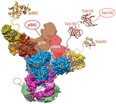
Telomerase and its components.

The image here is the “front view” of telomerase, with the enzyme’s components shown in greater detail than ever before. Credit: UCLA Department of Chemistry and Biochemistry.

Like the features of a cat in a dark alley, those of an important enzyme called telomerase have been elusive. Using a combination of imaging techniques, a research team led by Juli Feigon Exit icon of the University of California, Los Angeles, has now captured the clearest view ever of the enzyme.

Telomerase maintains the DNA at the ends of our chromosomes, known as telomeres, which act like the plastic tips on the ends of shoelaces. In the absence of telomerase activity, telomeres get shorter each time our cells divide. Eventually, the telomeres become so short that the cells stop dividing or die. On the other hand, cells with abnormally high levels of telomerase activity can constantly rebuild their protective chromosomal caps. Telomerase is particularly active within cancer cells. Continue reading “Seeing Telomerase’s ‘Whiskers’ and ‘Toes’”

Field Focus: Progress in RNA Interference Research

3 comments
Scientists first noticed what would later prove to be RNA interference when puzzling over an unexpected loss of color in petunia petals. Subsequent studies in roundworms revealed that double-stranded RNA can inactivate specific genes. Credit: Alisa Z. Machalek.

In less than two decades, RNA interference (RNAi)—a natural process cells use to inactivate, or silence, specific genes—has progressed from a fundamental finding to a powerful research tool and a potential therapeutic approach. To check in on this fast-moving field, I spoke to geneticists Craig Mello of the University of Massachusetts Medical School and Michael Bender of NIGMS. Mello shared the 2006 Nobel Prize in physiology or medicine with Andrew Fire Exit icon of Stanford University School of Medicine for the discovery of RNAi. Bender manages NIGMS grants in areas that include RNAi research.

How have researchers built on the initial discovery of RNAi?

A scientific floodgate opened after the 1998 discovery that it was possible to switch off specific genes by feeding microscopic worms called C. elegans double-stranded RNA that had the same sequence of genetic building blocks as a target gene. (Double-stranded RNA is a type of RNA molecule often found in, or produced by, viruses.) Scientists investigating gene function quickly began to test RNAi as a gene-silencing technique in other organisms and found that they could use it to manipulate gene activity in many different model systems. Additional studies led the way toward getting the technique to work in cells from mammals, which scientists first demonstrated in 2001. Soon, researchers were exploring the potential of RNAi to treat human disease.

Continue reading “Field Focus: Progress in RNA Interference Research”

Field Focus: Bringing Biology Into Sharper View with New Microscopy Techniques

0 comments
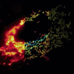
Composite image of mitochondria in a cell
In this composite image of mitochondria in a cell, the left panel shows a conventional optical microscopy image, the middle panel shows a three-dimensional (3-D) STORM image with color indicating depth, and the right panel shows a cross-section of the 3-D STORM image. Credit: Xiaowei Zhuang laboratory, Howard Hughes Medical Institute, Harvard University. View larger image.

Much as a photographer brings distant objects into focus with a telephoto lens, scientists can now see previously indistinct cellular components as small as a few billionths of a meter (nanometers). By overcoming some of the limitations of conventional optical microscopy, a set of techniques known as super-resolution fluorescence microscopy has changed once-blurry images of the nanoworld into well-resolved portraits of cellular architecture, with details never seen before in biology. Reflecting its importance, super-resolution microscopy was recognized with the 2014 Nobel Prize in chemistry.

Using the new techniques, scientists can observe processes in living cells across space and time and study the movements, interactions and roles of individual molecules. For instance, they can identify and track the proteins that allow a virus to invade a cell or those that enable tumor cells to migrate to distant parts of the body in metastatic cancer. The ability to analyze individual molecules, rather than collections of molecules, allows scientists to answer longstanding questions about cellular mechanisms and behavior, such as how cells move along a surface or how certain proteins interact with DNA to regulate gene activity. Continue reading “Field Focus: Bringing Biology Into Sharper View with New Microscopy Techniques”

Nobel Prize for Powerful Microscopy Technology

0 comments
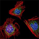
Fibroblasts
The cells shown here are fibroblasts, one of the most common cells in mammalian connective tissue. These particular cells were taken from a mouse. Scientists used them to test the power of a new microscopy technique that offers vivid views of the inside of a cell. The DNA within the nucleus (blue), mitochondria (green) and cellular skeleton (red) is clearly visible. Credit: Dylan Burnette and Jennifer Lippincott-Schwartz, Eunice Kennedy Shriver National Institute of Child Health and Human Development, National Institutes of Health.

William E. Moerner was at a conference in Brazil when he learned he’d be getting a Nobel Prize in chemistry. “I was incredibly excited and thrilled,” he said of his initial reaction.

An NIGMS grantee at Stanford University, Moerner received the honor for his role in achieving what was once thought impossible—developing super-resolution fluorescence microscopy, which is so powerful it allows researchers to see and track individual molecules in living organisms in real time.

Nobel recipients usually learn of the prize via a phone call from Stockholm, Sweden, in early October. For those in the United States, the call typically comes between 2:30 a.m. and 5:45 a.m.

Every year, the NIGMS communications office prepares for the Nobel Prize announcements in physiology or medicine and chemistry, the categories in which our grantees are most likely to be recognized. If the Institute played a significant role in funding the prize-winning research, we work quickly to provide information and context to reporters covering the story on tight deadlines. We issue a statement, identify an in-house expert on the research and arrange interviews with reporters. It’s all to help get the word out about the research and the taxpayers’ role in supporting it.

This year’s in-house expert, Cathy Lewis, shared her thoughts on the prize to Moerner in an NIGMS Feedback Loop post. You can also read this year’s statement and see a full list of NIGMS-supported Nobel laureates.

Cell Biology Advances and Computational Techniques Earn Nobels for NIGMS Grantees

0 comments
The winners of the 2013 Nobel Prize in physiology or medicine discovered that cells import and export materials using fluid-filled sacs called vesicles. Credit: Judith Stoffer.

Every October, a few scientists receive a call from Sweden that changes their lives. From that day forward, they will be known as Nobel laureates. This year, five of the new Nobelists have received funding from NIGMS.

In physiology or medicine, NIGMS grantees James Rothman (Yale University) and Randy Schekman (University of California, Berkeley) were honored “for their discoveries of machinery regulating vesicle traffic, a major transport system in our cells.” They share the prize with Thomas C. Südhof of Stanford University.

Rothman and Schekman started out working separately and in different systems—Schekman in yeast and Rothman in reconstituted mammalian cells—and their conclusions validated each others’. It’s yet another example of the power of investigator-initiated research and the value of model systems.

Highly accurate molecular models like this one are based on computational techniques developed by the winners of the 2013 Nobel Prize in chemistry. Credit: Rommie E. Amaro, University of California, San Diego.

The three Nobelists in chemistry are NIGMS grantees Martin Karplus (Harvard University), Michael Levitt (Stanford University) and Arieh Warshel (University of Southern California), who developed “multiscale models for complex chemical systems.” They used computational techniques to obtain, for the first time, detailed structural information about proteins and other large molecules. Because of that work, scientists around the world are now able to access, with a few keystrokes, highly accurate models of nearly 100,000 molecular structures. Studying these structures has advanced our understanding of countless diseases, pharmaceuticals and basic biological processes.

Learn more:

NIGMS Nobel Prize News Announcement
NIGMS Nobelists Fact Sheet