Tag: RNA

A Scientist’s Exploration of Regeneration

3 comments
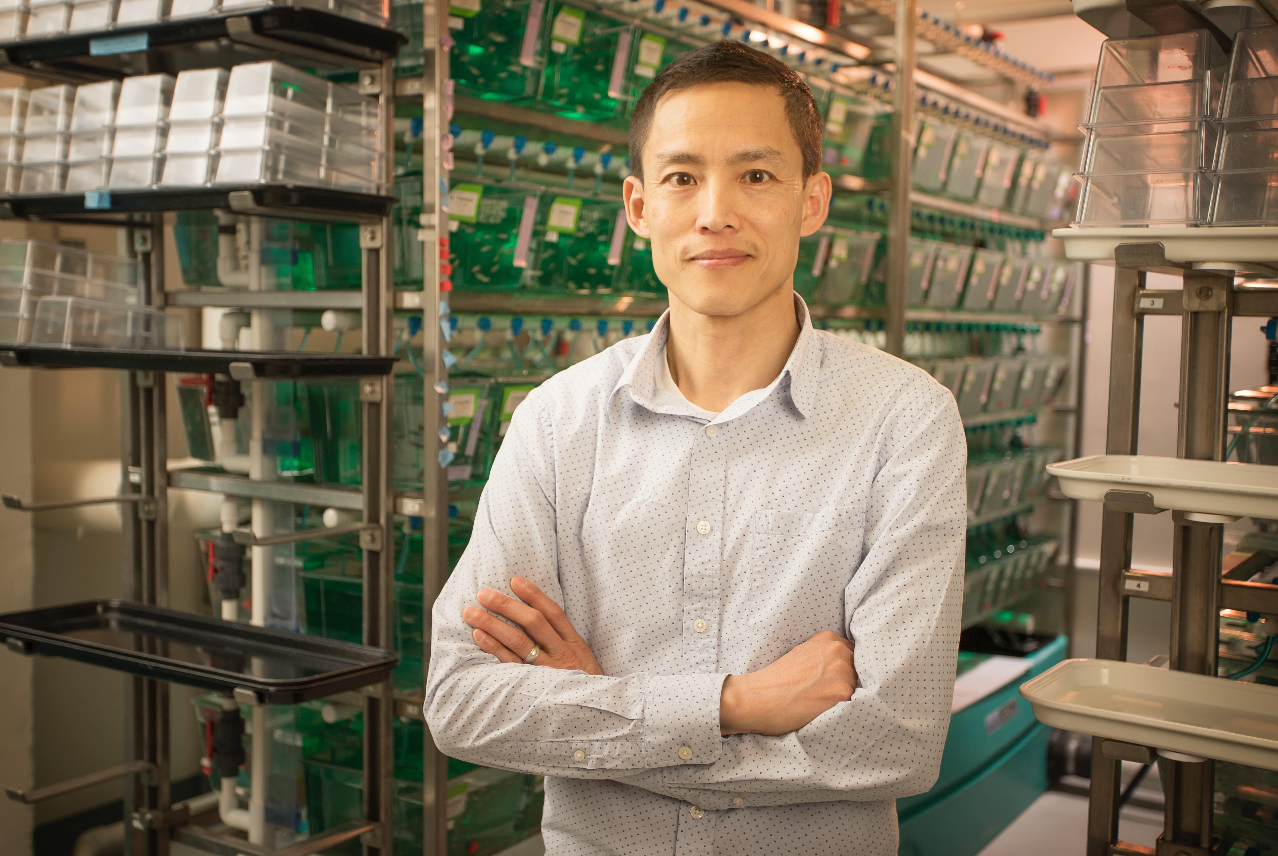
Viravuth (“Voot”) Yin, standing with arms crossed and smiling in front of a shelves holding tanks of zebrafish in his lab. Viravuth (“Voot”) Yin, associate professor of regenerative biology and medicine at MDI Biological Laboratory and chief scientific officer at Novo Biosciences, Inc., in Bar Harbor, Maine. Credit: MDI Biological Laboratory.

In 1980, a week after his 6th birthday, Viravuth (“Voot”) Yin immigrated with his mother, grandfather, and three siblings from Cambodia to the United States. Everything they owned fit into a single, 18-inch carry-on bag. They had to build new lives from almost nothing. So, it’s perhaps fitting that Yin studies regeneration, the fascinating ability of some animals, such as salamanders, sea stars, and zebrafish, to regrow damaged body parts, essentially from scratch.

Yin’s path wasn’t always smooth. His family settled in Hartford, Connecticut, near an uncle who had been granted asylum during the Vietnam War. Yin got into a lot of trouble in school, trying to learn a new culture and fit in. Things improved when his mother moved him and his siblings to West Hartford, well known for its strong schools.

Continue reading “A Scientist’s Exploration of Regeneration”

Interview With a Scientist—Julius Lucks: Shape Seeker

0 comments

While DNA acts as the hard drive of the cell, storing the instructions to make all of the proteins the cell needs to carry out its various duties, another type of genetic material, RNA, takes on a wide variety of tasks, including gene regulation, protein synthesis, and sensing of metals and metabolites. Each of these jobs is handled by a slightly different molecule of RNA. But what determines which job a certain RNA molecule is tasked with? Primarily its shape. Julius LucksLink to external web site, a biological and chemical engineer at Northwestern University, and his team study the many ways in which RNA can bend itself into new shapes and how those shapes dictate the jobs the RNA molecule can take on.

Continue reading “Interview With a Scientist—Julius Lucks: Shape Seeker”

Genomic Gymnastics of a Single-Celled Ciliate and How It Relates to Humans

0 comments
Laura Landweber
Credit: Denise Applewhite.
Laura Landweber
Grew up in: Princeton, New Jersey
Job site: Columbia University, New York City
Favorite food: Dark chocolate and dark leafy greens
Favorite music: 1940’s style big band jazz
Favorite hobby: Swing dancing
If I weren’t a scientist I would be a: Chocolatier (see “Experiments in Chocolate” sidebar at bottom of story)

One day last fall, molecular biologist Laura Landweber surveyed the Princeton University lab where she’d worked for 22 years. She and her team members had spent many hours that day laboriously affixing yellow Post-it notes to the laboratory equipment—microscopes, centrifuges, computers—they would bring with them to Columbia University, where Landweber had just been appointed full professor. Each Post-it specified the machinery’s location in the new lab. Items that would be left behind—glassware, chemical solutions, furniture, office supplies—were left unlabeled.

As Landweber viewed the lab, decorated with a field of sunny squares, her thoughts turned to another sorting process—the one used by her primary research subject, a microscopic organism, to sift through excess DNA following mating. Rather than using Post-it notes, the creature, a type of single-celled organism called a ciliate, uses small pieces of RNA to tag which bits of genetic material to keep and which to toss.

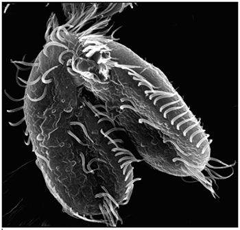
Landweber is particularly fond of Oxytricha trifallax, a ciliate with relatives that live in soil, ponds and oceans all over the world. The kidney-shaped cell is covered with hair-like projections called cilia that help it move around and devour bacteria and algae. Oxytricha is not only bizarre in appearance, it’s also genetically creative.

Unlike humans, whose cells are programmed to die rather than pass on genomic errors, Oxytricha cells appear to delight in genomic chaos. During sexual reproduction, the ciliate shatters the DNA in one of its two nuclei into hundreds of thousands of pieces, descrambles the DNA letters, throws most away, then recombines the rest to create a new genome.

Landweber has set out to understand how—and possibly why—Oxytricha performs these unusual genomic acrobatics. Ultimately, she hopes that learning how Oxytricha rearranges its genome can illuminate some of the events that go awry during cancer, a disease in which the genome often suffers significant reorganization and damage.

Oxytricha’s Unique Features

Oxytricha carries two separate nuclei—a macronucleus and a micronucleus. The macronucleus, by far the larger of the two, functions like a typical genome, the source of gene transcription for proteins. The tiny micronucleus only sees action occasionally, when Oxytricha reproduces sexually.

Oxytricha trifallax cells in the process of mating
Two Oxytricha trifallax cells in the process of mating. Credit, Robert Hammersmith.

What really makes Oxytricha stand out is what it does with its DNA during the rare occasions that it has sex. When food is readily available, Oxytricha procreates without a partner, like a plant grown from a cutting. But when food is scarce, or the cell is stressed, it seeks a mate. When two Oxytricha cells mate, the micronuclear genomes in each cell swap DNA, then replicate. One copy of the new hybrid micronucleus remains intact, while the other breaks its DNA into hundreds of thousands of pieces, some of which are tagged, recombined, then copied another thousand-fold to form a new macronucleus. Continue reading “Genomic Gymnastics of a Single-Celled Ciliate and How It Relates to Humans”

The Changing Needs of a Cell: No Membrane? No Problem!

0 comments

Russian nesting dolls. Credit: iStock.

How “membrane-less” organelles help with key cellular functions

Scientists have long known that animal and plant cells have specialized subdivisions called organelles. These organelles are surrounded by a semi-permeable barrier, called a membrane, that both organizes the organelles and insulates them from the rest of the cell’s mix of proteins, salt, and water. This set-up helps to make cells efficient and productive, aiding in energy production and other specialized functions. But, because of their semi-permeable membranes, organelles can’t regroup and reform in response to stress or other outside changes. Cells need a rapid response team working alongside the membrane-bound organelles to meet these fluctuating needs. Until recently, who those rapid responders were and how they worked has been a mystery.

Recent research has led biologists to learn that the inside of a cell or an organelle is not just a lot of different molecules dissolved in water. Instead, we now know that cells contain many pockets of liquid droplets (one type of liquid surrounded by a liquid of different density) with specialized composition and function that are not surrounded by membranes. Because these “membrane-less organelles” are not confined, they can rapidly come together in response to chemical signals, such as those that indicate stress, and equally rapidly fall apart when they are no longer needed, or when the cell is about to divide. This enables membrane-less organelles to be “rapid responders.” They can have complex, multilayered structures that help them to perform many critical cell functions with multiple steps, just like membrane-bound organelles. Scientists even suspect that the way these organelles form as droplets may shed light on how life on Earth first took shape (see sidebar “Could This Be How Life First Took Shape?” at bottom of page).

The Many Membrane-less Organelles

Scientists have identified more than a dozen membrane-less organelles at work in mammalian cells. Several kinds found inside the nucleus—including nuclear speckles, paraspeckles, and Cajal bodies—help with cell growth, stress response, the metabolizing (breaking down) of RNA, and the control of gene expression—the process by which information in a gene is used in the synthesis of a protein. Out in the cytoplasm, P-bodies, germ granules, and stress granules are membrane-less organelles that are involved in metabolizing or protecting messenger RNA (mRNA), controlling which mRNAs are made into proteins, and in maintaining balance, or homeostasis, of the cell’s overall health.

The nucleolus, located inside the nucleus, is probably the largest of the membrane-less organelles. It acts as a factory to assemble ribosomes, the giant molecular machines that “translate” messenger RNAs to make all cellular proteins.

Continue reading “The Changing Needs of a Cell: No Membrane? No Problem!”

The Endoplasmic Reticulum: Networking Inside the Cell

5 comments

Like a successful business networker, a cell’s endoplasmic reticulum (ER) is the structure that reaches out—quite literally—to form connections with many different parts of a cell. In several important ways, the ER enables those other parts, or organelles, to do their jobs. Exciting new images of this key member of the cellular workforce may clarify how it performs its roles. Such knowledge will also help studies of motor neuron and other disorders, such as amyotrophic lateral sclerosis (ALS), that are associated with abnormalities in ER functioning.

Structure Follows Function

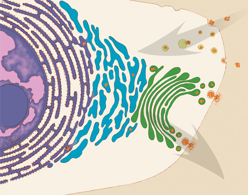
Illustration of some of the jobs that the ER performs in the cell.

An illustration of some of the jobs that the endoplasmic reticulum (ER) performs in the cell. Some ER membranes (purple) host ribosomes on their surface. Other ER membranes (blue) extending into the cytoplasm are the site of lipid synthesis and protein folding. The ER passes on newly created lipids and proteins to the Golgi apparatus (green), which packages them into vesicles for distribution throughout the cell. Credit: Judith Stoffer.

Initiated in 1965, the Postdoctoral Research Associate Program (PRAT) is a competitive postdoctoral fellowship program to pursue research in one of the laboratories of the National Institutes of Health. PRAT is a 3-year program providing outstanding laboratory experiences, access to NIH’s extensive resources, mentorship, career development activities and networking. The program places special emphasis on training fellows in all areas supported by NIGMS, including cell biology, biophysics, genetics, developmental biology, pharmacology, physiology, biological chemistry, computational biology, immunology, neuroscience, technology development and bioinformatics

The ER is a continuous membrane that extends like a net from the envelope of the nucleus outward to the cell membrane. Tiny RNA- and protein-laden particles called ribosomes sit on its surface in some places, translating genetic code from the nucleus into amino acid chains. The chains then get folded inside the ER into their three-dimensional protein structures and delivered to the ER membrane or to other organelles to start their work. The ER is also the site where lipids—essential elements of the membranes within and surrounding a cell—are made. The ER interacts with the cytoskeleton—a network of protein fibers that gives the cell its shape—when a cell divides, moves or changes shape. Further, the ER stores calcium ions in cells, which are vital for signaling and other work.

To do so many jobs, the ER needs a flexible structure that can adapt quickly in response to changing situations. It also needs a lot of surface area where lipids and proteins can be made and stored. Scientists have thought that ER structure combined nets of tubules, or small tubes, with areas of membrane sheets. However, recent NIGMS PRAT (Postdoctoral Research Associate; see side bar) fellow Aubrey Weigel, working with her mentor and former PRAT fellow Jennifer Lippincott-Schwartz of the Eunice Kennedy Shriver National Institute of Child Health and Human Development (currently at the Howard Hughes Medical Institute in Virginia) and colleagues, including Nobel laureate Eric Betzig, wondered whether limitations in existing imaging technologies were hiding a better answer to how the ER meets its surface-area structural needs in the periphery, the portion of the cell not immediately surrounding the nucleus. Continue reading “The Endoplasmic Reticulum: Networking Inside the Cell”

CRISPR Serves Up More than DNA

0 comments
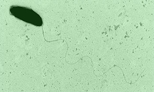
Marine bacterium Marinomonas mediterranea
The marine bacterium Marinomonas mediterranea uses a CRISPR system to spot invading RNAs and store a memory of the invasion event in its genome. Research team member Antonio Sanchez-Amat was the first to isolate and characterize this bacterial species. Credit: Antonio Sanchez-Amat, University of Murcia.

A new study has added another twist to the CRISPR story. As we’ve highlighted in several recent posts, CRISPR is an immune system in bacteria that recognizes and destroys viral DNA and other invading DNA elements, such as transposons. Scientists have adapted CRISPR into an indispensable gene-editing tool now widely used in both basic and applied research.

Many previously described CRISPR systems detect and cut viral DNA, insert the DNA pieces into the bacterial genome and then use them as molecular “mug shots” to flag and destroy the virus if it attacks again. But various viruses use RNA, not DNA, as genetic material. Although research has shown that some CRISPR systems also can target RNA, how these systems can archive harmful RNA encounters in the bacterial genome was unknown. Continue reading “CRISPR Serves Up More than DNA”

How Cells Manage Chance

3 comments

We asked the heads of our scientific divisions to tell us about some of the big questions in fundamental biomedical science that researchers are investigating with NIGMS support. This article is the second in an occasional series that explores these questions and explains how pursuing the answers could advance understanding of important biological processes.

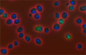
Sample slide, variability of mRNA in yeast cells
The number of copies of mRNA molecules (bright green) observed here in yeast cells (dark blue) fluctuates randomly. Credit: David Ball, Virginia Tech.

For some health conditions, the cause is clear: A single altered gene is responsible. But for many others, the path to disease is more complex. Scientists are working to understand how factors like genetics, lifestyle and environmental exposures all contribute to disease. Another important, but less well-known, area of investigation is the role of chance at the molecular level.

One team working in this field is led by John Tyson at Virginia Tech. The group focuses on how chance events affect the cell division cycle, in which a cell duplicates its contents and splits into two. This cycle is the basis for normal growth, reproduction and the replenishment of skin, blood and other cells throughout the body. Errors in the cycle are associated with a number of conditions, including birth defects and cancer. Continue reading “How Cells Manage Chance”

Field Focus: Progress in RNA Interference Research

3 comments
Scientists first noticed what would later prove to be RNA interference when puzzling over an unexpected loss of color in petunia petals. Subsequent studies in roundworms revealed that double-stranded RNA can inactivate specific genes. Credit: Alisa Z. Machalek.

In less than two decades, RNA interference (RNAi)—a natural process cells use to inactivate, or silence, specific genes—has progressed from a fundamental finding to a powerful research tool and a potential therapeutic approach. To check in on this fast-moving field, I spoke to geneticists Craig Mello of the University of Massachusetts Medical School and Michael Bender of NIGMS. Mello shared the 2006 Nobel Prize in physiology or medicine with Andrew Fire Exit icon of Stanford University School of Medicine for the discovery of RNAi. Bender manages NIGMS grants in areas that include RNAi research.

How have researchers built on the initial discovery of RNAi?

A scientific floodgate opened after the 1998 discovery that it was possible to switch off specific genes by feeding microscopic worms called C. elegans double-stranded RNA that had the same sequence of genetic building blocks as a target gene. (Double-stranded RNA is a type of RNA molecule often found in, or produced by, viruses.) Scientists investigating gene function quickly began to test RNAi as a gene-silencing technique in other organisms and found that they could use it to manipulate gene activity in many different model systems. Additional studies led the way toward getting the technique to work in cells from mammals, which scientists first demonstrated in 2001. Soon, researchers were exploring the potential of RNAi to treat human disease.

Continue reading “Field Focus: Progress in RNA Interference Research”

Meet Jennifer Doudna

3 comments
Jennifer Doudna
Credit: Jennifer Doudna
Jennifer Doudna
Fields: Biochemistry and structural biology
Studies: New genome editing tool called CRISPR
Works at: University of California, Berkeley
Raised in: Hilo, Hawaii
Studied at: Pomona College, Harvard University
Recent honors: Winner of the Lurie Prize in the Biomedical Sciences Exit icon, an annual award that recognizes outstanding achievement by promising scientists age 52 or younger
If she couldn’t be a scientist, she’d like to be: A papaya farmer or an architect

Jennifer Doudna likes to get her hands dirty. Literally. When she’s not in her laboratory, she can often be found amid glossy green leaves and brightly colored fruit in her Berkeley garden. She recently harvested her first three strawberry guavas.

Coaxing tropical fruit plants from her childhood home in Hawaii to grow in Northern California is more than just a hobby—it’s an intellectual challenge.

“I like solving puzzles, I like the process of figuring things out, and I enjoy working with my hands,” says Doudna. “Those things were what really drew me to science in the beginning.”

Since she was a graduate student, Doudna’s professional puzzle has been RNA, a type of genetic material inside our cells. Recently, there has been an explosion of discoveries about the many roles RNA molecules play in the body. Doudna’s work probes into how RNA molecules work, what 3-D shapes they form and how their structures drive their functions.

“I’ve been fascinated by understanding RNA at a mechanistic level,” Doudna says.

While teasing out answers to these fundamental questions, Doudna’s lab has played a leading role in a discovery that is upending the field of genetic engineering, with exciting implications for human health.

Her Findings

The discovery started with bacteriophages—viruses that infect bacteria, just like the common cold infects humans. About 10 years ago, researchers using high-powered computing to sift through bacterial genomes began to find mysterious repetitive gene sequences that matched those from viruses known to infect the bacteria. The researchers named these sequences “clustered regularly interspaced short palindromic repeats,” or CRISPRs for short.

Over the next few years, scientists came to understand that these CRISPR sequences are part of something not previously thought to exist—an adaptive bacterial immune system, which remembers viruses fought off before and raises a response to fight them when exposed again. CRISPRs were this immune system’s reference library, holding records of viral exposure.

Somehow, bacteria with a CRISPR-based immune system (there are three types now known to scientists) use these records to command certain proteins to recognize and chop up DNA from returning viruses.

Wanting to know more about this process, Doudna’s team picked one protein in a CRISPR-based defense system to study. This protein, called Cas9, had been identified by other researchers as being essential for protection against viral invasion.

To their delight, Doudna’s group had hit the jackpot. Cas9 turned out to be the system’s scalpel. Once CRISPR identifies a DNA sequence from the invading virus, Cas9 slices the sequence out of the viral genome, destroying the virus’s ability to copy itself.

Doudna’s lab and their European collaborators also identified the other key components of the CRISPR-Cas9 system—two RNA molecules that guide Cas9 to the piece of viral DNA identified by CRISPR.

Even more importantly, the researchers showed that the two guide RNAs could be manipulated in the lab to create a tool that both recognizes any specified DNA sequence and carries Cas9 there to make its cut.

“That was really where we made the connection between the basic, curiosity-driven research that we were doing and recognizing that we had in our hands something that could be a very powerful technology for genome editing,” remembers Doudna.

She was right. After publication of their 2012 paper, the field of CRISPR-guided genetic manipulation exploded. Labs around the world now use the tool Doudna’s team developed to cut target gene sequences in organisms ranging from plants to humans. The technique is already replacing more time-consuming, less-reliable methods of creating ‘knock-out’ model organisms (those missing a specific gene) for laboratory research. CRISPR-based editing even allows more than one gene to be knocked out at the same time, something that was not possible with previous genome-editing techniques.

The ability of CRISPR systems to recognize DNA sequences with extraordinary precision also holds potential for human therapeutics. For example, a paper from another laboratory published early this year showed that, in a mouse model, CRISPR-based editing could cut out and replace a defective gene responsible for a type of muscular dystrophy. Researchers are testing similar CRISPR-based techniques in models of human diseases ranging from cystic fibrosis to blood disorders.

Doudna is a co-founder of two biotechnology companies hoping to harness the potential of CRISPR-based genome editing. Although the technology holds great promise, she acknowledges that much work needs to be done before CRISPR can be considered safe for human trials. Major challenges include assuring that no off-target cuts are made in the genome and finding a safe way to deliver the editing system to living tissues.

She is also excited to continue working with her research team, advancing the basic understanding of the CRISPR-based system.

“I’m very interested in seeing what we can contribute to the whole question about how you deliver a technology like this, how you can use it therapeutically in an organism. That’s an area where we hope that our biochemical understanding of this system will be able to contribute,” she concludes.
An RNA Molecule That Cues the Internal Clock

0 comments
Clock
Dysfunction in our internal clocks may lead to insufficient sleep, which has been linked to an increased risk for chronic diseases. Credit: Stock image.

Our internal clocks tell us when to sleep and when to eat. Because they are sensitive to changes in daytime and nighttime cues, they can get thrown off by activities like traveling across time zones or working the late shift. Dysfunction in our internal clocks may lead to insufficient sleep, which has been linked to an increased risk for chronic diseases like high blood pressure, diabetes, depression and cancer.

Researchers led by Yi Liu Exit icon of the University of Texas Southwestern Medical Center have uncovered a previously unknown mechanism by which internal clocks run and are tuned to light cues. Using the model organism Neurospora crassa (a.k.a., bread mold), the scientists identified a type of RNA molecule called long non-coding RNA (lncRNA) that helps wind the internal clock by regulating how genes are expressed. When it’s produced, the lncRNA identified by Liu and his colleagues blocks a gene that makes a specific clock protein.

This inhibition works the other way, too: The production of the clock protein blocks the production of the lncRNA. This rhythmic gene expression helps the body stay tuned to whether it’s day or night.

The researchers suggest that a similar mechanism likely exists in the internal clocks of other organisms, including mammals. They also think that lncRNA-protein pairs may contribute to the regulation of other biologic processes.

Learn more:
University of Texas Southwestern Medical Center News Release Exit icon
Circadian Rhythms Fact Sheet