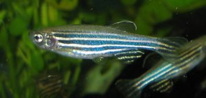
Name: Danio rerio Hometown: Freshwater ponds and rivers of India, Nepal, and neighboring countries Occupation: Research Long-term goal: Solving the basic mysteries of life Work site: More than 600 science labs worldwide
That’s me and some other zebrafish, swimming in a tank in one of the more than 600 labs around the world that use us to study embryo development, genetics, and all kinds of human diseases. Credit: Wikimedia Commons, Azul.
Apart from the tell-tale stripes that give me my nickname, zebrafish, I look a lot like your standard minnow swimming in the shallows of any pond, lake, or river. But I like to think I’m more important than that. In fact, researchers around the world have turned to me and my extended family to understand some of the most basic mysteries of life. From studying us, they’re learning about how embryos develop, how cancer works, and whether someday humans might be able to rebuild a heart, repair a spinal cord injury, or regrow a severed limb.
Why us? Because zebrafish are pretty special and researchers think we’re easy to work with. First, unlike your standard lab mouse or rat, we lay lots of eggs, producing baby fish that grow up fast. We develop outside our mothers and go from egg to embryo to free-swimming larva in just 3 days (check out this video of how we grow, cell by cell, during the first 24 hours). Within 3 months, we’re fully mature.
Not only do zebrafish moms have many babies at the same time, and not only do these babies grow up quickly, but our eggs and embryos are see-through, so scientists can literally watch us grow one cell at a time. We stay mostly transparent for a few weeks after hatching. That makes it super easy for scientists to monitor us for both normal and abnormal development. In fact, scientists have learned how to turn off the genes that give our skin its color. These zebrafish, named casper, after the “friendly ghost” of cartoon fame, stay semi-transparent, or translucent, through adulthood.
And last, but certainly not least, did I mention that we can regenerate? If parts of my body are damaged, even to a significant degree, they can regrow. This holds true for my heart, fins, spinal cord, and even brain tissue. Our regenerative capacity is seemingly unlimited; my caudal fin, for example, can grow back dozens of times. Continue reading “Zebrafish Scrapbook”
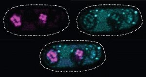
These glowing images of yeast (Schizosaccharomyces kambucha) reproductive cells show an example of a selfish gene at work. Here, the selfish gene boosts its chances of being passed to the next generation by producing both a toxin (stained cyan) and an antitoxin (stained magenta). Cells with a copy of the selfish gene are protected by the antitoxin, left and bottom ovals. Those without the selfish gene are destroyed by the toxin. Scientists suspect that selfish genes could be operating throughout many organisms’ genomes, possibly having a major impact on how genetic material is inherited over generations. Credit: Image courtesy of María Angélica Bravo Núñez and Nicole Nuckolls.
There’s an old saying that rules are meant to be broken. In the 1860s, Gregor Mendel found that each copy of a gene in an organism has an equal chance of being passed to the next generation. According to this simple rule, each version of a gene gets passed to offspring with the same frequency. The natural selection process can then operate efficiently, favoring the genes that produce an advantage for an organism’s survival or reproductive success and, over successive generations, eliminating genes from the gene pool that bring a disadvantage.
Of course, the way organisms inherit genes is not as straightforward as Mendel’s work predicted. In natural systems, inheritance often fails to follow the rules. One culprit scientists are identifying again and again are what are called “selfish genes”: one or more genes that have evolved a method of skewing inheritance in their favor.
Scientists refer to these selfish genes—which are often complexes of multiple genes working together—as “selfish” because they enhance their own transmission to the next generation, sometimes by killing off any of the organism’s reproductive cells that lack copies of those genes. Using a variety of techniques, the genes are effective at passing themselves on to future generations. However, their methods set up a conflict within the organism by damaging its fertility; overall, fewer reproductive cells or offspring survive to produce a new generation.
Selfish genes are a challenge for scientists to identify, but researchers say that knowing more about these genes could help explain a range of genetic mysteries, from causes of infertility to details on how species evolve. The methods these genes use could also be harnessed to help control the reproduction of certain populations such as mosquitos that spread disease.
One recently described selfish gene system is found in the yeast cells pictured above. Sarah Zanders and her colleagues at the Stowers Institute for Medical Research in Kansas City, Missouri, and the Fred Hutchinson Cancer Research Center in Seattle, Washington, study selfish gene systems in yeast to understand how common they are and how they affect a species’ fertility and evolution. “Usually we think about infertility stemming from the good guys failing. For example, a gene that normally promotes fertility could be mutated so that it can no longer do its job,” says Zanders. “But selfish genes are another potential source of infertility. Learning general principles about selfish genes in simple models will guide future searches for selfish genes that could be contributing to human infertility.”
Recently, the scientists discovered a single selfish gene, wtf4, that encodes both a toxin and an antitoxin protein. When yeast produce their reproductive cells, called spores, the wtf4 toxin protein is released into the immediate vicinity, but the antitoxin remains inside spores that contain a copy of wtf4. The toxin destroys all the spores that don’t have the antitoxin protein. Although the yeast has fewer spores—and therefore reduced fertility—each spore carries wtf4, ensuring that the gene will be passed to the next generation of yeast.
How do you measure pain? A patient’s furrowed brow, a child’s cries or tears—all are signs of pain. But what if the patient suffers from severe dementia and can’t describe what she is feeling or is a young child who can’t yet talk? Caregivers can help read the signs of pain, but their interpretations may differ greatly from patient to patient, because people have different ways of showing discomfort. And when the patient is unconscious, such as during surgery or while in intensive care, the caregiving team has even fewer ways to measure pain.
Assessing pain is an inexact science. It includes both subjective and objective measures. A patient might be asked during a subjective assessment (performed, perhaps, with a caregiver showing a pain-rating scale such as the one in the figure), “How much pain are you feeling today?” That feedback is coupled with biological markers such as an increased heart rate, dilated pupils, sweating, and inflammation as well as blood tests to monitor high levels of the stress hormone cortisol. Combined, these measurements can give doctors a fairly clear picture of how much pain a patient feels.
Patients can point to one of the faces on this subjective pain scale to show caregivers the level of pain they are experiencing. Credit: Wong-Baker Faces Foundation.
But imagine if members of the surgical or caregiving team could actually “see” how the patient is feeling? Such insight would let them select better drugs to use during and after surgery, tailoring care to each patient. That tool could be put into service in the operating room and by the bedside in intensive care, giving nonstop reports of pain as the patient experiences it.
An objective measure of pain also has uses beyond the operating room and intensive care unit. Given the high risk for opioid misuse, such a measure could take the guesswork out of pain management and give doctors a more accurate indication of pain levels to prevent over-prescribing opioid pain relievers.
Although not as well-known as other medical conditions, sepsis kills more people in the United States than AIDS, breast cancer, or prostate cancer combined. Sepsis is body-wide inflammation, usually triggered by an overwhelming immune response to infection. Though doctors and medical staff are well-aware of the condition—it is involved in 1 in 10 hospital deaths—the condition is notoriously hard to diagnose. In this video, sepsis expert Sarah Dunsmore, a program director with the National Institute of General Medical Sciences (NIGMS), describes what sepsis is and how to recognize it, what kinds of patients are most at risk, and what NIGMS is doing to reduce the impact of this deadly condition.
One of NIGMS’ primary goals is to provide support to train the next generation of biomedical research scientists. In pursuit of this goal, NIGMS aims to enhance the diversity of the scientific workforce and develop research capacities throughout the country. NIGMS-administered training programs at the undergraduate level provide support for trainees underrepresented in the biomedical sciences to develop skills to successfully transition into doctoral programs. Three unique NIGMS-administered undergraduate-focused programs are highlighted below.
Building Infrastructure Leading to Diversity (BUILD) grant awards help undergraduate institutions implement and study ways to engage and retain students from diverse backgrounds in biomedical research. The program aims to help these students on the pathway to becoming scientists. Primary institutions eligible for BUILD awards have fewer than $7.5 million in total NIH research project grant funding and a student population with at least 25 percent Pell Grant recipients. BUILD is part of the Common Fund Diversity Program Consortium, a national collaborative dedicated to enhancing diversity in the biomedical research workforce.
Maximizing Access to Research Careers Undergraduate Student Training in Academic Research (MARC U-STAR) awards provide support for undergraduate trainees from underrepresented backgrounds to gain skills and improve their preparation for high-caliber graduate training at the doctoral level. Awards are made to colleges and universities that offer the baccalaureate degree.
The Research Initiative for Scientific Enhancement (RISE) program aims to help reduce the existing gap between underrepresented and well-represented students in completing doctoral degrees. RISE supports institutions that award the baccalaureate, master’s, or doctoral degree in biomedical science fields; programs include well-integrated developmental activities designed to strengthen students’ academic preparation, research training, and professional skills.
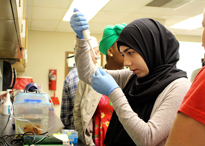
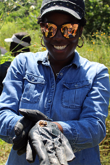
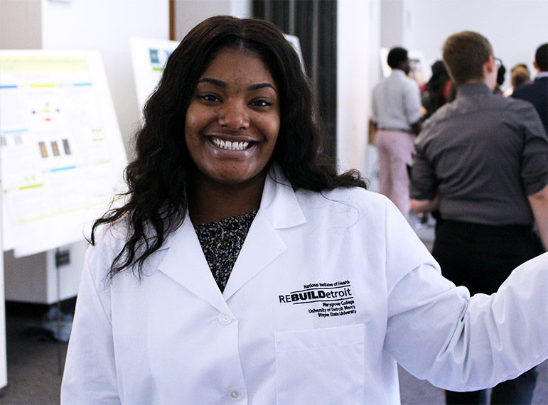
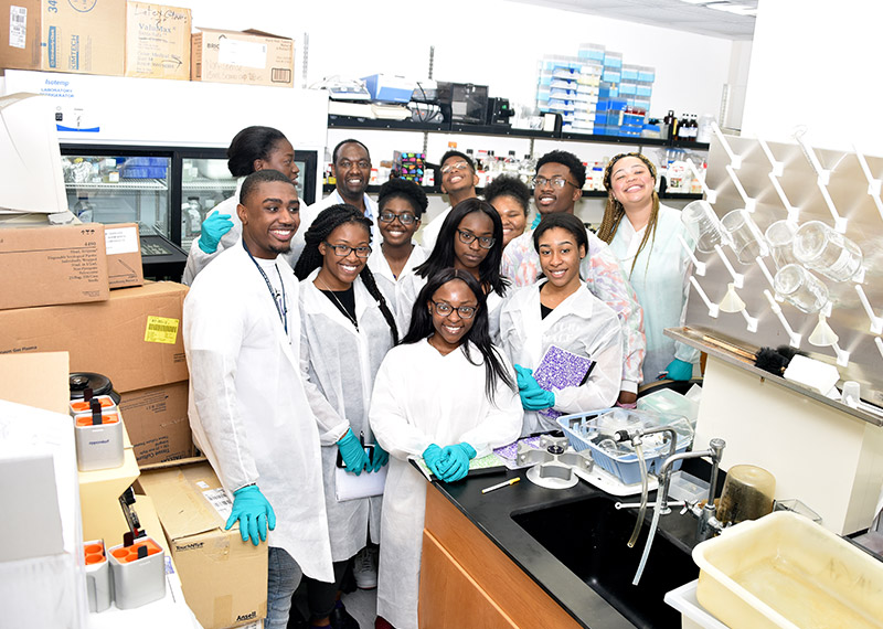
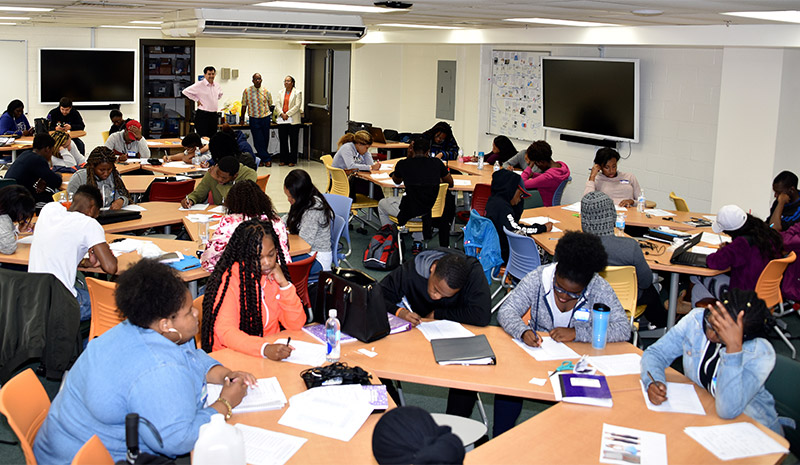
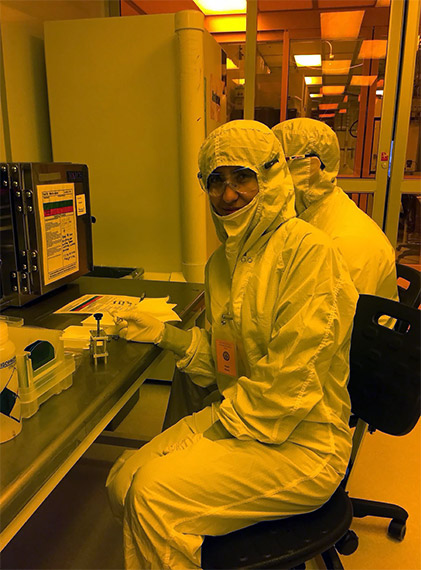
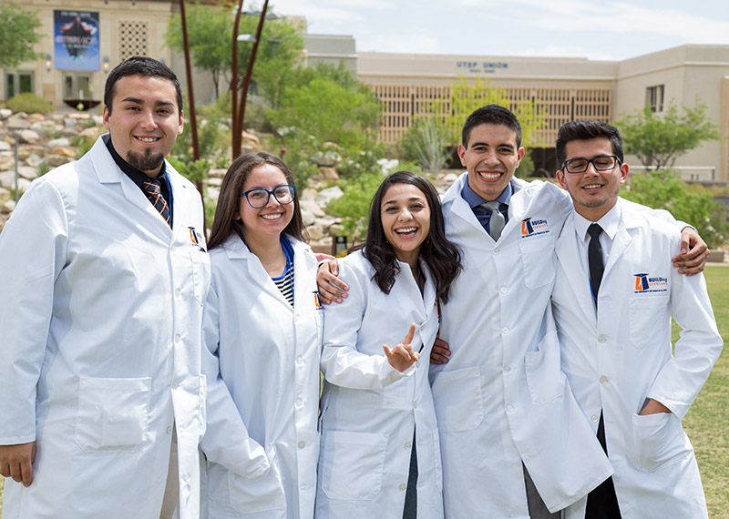
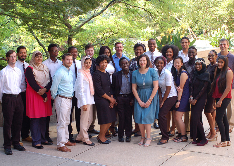
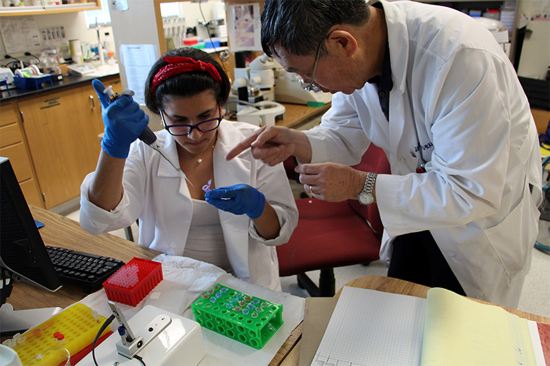
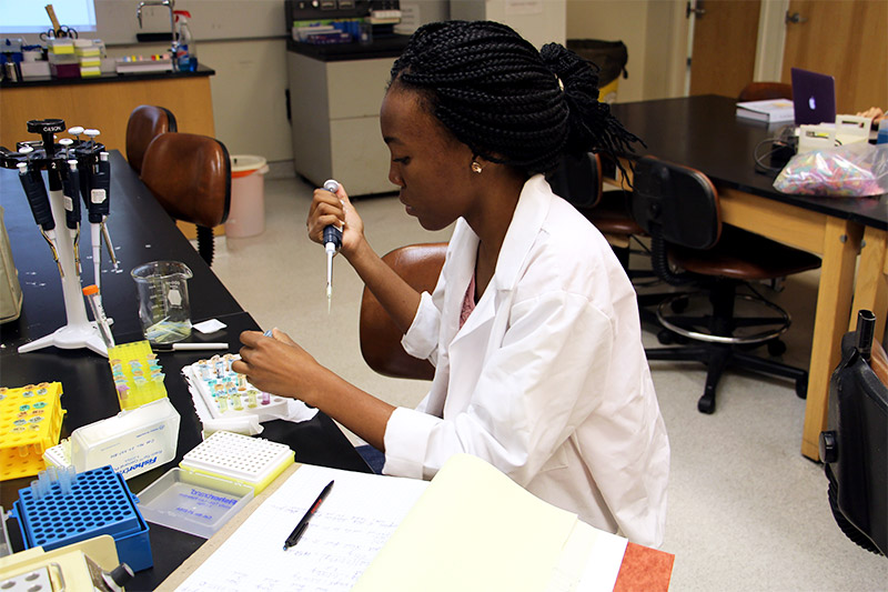
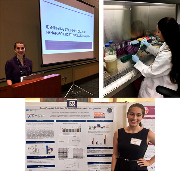
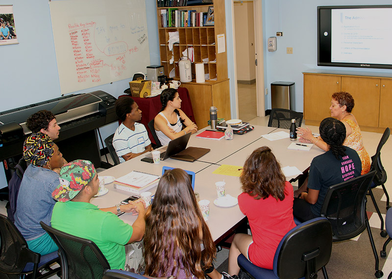
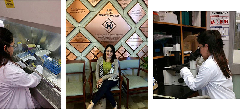
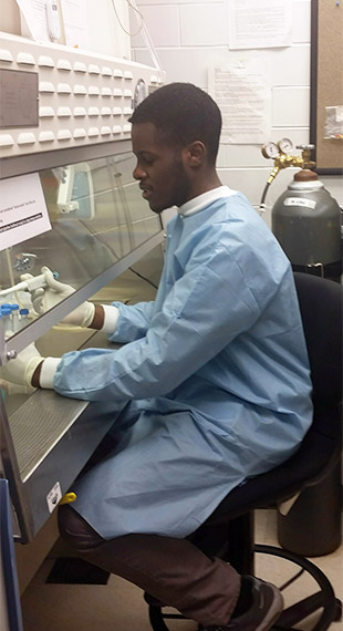
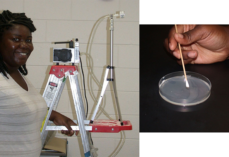
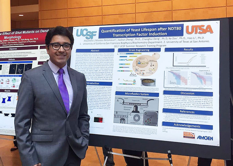
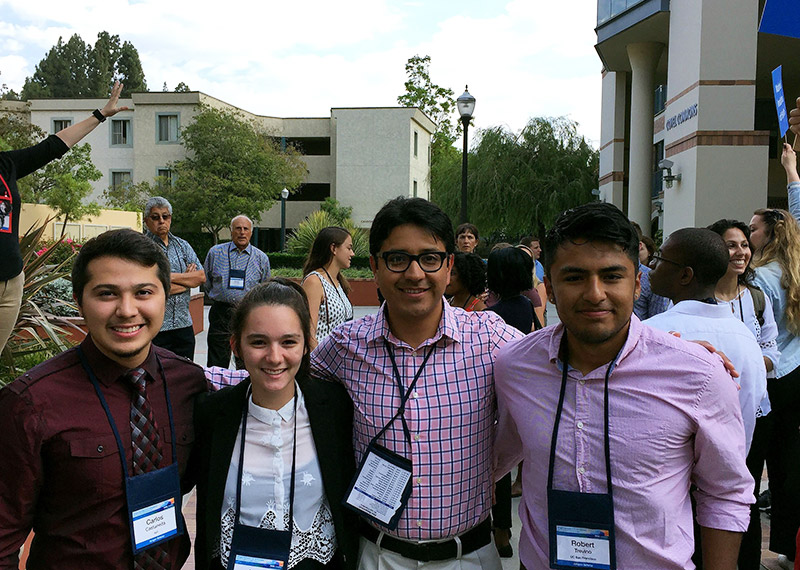
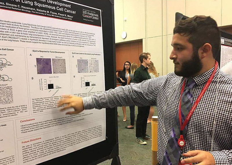
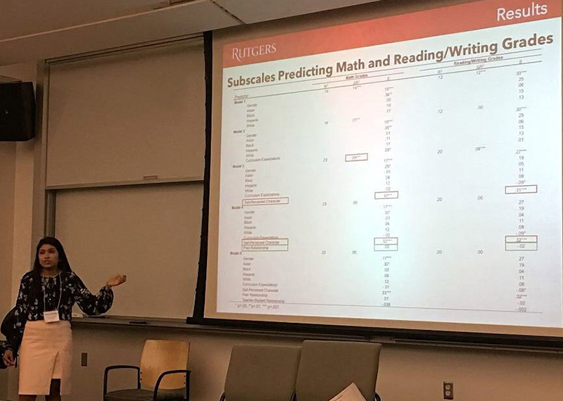
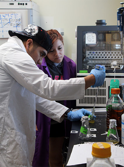
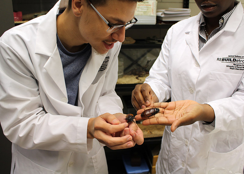
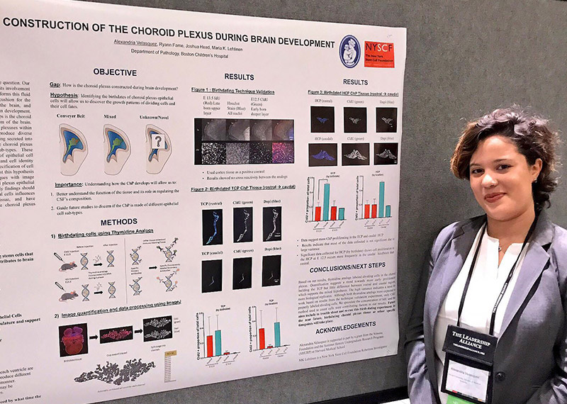
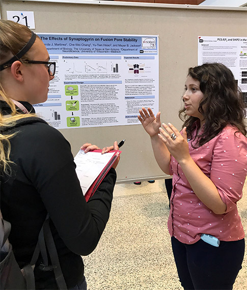
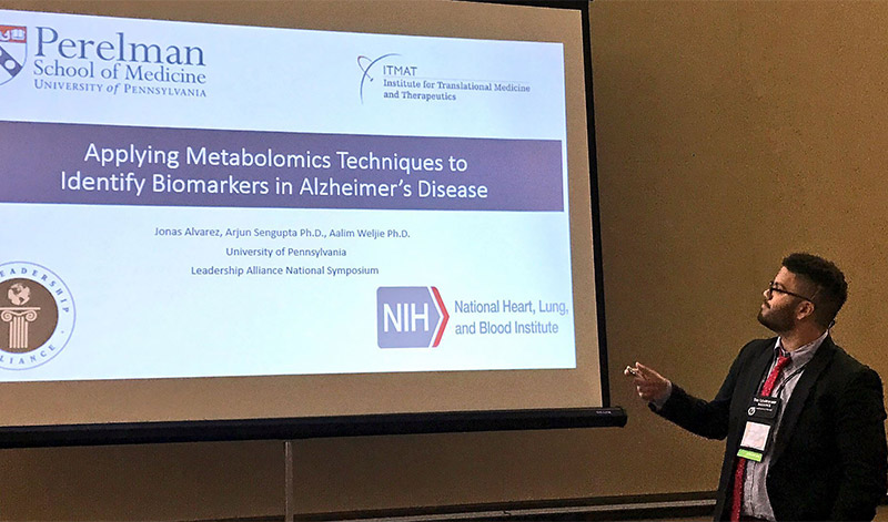
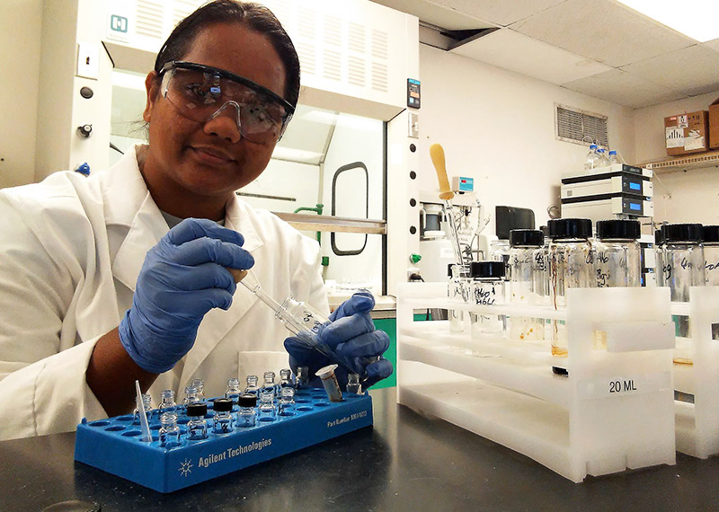
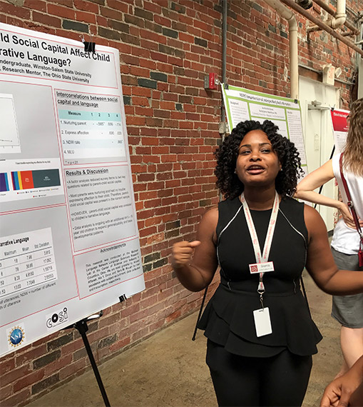
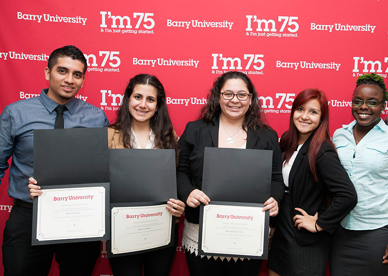
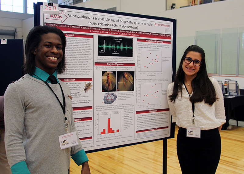
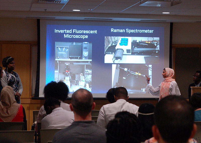
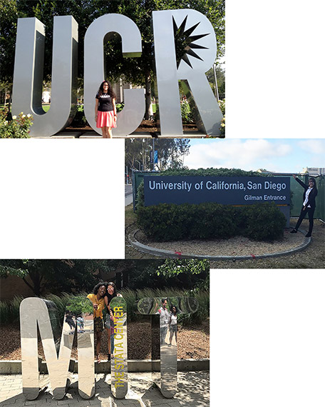
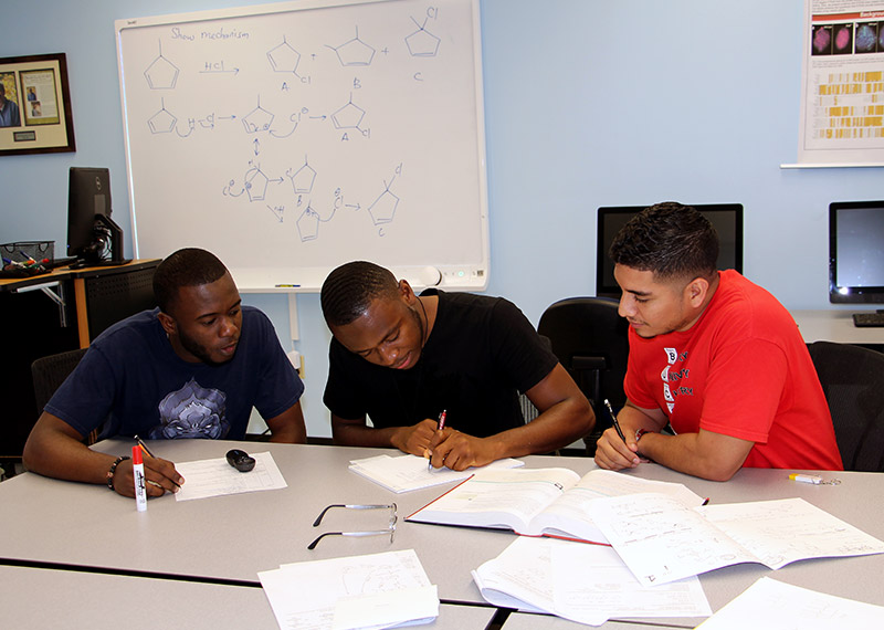
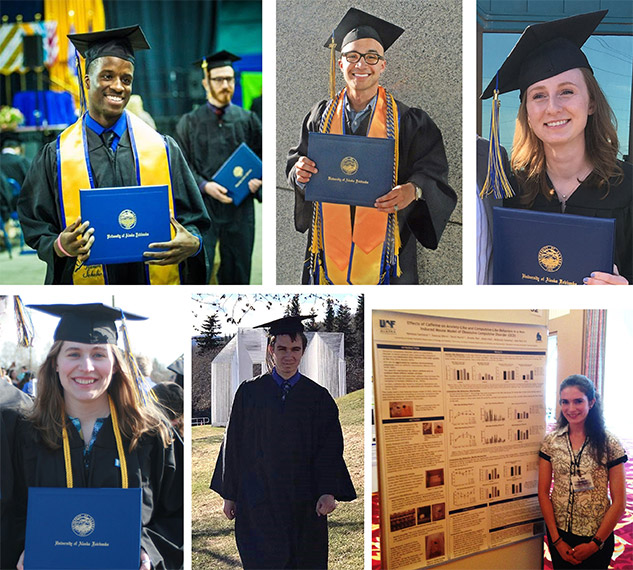
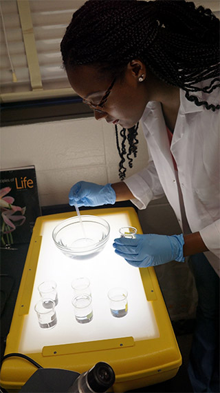
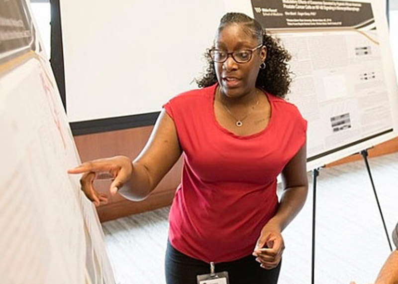
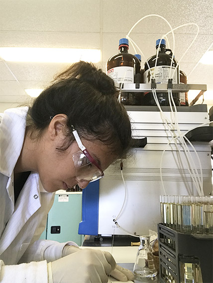
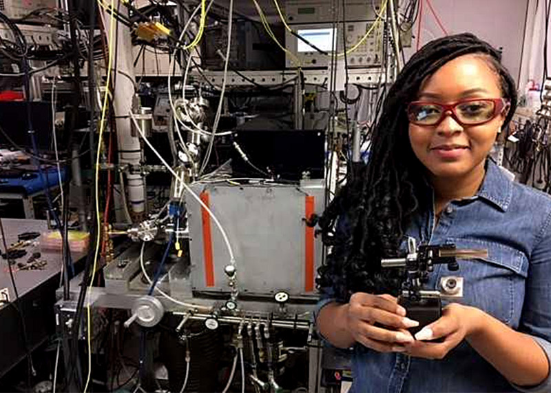
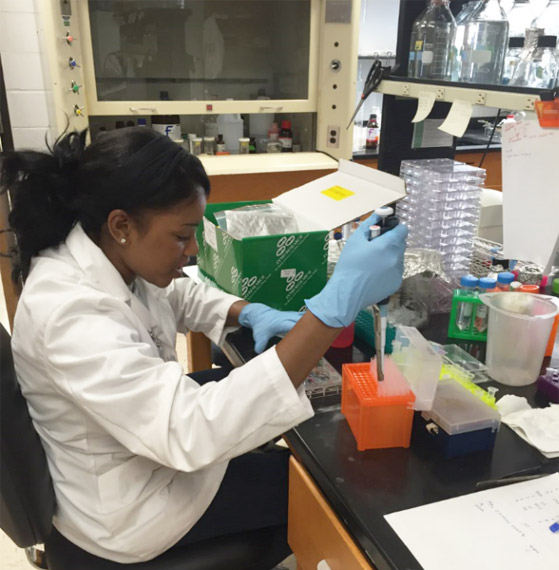
Although BUILD, MARC, and RISE offer a variety of activities at more than 100 supported institutions during the school year—including laboratory research opportunities, faculty mentoring, seminars, and workshops—the programs also provide training experiences throughout the summer. The slideshow below gives a quick peek into what several students participating in MARC, RISE, and BUILD activities did over the summer.
Sepsis is a serious medical condition caused by an overwhelming immune response to infection. The body’s infection-fighting chemicals trigger widespread inflammation, which can lead to blood clots and leaky blood vessels. As a result, blood flow is impaired, depriving organs of nutrients and oxygen. In severe cases, one or more organs fail. In the worst cases, blood pressure drops, the heart weakens, and the patient spirals toward septic shock. Once this happens, multiple organs—lungs, kidneys, liver—may quickly fail, and the patient can die.
Because sepsis is traditionally hard to diagnose, doctors do not always recognize the condition in its early stages. In the past, it has been unclear how quickly sepsis needs to be diagnosed and treated to provide patients with the best chance of surviving.
Credit: University of Pittsburgh.
Now we may have an answer: A large-scale clinical study, published recently in the New England Journal of Medicine, found that for every hour treatment is delayed, the odds of a patient’s survival are reduced by 4 percent. Christopher Seymour, assistant professor of critical care and emergency medicine at the University of Pittsburgh, and his team analyzed the medical records of nearly 50,000 sepsis patients at 149 clinical centers to determine whether administering the standard sepsis treatment—antibiotics and intravenously administered fluids—sooner would save more lives.
I spoke with Seymour about his experience treating sepsis patients and his research on the condition, including the new study.
CP: How big a public health problem is sepsis?
CS: Our recent work with the Centers for Disease Control and Prevention suggests there might be as many as 2 million sepsis cases in the United States each year. I can share personally that sepsis, or septic shock, is far and away the most common life-threatening condition that I treat in the ICU (intensive care unit). It’s quite devastating, particularly among our elders, and it requires prompt care. Although the mortality rate may be decreasing, it’s still quite high. About 1 in 10 patients with sepsis don’t survive their hospital stay. Even young, healthy people can succumb from sepsis. And if you’re fortunate to survive, you can have significant problems with cognitive and physical function for many months to years down the line.
Unfortunately, the incidence of sepsis may even be increasing. More patients are surviving serious illnesses that used to be fatal. They’re alive, but their health is compromised, so they are at higher risk for sepsis. Also—and this is a positive—we are seeing greater recognition and increased reporting of sepsis. Both factors probably contribute to the higher numbers of reported sepsis cases.
CP: What are some of the biggest challenges in fighting sepsis?
CS: The first challenge is public awareness. It’s important that the public knows the word sepsis, that they’re familiar with sepsis being a life-threatening condition that results from an infection, and that they know it can strike anyone—young, old, healthy, or sick. But it’s also important to know that not every infection is septic, nor will every cut or abrasion lead to life-threatening organ dysfunction.
Another part of the problem is that sepsis is not as easy for patients to recognize as, say, myocardial infarction (heart attack). When patients clutch their chest in pain, they intuitively recognize what’s happening. Patients frequently don’t recognize that they’re septic. People should know that when they have an infection or take antibiotics as an outpatient, and they’re starting to feel worse or having other new symptoms, they may be at risk of sepsis. They should go to the emergency department or seek medical help.
The second challenge in fighting sepsis is that it’s just hard to diagnose, even for well-trained clinicians. Both issues can lead to delays in care, the most important of which is the delay in treatment with antibiotics.
CP: Tell me about your recent clinical trial. What question did you set out to answer?
CS: There’s been a lot of interest in the early recognition and treatment of sepsis over the past decade. Recently, the National Institutes of Health/National Institute of General Medical Sciences funded a large, multicenter trial called ProCESS, which tested various strategies for treating sepsis. This trial told us that a standardized sepsis protocol among people who had already received antibiotics didn’t necessarily change survival rates. But what it left unanswered was the very important question of when the patient first arrives at the emergency department, how fast do we need to provide antibiotics and fluids for the best possible outcome?
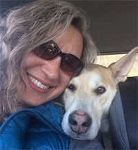
Lori Gildehaus and her lovable, mischievous dog, Charley. Credit: Lori Gildehaus.
Lori Gildehaus loves her job because she’s almost always doing something different. Some days, she leads professional development sessions for undergraduate students at the University of Alaska, Fairbanks (UAF). Other days, she’s weathered down in isolated communities along Alaska’s coast while leading community science and outreach events. These activities are just a few of her many responsibilities. Gildehaus is a laboratory research and teaching technician for UAF’s Biomedical Learning and Student Training (BLaST) program.
UAF’s BLaST program is one of 10 sites across the country in the Building Infrastructure Leading to Diversity (BUILD) initiative. As a component of the NIH Diversity Program Consortium, BUILD aims to find the best ways to engage and retain students from diverse backgrounds in biomedical research. Each BUILD site is as unique as the community it serves. UAF’s BLaST program embraces Alaska Native culture and the unique landscape that its students, faculty, and staff call home.
UAF attracts students from across Alaska, making for a diverse student body. BLaST serves not only UAF but also seven other campuses throughout Alaska, ranging from IỊisaġvik College in Utqiaġvik (formerly Barrow) at the northern tip of the state, to the University of Alaska Southeast in Sitka, more than 1,000 miles away. In any area that large, it would be difficult to organize community science outreach and foster connections between institutions. But in Alaska, there aren’t even roads connecting most rural campuses to Fairbanks.
Bridging gaps
Gildehaus and BLaST’s four other laboratory research and teaching technicians help bridge these gaps and bring science to local communities. They also serve as intermediaries between undergraduate students doing research and their professors. For undergraduates, talking to professors can be intimidating, and navigating the university landscape can be overwhelming. One of Gildehaus’ responsibilities is providing guidance to students.
“We want undergraduates to have a really good opportunity to explore their interests and have a good experience on their research projects,” Gildehaus says.
Gildehaus has a broad background, including biological sciences, human anatomy and physiology, science outreach, and mentoring. This experience helps her develop BLaST’s mentoring component. BLaST uses a tiered mentoring approach to provide opportunities for undergraduate and graduate students to share experiences and participate in mentoring.
Gildehaus has planned three mentoring workshops for fall 2017. One of these workshops, organized with assistance from the National Research Mentoring Network, will focus on culturally aware mentoring. Another will teach attendees how to navigate conversations, share stories, and increase awareness and understanding of Alaska Native and other cultures.
Bringing science outside the lab
BLaST’s diverse group of students includes many people who reside in rural areas and live a subsistence lifestyle. Traditional lab work schedules and science education can often seem disconnected from these communities. To better engage students, BLaST implements the One Health Approach, which emphasizes the interconnectedness between human, animal, and environmental health by promoting ways to expand interdisciplinary collaborations to attain optimal health for all. The program helps students recognize that there are opportunities to be involved in biomedical research in their communities, such as researching the natural vegetation of the Alaskan tundra, studying marine mammals, or finding cures for illnesses. Continue reading “Having a BLaST in Alaska … and Beyond”
It’s back! Check out the new issue of Findings magazine.
Findings presents cutting-edge research from scientists in diverse biomedical fields. The articles are aimed at high school students with the goal of making science—and the people who do it—interesting and exciting, and to inspire young readers to pursue careers in biomedical research. In addition to putting a face on science, Findings offers activities such as quizzes and crossword puzzles and, in its online version, video interviews with scientists.
As school starts up again, we look forward to a year that further enhances health and science literacy and brings students closer to pursuing science as an exciting future career. The National Institutes of Health continues to help both educators and students toward these goals through its Science Education Partnership Award (SEPA) Program.
What Is SEPA?
SEPA funds innovative science, technology, engineering, and mathematics (STEM), and informal science education (ISE) projects for students in pre-kindergarten through grade 12 (P-12), as well as public outreach activities such as science museum exhibits. Its goal is to invest in educational activities, including interactive online resources, that improve the training of a future workforce to meet the country’s biomedical research needs. SEPA encourages partnerships between biomedical researchers and P-12 teachers, schools, and other interested groups. SEPA provides:
Opportunities for students from underserved communities to learn about careers in basic or clinical research.
Professional development, skills, and knowledge building for science teachers.
Support for science centers and museum exhibits on health and medicine to improve community health literacy.
In March 2017, SEPA found its new home with the National Institute of General Medical Sciences (NIGMS). Congress mandated the move so that SEPA could more efficiently integrate with our other institution-building and research training programs and increase collaboration opportunities between them.
SEPA-Funded Resources
The following are just some of the various SEPA-funded resources that educators can use to engage their students in science:
The Partnership in Education at Duquesne University specializes in using cutting-edge technologies and creative media platforms—including videos, apps, posters, and lesson plans—to bring science to life and inspire lifelong learning. Topics include development, evolution, the science of sleep, and regenerative medicine.
It’s back-to-school time. That means learning lots of new facts and figures. In science, terms tend to be several syllables, sometimes with a Latin word thrown into the mix. As a result, many are referred to by their acronyms, such as DNA—short for deoxyribonucleic acid. This makes them easier both to remember and to say.
Researcher Mark Howarth of Oxford University, has taken this a step further. Searching through information stored in the NIGMS-funded Protein Data Bank, he curated a 3-D protein alphabet. It’s a set of 26 protein crystal structures that look like they were fashioned from bits of rainbow-colored curly ribbon. This 3-D alphabet helps us see what different protein strands look like, and explains terms and concepts relating to protein structure and function.
Proteins are molecules that play important roles in virtually every activity in the body. They form hair and fingernails, carry oxygen in the blood, enable muscle movement and much more. Although proteins are made of long strands of small molecules called amino acids, they do not remain as a straight chain. Some proteins are composed of multiple amino acid strands that wind together in the completed protein. The strands twist, bend and fold into a specific shape, and the protein’s structure enables it to perform its task. For instance, the “Y” shape of antibodies helps these immune system proteins bind to foreign molecules such as bacteria or viruses while also drawing in other immune system molecules.

 Patients can point to one of the faces on this subjective pain scale to show caregivers the level of pain they are experiencing. Credit: Wong-Baker Faces Foundation.