
The tiny roundworm Caenorhabditis elegans is one of the most common research organisms—creatures scientists use to study life. While C. elegans may seem drastically different from humans, it shares many genes and molecular pathways with us. Viewed with a microscope, the worm can also be surprisingly beautiful. Aside from the stunning imagery, these examples from our Image and Video Gallery show how C. elegans helps scientists advance our understanding of living systems and find new ways to improve our health.
 Credit: Keir Balla and Emily Troemel, University of California San Diego.
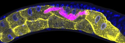
This C. elegans has been infected with microsporidia (purple), parasites closely related to fungi. The yellow shapes are the worm’s gut cells, and the blue dots are nuclei. Some microsporidia can infect people, so studying the parasites in worms could help researchers devise strategies to prevent or treat infections.
 Credit: Snusha Ravikumar, Ph.D., University of Maryland, College Park, and Antony M. Jose, Ph.D., University of Maryland, College Park.
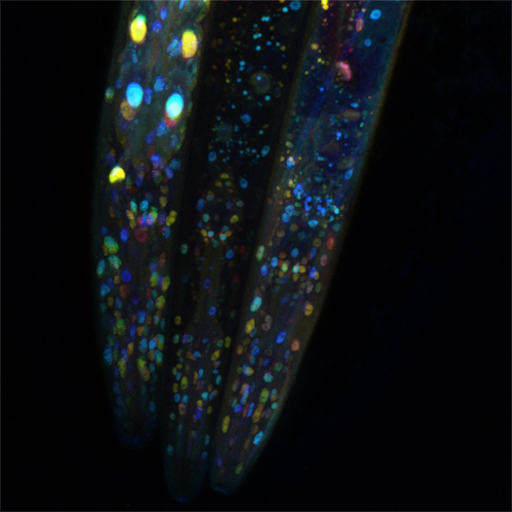
Seen here are the heads of three worms that were genetically modified to express a fluorescent protein. Researchers used these C. elegans to show that seemingly identical cells can use different proteins to carry out the same function in an important cellular process called functional mosaicism. This may explain why some medicines, which are often designed to target a specific molecule, aren’t completely effective.
 Credit: Shawn Xu, University of Michigan.
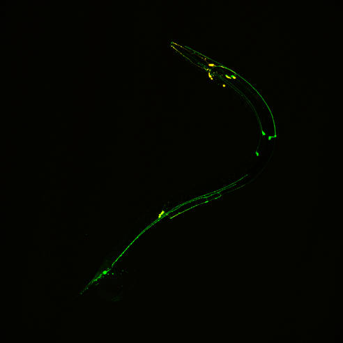
The neon green and yellow in this image highlight the processes and cell bodies of some C. elegans neurons. Findings have revealed that the strategies the worms use to control their motions are similar to those the human brain uses to command movement of our eyes, arms, and legs.
 Credit: Hannah Somers, Mount Desert Island Biological Laboratory.
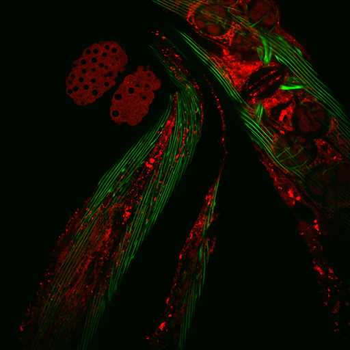
Researchers used the C. elegans in this photo to examine a molecular pathway that affects aging. The green structures are muscle fibers, and the red ones are a ribosomal protein that’s involved in protein translation and may play a role in dietary restriction-induced longevity.
Visit our Image and Video Gallery for more C. elegans images as well as other scientific photos, illustrations, and videos.