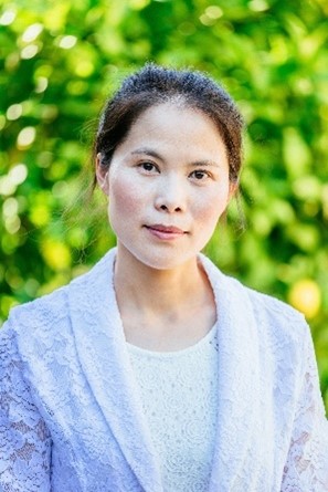
The brain is a large and complex organ, but some very small structures guide its development. Xuecai Ge, Ph.D., an associate professor of molecular and cell biology at the University of California, Merced (UC Merced), has devoted her career to understanding one of these structures called the primary cilium. In an interview, Dr. Ge shared how her childhood experience inspired her to study science and what makes the primary cilium fascinating.
Q: How did you first become interested in science?
A: When I was a little kid, my mom was a primary care doctor, and I saw her treat patients in our community. I noticed that no matter who got a particular illness, she could use the same medicine to treat them. My little mind was amazed that the same medicine could work for so many different people! I think this early experience planted the original seed of my interest in life science.
Q: What was your path to becoming a researcher?
A: I majored in pharmaceutics as an undergraduate student at Peking University Health Science Center in Beijing, China, but found it more interesting to study the biology of the human body than medicine itself. So, after earning my bachelor’s degree, I entered the graduate program at Tsinghua University in China. During that period, I studied learning and memory using the fruit fly as a research organism and quickly became enthralled by neuroscience.
I went on to enter a neuroscience program at Harvard Medical School in Boston, Massachusetts, as a Ph.D. student. During my first year there, I learned how the brain’s cortex—the outermost layer that’s responsible for cognitive function or thought—is built at the molecular and cellular level, and what types of disorders can occur if it doesn’t develop normally. I was intrigued because the brain is so complicated, but you can understand how it’s built cell by cell at the beginning of its development. This fascination led me to join the lab of Li-Huei Tsai,
Ph.D., and conduct my dissertation research on the regulation of neurogenesis—the formation of neurons—in the embryonic brain.
After completing my Ph.D., I eventually ended up working as a postdoctoral researcher with Matthew Scott, Ph.D., at Stanford University in California, studying an important signaling pathway in the central nervous system called the Hedgehog pathway. It relies heavily on the primary cilium—a tiny sensory organelle that functions like an antenna for the cells. In Dr. Scott’s lab, I studied how Hedgehog signaling was regulated during brain development as well as in pediatric brain tumors. During my research, I recognized that we all knew very little about the primary cilium in the developing brain, so when I started my lab at UC Merced in 2017, I decided to investigate primary cilia and ciliary signaling in the developing cortex.
Q: What is your lab studying?
A: My lab studies signaling pathways involving the primary cilium. Pretty much every cell in our bodies has one primary cilium. It’s like a thin, short hair that grows from the cell surface. For a long time, researchers overlooked its function, considering it an evolutionary vestige. But in the past 20 years or so, studies have shown that the primary cilium is essential for cells to sense signals from their environment and to activate signaling pathways within the cell. So we study the pathways it mediates to understand how signaling errors lead to ciliopathies—diseases that involve almost every organ in the body.
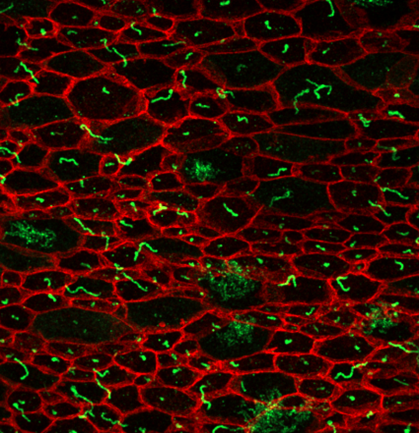
We’re identifying signaling proteins expressed in the cilium to try to understand how the primary cilium facilitates signaling and if cilia in different tissues respond to specific molecules. We’re looking at what proteins are expressed over the course of the Hedgehog signaling pathway as well as in various regions of the developing cortex. We use neural progenitor cells, the cells that produce neurons in the developing cortex, which each have a primary cilium that extends from the surface of the cell into a liquid called cerebral spinal fluid (CSF). It’s believed that the cilia may be sensing signals in the CSF that help regulate development, but we know very little about what molecules the cilia are detecting. To try to figure this out, my lab is cataloging the proteins expressed in the primary cilia in different brain regions and at various developmental stages using mice as research organisms. These projects will provide valuable information on basic mechanisms of signal transduction in the primary cilium and hopefully shed light on the molecular causes of ciliopathies.
Q: What do you consider your biggest accomplishment so far?
A: I think my biggest accomplishment has been establishing a platform to map all the proteins in the primary cilium. This project was very challenging because the cilium is so miniscule—its volume is only about one-10,000th of the entire cell. We selectively labeled proteins in the cilium with biotin—a special tag that can be used to easily separate molecules from other cell material. We then purified the tagged proteins and identified them through mass spectrometry.
We first used this method to identify ciliary proteins in cultured cells. We found known cilium proteins—which meant we were on the right track—as well as proteins that hadn’t been identified in the primary cilium before. We also applied this method to neural progenitor cells from mouse brain samples, which is time and labor intensive. But the hard work paid off! We discovered new ciliary proteins that are exclusively expressed in neural progenitors, including many that were previously reported to be involved in neurodegenerative diseases or developmental disorders. Our results highlight new ways to study the mechanisms underlying these neurological diseases.
Q: What goals do you ultimately hope to achieve with your research and career?
A: In research, my goal is to characterize currently unknown mechanisms in cell signaling in the cilium, ultimately contributing to a better understanding of ciliopathic diseases. Additionally, I aim to use my research as a platform to inspire and mentor the next generation of scientists. Throughout my career, I’ve been fortunate to have many inspiring mentors who have supported and guided me. I’m very excited to now have an opportunity to pay that forward to my own students.
Q: What kinds of challenges have you faced in your career?
A: I’ve faced many challenges throughout my career, and I try to view them as opportunities for personal and professional growth rather than barriers. At the beginning of my career, I had to overcome cultural and language hurdles. As my career progressed, I experienced stereotyping because of my gender and race, in both working and nonworking environments.
Like many people, particularly women in the academic setting with children, I’ve also struggled with work-life balance. As a professor, you have to juggle many tasks, including leading a research team, teaching, and raising a family.
Q: What advice would you give students who are interested in pursuing a career in science?
A: Find a question that you’re truly passionate about and then persevere in pursuing your scientific dream. Scientific careers have ups and downs, so be resilient in the face of setbacks and embrace every challenge as an opportunity to grow stronger.
Dr. Ge’s research is supported by NIGMS through grant R01GM143276.