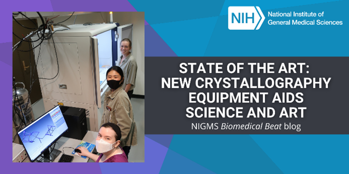
Upgrading X-ray crystallography equipment at the University of Arkansas in Fayetteville has had an unexpected benefit: enabling analyses that could help art museums authenticate, restore, and learn more about their pieces.
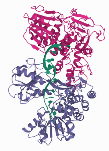
Scientists use X-ray crystallography to determine the detailed 3D structures of molecules. In biomedical contexts, researchers often apply X-ray crystallography to map the structures of proteins and other biomolecules like DNA and RNA. A molecule’s structure can shed light on its function and help answer scientific questions. For example, knowing the structures of proteins involved in antibiotic resistance can help researchers determine how those molecules work and how to combat bacteria that produce them.
Continue reading “State of the Art: New Crystallography Equipment Aids Science and the Study of Artifacts”

