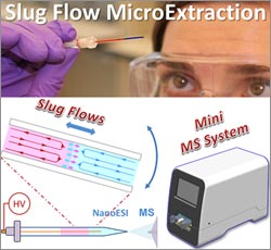
It’s not unusual for the standard dose of a drug to work well for one person but be less effective for another. One reason for such differences is that individuals can break down drugs at different rates, leading to different concentrations of drugs and of their breakdown products (metabolites) in the bloodstream. A promising new process called slug-flow microextraction could make it faster, easier and more affordable to regularly monitor drug metabolites so that medication dosages could be tailored to each patient’s needs, an approach known as personalized medicine. This technique could also allow researchers to better monitor people’s responses to new drug treatments during clinical trials. Continue reading “New Streamlined Technique for Processing Biological Samples”
Tag: Cool Tools/Techniques
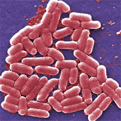
E. Coli Bacteria as Medical Sensors and Hard Drives?
E. coli bacteria help us digest our food, produce vitamin K and have served as a model organism in research for decades. Now, they might one day be harnessed as environmental or medical sensors and long-term data storage devices .
MIT researchers Timothy Lu and Fahim Farzadfard modified the DNA of E. coli cells so that the cells could be deployed to detect a signal (for example, a small molecule, a drug or the presence of light) in their surroundings. To create the modified E. coli, the scientists inserted into the bacteria a custom-designed genetic tool.
When exposed to the specified signal, the tool triggers a series of biochemical processes that work together to introduce a single mutation at a specific site in the E. coli’s DNA. This genetic change serves to record exposure to the signal, and it’s passed on to subsequent generations of bacteria, providing a continued record of exposure to the signal. In essence, the modified bacteria act like a hard drive, storing biochemical memory for long periods of time. The memory can be retrieved by sequencing the bacteria or through a number of other laboratory techniques.
Continue reading “E. Coli Bacteria as Medical Sensors and Hard Drives?”Cool Image: Snap-Together Laboratory
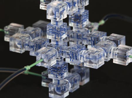
Like snapping Lego blocks together to build a fanciful space station, scientists have developed a new way to assemble a microfluidics system, a sophisticated laboratory tool for manipulating small volumes of fluids.
Microfluidics systems are used by scientists to perform tasks as diverse as DNA analysis, microbe detection and disease diagnosis. Traditionally, they have been slow and expensive to produce, as each individual “lab on a chip” had to be built from scratch in a special facility.
Now, researchers including Noah Malmstadt of the University of Southern California have harnessed 3-D printing technology to create a faster, cheaper, easier-to-use system . The team first identified the smallest functional pieces of a microfluidics system. Each of these pieces performs one simple task like detecting the size of fluid droplets or mixing two fluids together. After 3-D printing individual components, the team showed that they could be snapped together by hand into a working system in a matter of hours. The individual pieces can be pulled apart and re-assembled as needed before use in an actual experiment, which was impossible with the traditional microfluidics systems.
The researchers have created eight block-like components so far. They hope to start an online community where scientists will share designs for additional components in an open-source database, helping to speed further development of the technology.
This work was funded in part by NIH under grant R01GM093279.
Field Focus: Precision Gene Editing with CRISPR
Like humans, bacteria can be infected by viruses and have evolved ways to defend themselves. Researchers are now adapting this bacterial “immune system” to precisely and efficiently edit genes in cells from humans and a wide range of other organisms. Scientists are excited about the tremendous potential of this powerful tool for advancing biomedical research and treating diseases.
The bacterial defense system is called CRISPR, for clustered regularly interspaced short palindromic repeats. A breakthrough in understanding CRISPR came from examining bacteria used by the dairy industry for the production of yogurt and cheese. In a study published in 2007, researchers showed that these bacteria insert viral DNA sequences into their own genomes and use that information to disarm the virus when it attacks again. Subsequent research has shown that the CRISPR system consists of small RNA molecules that target specific viral DNA sequences and proteins that cut the DNA, thus destroying the virus.
Researchers have already adapted CRISPR into a gene-editing tool that’s quicker, cheaper and more precise than existing methods. Researchers can use CRISPR to add, delete, rev up or tone down certain genes as well as create animal models for studying human diseases. The ability to precisely target genes in human cells is expected to speed progress in the development of gene-based therapies.
Although much is known about CRISPR, we still have a lot to learn. For example, how do bacterial cells obtain and insert the viral DNA into their genome? What triggers production of the CRISPR RNA molecules? How are invading viral DNAs targeted for destruction? This last question is answered in part by a pair of findings described in an earlier post, A Crisper View of the CRISPR Gene-Editing Mechanism. We also want to figure out how we can make the CRISPR gene-editing tool even more versatile and precise.
The CRISPR story offers a good example of how studying basic biological processes leads to new—and sometimes unexpected—insights and applications.
Emily Carlson also contributed to this blog post.
Related advances:
CRISPR/Cas9 Protein Complex Can Be Programmed to Recognize and Cleave RNA
CRISPR System Adapted to Reversibly Regulate Gene Expression
The “Virtuous Cycle” of Technology and Science
Whether it’s a microscope, computer program or lab technique, technology is at the heart of biomedical research. Its central role is particularly clear from this month’s posts.
Some show how different tools led to basic discoveries with important health applications. For instance, a supercomputer unlocked the secrets of a drug-making enzyme, a software tool identified disease-causing variations among family members and high-powered microscopy revealed a mechanism allowing microtubules—and a cancer drug that targets them—to work.
Another theme featured in several posts is novel uses for established technologies. The scientists behind the cool image put a new spin on a long-standing imaging technology to gain surprising insights into how some brain cells dispose of old parts. Similarly, the finding related to sepsis demonstrates yet another application of a standard lab technique called polymerase chain reaction: assessing the immune state of people with this serious medical condition.
“We need tools to answer questions,” says NIGMS’ Doug Sheeley, who oversees biomedical technology research resource grants. “When we find the answers, we ask new questions that then require new or improved tools. It’s a virtuous cycle that keeps science moving forward.”
Good Vibrations
For people living with type 2 diabetes, wounds often heal slowly, sometimes even becoming chronic. Now, scientists have shown that low-intensity vibrations can speed up the healing process in a strain of diabetic mice commonly used to study delayed wound healing. The research team, led by Timothy Kohof the University of Illinois at Chicago, found that exposing the mice to barely perceptible vibrations five times a week for just 30 minutes promoted wound healing by increasing the formation of new blood vessels and of granulation tissue, a type of tissue critical in the early stages of healing. If researchers can show that the vibration technique also works in humans, this approach could one day offer a drug-free, non-invasive therapy for chronic wounds in people with diabetes.
This work also was funded by NIH’s National Institute of Dental and Craniofacial Research.
Learn more:
University of Illinois at Chicago News Release
Nanoparticles Developed to Stick to Damaged Blood Vessels, Deliver Drugs
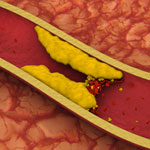
Heart disease is the leading cause of death for both men and women in the United States, according to the Centers for Disease Control and Prevention. One treatment challenge is developing non-invasive ways to direct medication to damaged or clogged arteries, which can block blood flow and increase the risk for heart attack and stroke. A team led by Naren Vyavahare at Clemson University has engineered extremely tiny particles—nanoparticles—that offer a promising step forward.
Healthy arteries have elastic fibers that make the arteries flexible. But, in most cardiovascular diseases, the fibers get damaged. The new nanoparticles, which can deliver drugs, attach only to damaged fibers and could enable site-specific drug delivery to minimize off-target side effects. The nanoparticles also allow drugs to be released over longer periods of time, potentially increasing the drugs’ effectiveness. The new biomaterial was tested in rodent models for studying vascular disease, so it is still in the early stages of development.
This work also was funded by NIH’s National Heart, Lung, and Blood Institute.
An Experimental Contact Lens to Prevent Glaucoma-Induced Blindness
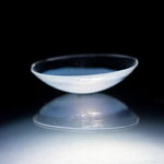
Like a miniature donut stuffed inside a tiny pita pocket, a common glaucoma medicine held within a biomaterial ring is sandwiched inside this contact lens. In laboratory experiments, the lens, which can also correct vision, releases the eyesight-saving medication at a steady rate for up to a month. Its construction offers numerous potential clinical advantages over the standard glaucoma treatment and may have additional applications, such as delivering anti-inflammatory drugs or antibiotics to the eye. Led by Daniel Kohane and Joseph Ciolino at Harvard Medical School, the researchers who developed the lens are now gearing up to test its effectiveness in additional laboratory studies. They hope a Phase I clinical trial to evaluate the safety and ability of the lens to reduce pressure in the human eye could begin in about a year.
This work also was funded by NIH’s National Eye Institute.
Learn more:
NEI Glaucoma Awareness Month Resources
Meet Shanta Dhar
Fields: Chemistry and cancer immunotherapy
Works at: University of Georgia, Athens
Born and raised in: Northern India
Studied at: Indian Institute of Science, Bangalore; Johns Hopkins University, Baltimore, Md.; and Massachusetts Institute of Technology, Cambridge, Mass.
To unwind: She hits the gym
Credit: Frankie Wylie, Stylized Portraiture
The human body is, at its most basic level, a giant collection of chemicals. Finding ways to direct the actions of those chemicals can lead to new treatments for human diseases.
Shanta Dhar, an assistant professor of chemistry at the University of Georgia, Athens (UGA), saw this potential when she was exposed to the field of cancer immunotherapy as a postdoctoral researcher at the Massachusetts Institute of Technology. (Broadly, cancer immunotherapy aims to direct the body’s natural immune response to kill cancer cells.) Dhar was fascinated by the idea and has pursued research in this area ever since. “I always wanted to use my chemistry for something that could be useful [in the clinic] down the line,” she said.
A major challenge in the field has been training the body’s immune system—specifically the T cells—to recognize and attack cancer cells. The process of training T cells to go after cancer is rather like training a rescue dog to find a lost person: First, you present the scent, then you command pursuit.
The type of immune cell chiefly responsible for training T cells to search for and destroy cancer is a called a dendritic cell. First, dendritic cells present T cells with the “scent” of cancer (proteins from a cancer cell). Then they activate the T cells using signaling molecules.
Dhar’s Findings
Dhar’s work focuses on creating the perfect trigger for cancer immunotherapy—one that would provide both the scent of cancer for T cells to recognize and a burst of immune signaling to activate the cells.
Using cells grown in the lab, Dhar’s team recently showed that they could kill most breast cancer cells using a new nanotechnology technique, then train T cells to eradicate the remaining cancer cells.
For the initial attack, the researchers used light-activated nanoparticles that target mitochondria in cancer cells. Mitochondria are the organelles that provide cellular energy. Their destruction sets off a signaling cascade that triggers dendritic cells to produce one of the proteins needed to activate T cells.
Because the strategy worked in laboratory cells, Dhar and her colleague Donald Harn of the UGA infectious diseases department are now testing it in a mouse model of breast cancer to see if it is similarly effective in a living organism.
For some reason, the approach works against breast cancer cells but not against cervical cancer cells. So the team is examining the nanoparticle technique to see if they can make it broadly applicable against other cancer types.
Someday, Dhar hopes to translate this work into a personalized cancer vaccine. To create such a vaccine, scientists would remove cancer cells from a patient’s body during surgery. Next, in a laboratory dish, they would train immune cells from the patient to kill the cancer cells, then inject the trained immune cells back into the patient’s body. If the strategy worked, the trained cells would alert and activate T cells to eliminate the cancer.
Silencing Extra Copy of Chromosome 21
Each year about 1 in 700 babies is born with Down syndrome, a condition that occurs when cells contain three copies of chromosome 21. A new technique offers a proof of principle for silencing the extra copy. Using induced pluripotent stem cells derived from a person with Down syndrome, a research team led by Jeanne Lawrence of the University of Massachusetts Medical School inserted a gene called XIST into the extra chromosome 21. The gene, which normally turns off one whole X chromosome in females, rendered the chromosome copy and most of its genes inactive. The researchers plan to test the approach in a mouse model of Down syndrome and use it to further explore the biology of chromosome errors. The findings could eventually aid the development of therapies to mitigate resulting medical problems.
This work also was funded by NIH’s National Cancer Institute and Eunice Kennedy Shriver National Institute of Child Health and Human Development.
Learn more:
University of Massachusetts Medical School News Release
Lawrence Lab