We seldom see microscopic objects next to one another, so it can be difficult to picture how they compare. For instance, it might surprise you that a thousand cold-virus particles could line up across one human skin cell! The largest objects that scientists view through microscopes are about a millimeter (roughly the size of a poppyseed), and they’re about 10 million times larger than the smallest molecules scientists can view: atoms.
Continue reading “Through the Looking Glass: Microscopic Structures in Many Sizes”Tag: Proteins
An Enlightening Protein
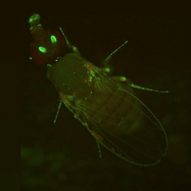
During the holiday season, twinkling lights are a common sight. But no matter what time of the year, you can see colorful glows in many biology labs. Scientists have enabled many organisms to light up in the dark—from cells to fruit flies and Mexican salamanders. These glowing organisms help researchers better understand basic cell processes because their DNA has been edited to express molecules such as green fluorescent protein.
Continue reading “An Enlightening Protein”See Your Name With Our Interactive Protein Alphabet!
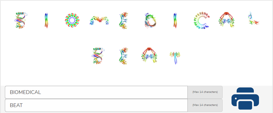
With our new interactive protein alphabet, you can type your first and last name, or any two words, and see them spelled out in colorful 3D shapes!
PECASE Honoree Michael Boyce on Sugar’s Role in Cell Signaling and on Diversity, Equity, and Inclusion in the Scientific Workforce
Sugars aren’t merely energy sources for our cells. They also play important signaling roles through a process called glycosylation, where they attach to proteins and lipids as tags. Although these sugar tags, called glycans, impact many cellular processes, they have long been understudied due to technical challenges. Now, advances in analytical tools like mass spectrometry are enabling scientists to examine the enormous complexity of glycans. Other advances also allow researchers to synthesize complex sugars, providing them with standards for analytical experiments.
Continue reading “PECASE Honoree Michael Boyce on Sugar’s Role in Cell Signaling and on Diversity, Equity, and Inclusion in the Scientific Workforce”RNA Polymerase: A Target for New Antibiotic Drugs?
DNA, with its double-helix shape, is the stuff of genes. But genes themselves are only “recipes” for protein molecules, which are molecules that do the real heavy lifting (or do much of the work) inside cells.
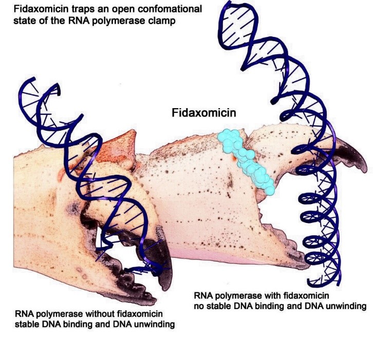
Here’s how it works. A molecular machine called RNA polymerase (RNAP) travels along DNA to find a place where a gene begins. RNAP uses a crab-claw-like structure to grasp and unwind the DNA double helix at that spot. RNAP then copies (“transcribes”) the gene into messenger RNA (mRNA), a molecule similar to DNA.
The mRNA molecule travels to one of the cell’s many protein-making factories (ribosomes), which use the mRNA message as instructions for making a specific protein.
Continue reading “RNA Polymerase: A Target for New Antibiotic Drugs?”Roses Are Red and So Is . . . Blood?
When you think of blood, chances are you think of the color red. But blood actually comes in a variety of colors, including red, blue, green, and purple. This rainbow of colors can be traced to the protein molecules that carry oxygen in the blood. Different proteins produce different colors.
Continue reading “Roses Are Red and So Is . . . Blood?”Interview With a Scientist – Rommie Amaro: Computational and Theoretical Model Builder
Many researchers who search for anti-cancer drugs have labs filled with chemicals and tissue samples. Not Rommie Amaro. Her work uses computers to analyze the shape and behavior of a protein called p53. Defective versions of p53 are associated with more human cancers than any other malfunctioning protein.
Optogenetics Sparks New Research Tools
Imagine if scientists could zap a single cell (or group of cells) with a pulse of light that makes the cell move, or even turns on or off the cell’s vital functions.
Scientists are working toward this goal using a technology called optogenetics. This tool draws on the power of light-sensitive molecules, called opsins and cryptochromes, which are naturally occurring molecules found in the cell membranes of a wide variety of species, from microscopic bacteria and algae to plants and humans. These light-reacting molecules change their shape or activity when they sense light, so they can be used to trigger cellular activity, such as turning on or off ion flow into the cell and other regulatory pathways. The ability to induce changes in cells has a broad range of practical applications, from enabling scientists to see how cells function to providing the basis for potential therapeutic applications for blindness, cancer, and epilepsy.
Opsins first gained a foothold in research about a decade ago when scientists began using them to study specific electrical networks in the brain. This research relied on channelrhodopsins, opsins that could be used to control the flow of charged ions into and out of the cell. Normally, when a neuron reaches a certain ion concentration, it is triggered to fire, but neuron firing can be changed by inserting opsins in the membrane. Neuroscientists figured out how to incorporate light-sensitive opsin proteins by inserting the opsin gene into the host’s DNA. The genetically encoded opsin proteins in the neuronal membranes could be turned on or off by shining light into the brain itself, using optical fibers or micro-LEDs, to switch on or off the flow of ions and neuron firing.
Since those early studies in the brain, the optogenetics field has come a long way. But the leap from brain cells to other cells has been challenging. Scientists first needed to find a way to deliver light into tissues deep in the body. And, unlike stationary brain cells, they needed a way to target cells that are on the move (such as immune cells). They also needed to develop a way to study not only cell networks but also individual cells and cell parts. The NIGMS-funded researchers highlighted below are among the scientists working to overcome these obstacles and are using optogenetics in new and inventive ways.
Building Bridges
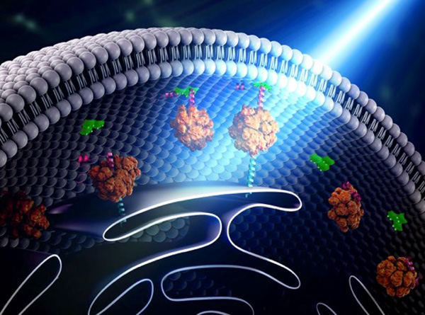
Yubin Zhou of Texas A&M is using optogenetics to control the way cells communicate and to study immune cell function. In one line of research, Zhou is using light to make it easier for calcium ions to enter cells. The ions carry instructions for the cell and also help tether small cellular structures (called organelles). Those inter-membrane tethers allow for the movement of proteins and lipids back and forth across the cell, and are critical for sending chemical messengers to communicate information (see illustration). When this process is disrupted, it can lead to extreme changes in cell function and even cell death. Using this technology to “switch on” normal pathways enables the scientists to better understand how such processes can be disrupted.
The Changing Needs of a Cell: No Membrane? No Problem!
Russian nesting dolls. Credit: iStock.
How “membrane-less” organelles help with key cellular functions
Scientists have long known that animal and plant cells have specialized subdivisions called organelles. These organelles are surrounded by a semi-permeable barrier, called a membrane, that both organizes the organelles and insulates them from the rest of the cell’s mix of proteins, salt, and water. This set-up helps to make cells efficient and productive, aiding in energy production and other specialized functions. But, because of their semi-permeable membranes, organelles can’t regroup and reform in response to stress or other outside changes. Cells need a rapid response team working alongside the membrane-bound organelles to meet these fluctuating needs. Until recently, who those rapid responders were and how they worked has been a mystery.
Recent research has led biologists to learn that the inside of a cell or an organelle is not just a lot of different molecules dissolved in water. Instead, we now know that cells contain many pockets of liquid droplets (one type of liquid surrounded by a liquid of different density) with specialized composition and function that are not surrounded by membranes. Because these “membrane-less organelles” are not confined, they can rapidly come together in response to chemical signals, such as those that indicate stress, and equally rapidly fall apart when they are no longer needed, or when the cell is about to divide. This enables membrane-less organelles to be “rapid responders.” They can have complex, multilayered structures that help them to perform many critical cell functions with multiple steps, just like membrane-bound organelles. Scientists even suspect that the way these organelles form as droplets may shed light on how life on Earth first took shape (see sidebar “Could This Be How Life First Took Shape?” at bottom of page).
The Many Membrane-less Organelles
Scientists have identified more than a dozen membrane-less organelles at work in mammalian cells. Several kinds found inside the nucleus—including nuclear speckles, paraspeckles, and Cajal bodies—help with cell growth, stress response, the metabolizing (breaking down) of RNA, and the control of gene expression—the process by which information in a gene is used in the synthesis of a protein. Out in the cytoplasm, P-bodies, germ granules, and stress granules are membrane-less organelles that are involved in metabolizing or protecting messenger RNA (mRNA), controlling which mRNAs are made into proteins, and in maintaining balance, or homeostasis, of the cell’s overall health.
The nucleolus, located inside the nucleus, is probably the largest of the membrane-less organelles. It acts as a factory to assemble ribosomes, the giant molecular machines that “translate” messenger RNAs to make all cellular proteins.
Continue reading “The Changing Needs of a Cell: No Membrane? No Problem!”Protein Alphabet: A Picture Is Worth One Letter
It’s back-to-school time. That means learning lots of new facts and figures. In science, terms tend to be several syllables, sometimes with a Latin word thrown into the mix. As a result, many are referred to by their acronyms, such as DNA—short for deoxyribonucleic acid. This makes them easier both to remember and to say.
Researcher Mark Howarth of Oxford University, has taken this a step further. Searching through information stored in the NIGMS-funded Protein Data Bank , he curated a 3-D protein alphabet. It’s a set of 26 protein crystal structures that look like they were fashioned from bits of rainbow-colored curly ribbon. This 3-D alphabet helps us see what different protein strands look like, and explains terms and concepts relating to protein structure and function.
Proteins are molecules that play important roles in virtually every activity in the body. They form hair and fingernails, carry oxygen in the blood, enable muscle movement and much more. Although proteins are made of long strands of small molecules called amino acids, they do not remain as a straight chain. Some proteins are composed of multiple amino acid strands that wind together in the completed protein. The strands twist, bend and fold into a specific shape, and the protein’s structure enables it to perform its task. For instance, the “Y” shape of antibodies helps these immune system proteins bind to foreign molecules such as bacteria or viruses while also drawing in other immune system molecules.
Continue reading “Protein Alphabet: A Picture Is Worth One Letter”