Category: Cells

Molecular Fireworks: How Microtubules Form Inside Cells

1 comment
A video depicting red strands of various lengths exploding outward from a focal point at the left. The strands are tipped in neon green.
       Microtubules sprout from one another. Credit: Petry lab, Princeton University.

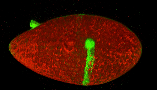
The red spray pictured here may look like fireworks erupting across the night sky on July 4th, but it’s actually a rare glimpse of tiny protein strands called microtubules sprouting and growing from one another in a lab. Microtubules are the largest of the molecules that form a cell’s skeleton. When a cell divides, microtubules help ensure that each daughter cell has a complete set of genetic information from the parent. They also help organize the cell’s interior and even act as miniature highways for certain proteins to travel along.

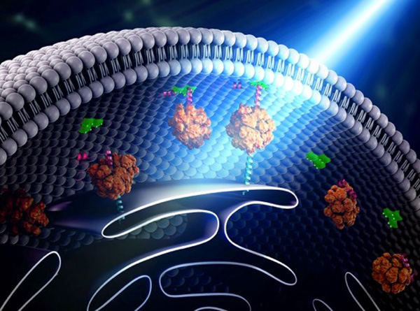
As their name suggests, microtubules are hollow tubes made of building blocks called tubulins. Scientists know that a protein called XMAP215 adds tubulin proteins to the ends of microtubules to make them grow, but until recently, the way that a new microtubule starts forming remained a mystery.

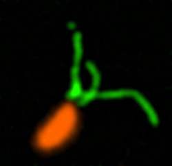
Sabine Petry and her colleagues at Princeton University developed a new imaging method for watching microtubules as they develop and found an important clue to the mystery. They adapted a technique called total internal reflection fluorescence (TIRF) microscopy, which lit up only a tiny sliver of a sample from frog egg (Xenopus) tissue. This allowed the scientists to focus clearly on a few of the thousands of microtubules in a normal cell. They could then see what happened when they added certain proteins to the sample.

Continue reading “Molecular Fireworks: How Microtubules Form Inside Cells”

Interview With a Scientist: Andrew Goodman, Separating Causation and Correlation in the Microbiome

0 comments

You’ve likely heard some variation of the statistic that there are at least as many microbial cells in our body as human cells. You may have also heard that the microscopic bugs that live in our guts, on our skins, and every crevice they can find, collectively referred to as the human microbiome, are implicated in human health. But do these bacteria, fungi, archaea, protists, and viruses cause disease, or are the specific populations of microbes inside us a result of our state of health? That’s the question that drives the research in the lab of Andrew Goodman, associate professor of microbial pathogenesis at Yale University.

Continue reading “Interview With a Scientist: Andrew Goodman, Separating Causation and Correlation in the Microbiome”

CLAMP Helps Lung Cells Pull Together

0 comments
ALT TEXT
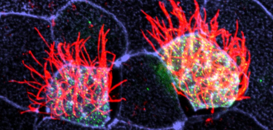
Cells covered with cilia (red strands) on the surface of frog embryos. Credit: The Mitchell Lab.

The outermost cells that line blood vessels, lungs, and other organs also act like guards, alert and ready to thwart pathogens, toxins, and other invaders that can do us harm. Called epithelial cells, they don’t just lie passively in place. Instead, they communicate with each other and organize their internal structures in a single direction, like a precisely drilled platoon of soldiers lining up together and facing the same way.

Lining up this way is crucial during early development, when tissues and organs are forming and settling into their final positions in the developing body. In fact, failure to line up in the correct way is linked to numerous birth defects. In the lungs, for instance, epithelial cells’ ability to synchronize with one another is important since these cells have special responsibilities such as carrying mucus up and out of lung tissue. When these cells can’t coordinate their functions, disease results.

Some lung epithelial cells are covered in many tiny, hair-like structures called cilia. All the cilia on lung epithelial cells must move uniformly in a tightly choreographed way to be effective in their mucus-clearing job. This is a unique example of a process called planar cell polarity (PCP) that occurs in cells throughout the body. Researchers are seeking to identify the signals cells use to implement PCP. Continue reading “CLAMP Helps Lung Cells Pull Together”

Pericytes: Capillary Guardians in the Brain

0 comments
ALT TEXT
The long arms of pericytes cells (red) stretch along capillaries (blue) in a mouse brain. Credit: Andy Shih.

Nerve cells, or neurons, in our brains do amazing work, from telling our hearts to beat to storing our memories. But neurons cannot operate alone. Many kinds of cells support and regulate neurons and—like neurons—they can come under attack due to injuries or disorders, such as stroke or Alzheimer’s disease. Learning what jobs these cells do and how they respond to disease may show researchers new ways to treat central nervous system disorders. One type of support cell, the pericyte, plays some key roles in brain health. These cells are readily adaptable, even in adult brains, and can support a variety of functions.

Pericytes help with blood flow to nerve cells in the brain. They lie wrapped all along the huge networks of capillaries—the tiniest blood vessels—that both feed neurons and form the blood-brain barrier, which filters out certain substances from blood to protect the brain. Pericytes have a body that appears as a bump protruding from a capillary surface. Pericytes also have long thin arms that stretch along each capillary like a snake on a tree branch. These arms, called processes, reach almost to where the next pericyte process begins, without overlapping. This creates a pericyte chain that covers nearly the entire capillary network.

Pericytes are critical for blood vessel stability and blood-brain barrier function. They’re also known to die off as a result of trauma and disease. Andy Shih, Andree-Ann Berthiaume, and colleagues at the Medical University of South Carolina in Charleston, set up an imaging technique in mouse brains that allowed them to see what pericytes do under normal conditions as well as how these cells respond when some are damaged.

Continue reading “Pericytes: Capillary Guardians in the Brain”

Optogenetics Sparks New Research Tools

1 comment

Imagine if scientists could zap a single cell (or group of cells) with a pulse of light that makes the cell move, or even turns on or off the cell’s vital functions.

Scientists are working toward this goal using a technology called optogenetics. This tool draws on the power of light-sensitive molecules, called opsins and cryptochromes, which are naturally occurring molecules found in the cell membranes of a wide variety of species, from microscopic bacteria and algae to plants and humans. These light-reacting molecules change their shape or activity when they sense light, so they can be used to trigger cellular activity, such as turning on or off ion flow into the cell and other regulatory pathways. The ability to induce changes in cells has a broad range of practical applications, from enabling scientists to see how cells function to providing the basis for potential therapeutic applications for blindness, cancer, and epilepsy.

Opsins first gained a foothold in research about a decade ago when scientists began using them to study specific electrical networks in the brain. This research relied on channelrhodopsins, opsins that could be used to control the flow of charged ions into and out of the cell. Normally, when a neuron reaches a certain ion concentration, it is triggered to fire, but neuron firing can be changed by inserting opsins in the membrane. Neuroscientists figured out how to incorporate light-sensitive opsin proteins by inserting the opsin gene into the host’s DNA. The genetically encoded opsin proteins in the neuronal membranes could be turned on or off by shining light into the brain itself, using optical fibers or micro-LEDs, to switch on or off the flow of ions and neuron firing.

Since those early studies in the brain, the optogenetics field has come a long way. But the leap from brain cells to other cells has been challenging. Scientists first needed to find a way to deliver light into tissues deep in the body. And, unlike stationary brain cells, they needed a way to target cells that are on the move (such as immune cells). They also needed to develop a way to study not only cell networks but also individual cells and cell parts. The NIGMS-funded researchers highlighted below are among the scientists working to overcome these obstacles and are using optogenetics in new and inventive ways.

Illustration showing how bridges can be built within a cell using light-reacting molecules
Illustration shows how “bridges” can be built within a cell through the use of light-reacting molecules. The light triggers proteins to line up within the cell, making it easier to shuttle molecules between the membranes of two subcellular organelles. This optogenetic strategy is helping scientists to control cell function with a simple beam of light. Illustration courtesy of Yubin Zhou.

Building Bridges

Yubin Zhou of Texas A&M is using optogenetics to control the way cells communicate and to study immune cell function. In one line of research, Zhou is using light to make it easier for calcium ions to enter cells. The ions carry instructions for the cell and also help tether small cellular structures (called organelles).  Those inter-membrane tethers allow for the movement of  proteins and lipids back and forth across the cell, and are critical for sending chemical messengers to communicate information (see illustration). When this process is disrupted, it can lead to extreme changes in cell function and even cell death. Using this technology to “switch on” normal pathways enables the scientists to better understand how such processes can be disrupted.

Continue reading “Optogenetics Sparks New Research Tools”

Cellular Footprints: Tracing How Cells Move

0 comments
ALT TEXT
An engineered cell (green) in a fruit fly follicle (red), or egg case, leaves a trail of fluorescent material as it moves across a fruit fly egg chamber, allowing scientists to trace its path and measure how long it took to complete its journey. Credit: David Bilder, University of California, Berkeley.

Cells are the basis of the living world. Our cells make up the tissues and organs of our bodies. Bacteria are also cells, living sometimes alone and sometimes in groups called biofilms. We think of cells mostly as staying in one spot, quietly doing their work. But in many situations, cells move, often very quickly. For example, when you get a cut, infection-fighting cells rally to the site, ready to gobble up bacterial intruders. Then, platelet cells along with proteins from blood gather and form a clot to stop any bleeding. And finally, skin cells surrounding the wound lay down scaffolding before gliding across the cut to close the wound.

This remarkable organization and timing is evident right from the start. Cells migrate within the embryo as it develops so that body tissues and organs end up in the right places. Harmful cells use movement as well, as when cells move and spread (metastasize) from an original cancer tumor to other parts of the body. Learning how and why cells move could give scientists new ways to guide those cells or turn off or slow down the movement when needed.

Glowing Breadcrumbs

Scientists studying how humans and animals form, from a single cell at conception to a complex body at birth, are particularly interested in how and when cells move. They use research organisms like the fruit fly, Drosophila, to watch movements by small populations of cells. Still, watching cells migrate inside a living fly is challenging because the tissue is too dense to see individual cell movement. But moving those cells to a dish in the lab might cause them to behave differently than they do inside the fly. To solve this problem, NIGMS-funded researcher David Bilder and colleagues at the University of California, Berkeley, came up with a way to alter fly cells so they could track how the cells behave without removing them from the fly. They engineered the cells to lay down a glowing track of proteins behind them as they moved, leaving a traceable path through the fly’s tissue. The technique, called M-TRAIL (matrix-labeling technique for real-time and inferred location), allows the researchers to see where a cell travels and how long it takes to get there.

Bilder and his team first used M-TRAIL in flies to confirm the results of past studies of Drosophila ovaries in the lab using other imaging techniques. In addition, they found that M-TRAIL could be used to study a variety of cell types. The new technique also could allow a cell’s movement to be tracked over a longer period than other imaging techniques, which become toxic to cells in just a few hours. This is important, because cells often migrate for days to reach their final destinations.

Continue reading “Cellular Footprints: Tracing How Cells Move”

Have Nucleus, Will Travel (in Three Dimensions)

0 comments
A closeup of two human cells with the cells dyed green and the necleaus dyed red.
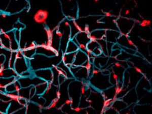
These two human cells are nearly identical, except that the cell on the left had its nucleus (dyed red) removed. The structures dyed green are protein strands that give cells their shape and coherence. Credit: David Graham, UNC-Chapel Hill.

Both of the cells above can scoot across a microscope slide equally well. But when it comes to moving in 3D, the one without the red blob in the center (the nucleus) stalls out. That’s sort of like an Olympic speed skater who wouldn’t be able to perform even a single leap in a figure skating competition.

Scientists have known for some time that the nucleus is involved in moving cells across a flat surface—it slides to one side of the cell and “pushes” from behind. However, scientists have also shown that cells with their nuclei removed can migrate along a flat surface just as well as their brethren with intact nuclei. But they had no idea that, without a nucleus, a cell could no longer move in three dimensions.

This discovery was made by UNC-Chapel Hill biologists Keith Burridge and James Bear and their colleagues. These NIGMS-funded researchers also observed that cells whose nuclei had been disconnected from the cytoskeleton could not move through 3D matrices. The cytoskeleton is the microscopic network of actin protein filaments and tubules in the cytoplasm of many cells that provides the cell’s shape and coherence. It has also has been thought to play a major role in cell movement.

Two views of cells one on top of the other. The top animation shows a cell moving across the frame while the cells in the bottom box are static.The gray, stringy background of these videos is a 3D jello-like matrix. The cell in the top half of this video has a nucleus and can migrate through the matrix. Both cells in the bottom half have been enucleated (a fancy term for having its nucleus removed) and cannot travel through the matrix. Credit: Graham et al., Journal of Cell Biology, 2018.

The researchers speculate that the reason cells without nuclei (or those whose nuclei have been disconnected from the cytoskeleton) don’t navigate in 3D has to do with complex mechanical interactions between the cytoskeleton and the nucleoskeleton. The nucleoskeleton is a molecular scaffold within the nucleus supporting many functions such as DNA replication and transcription, chromatin remodeling, and mRNA synthesis. The interface between the cytoskeleton and nucleoskeleton consists of interlocking proteins that together provide the physical traction that cells need to push their way through 3D environments. Disrupting this interface is the equivalent of breaking the clutch in a car: the motor revs, but the wheels don’t spin, and the car goes nowhere.

A better understanding of the physical connections between the nucleus and the cytoskeleton and how they influence cell migration may provide additional insight into the role of the nucleus in diseases, such as cancer, in which the DNA-containing organelle is damaged or corrupted.

This research was funded in part by NIGMS grants 5R01GM029860-35, 5P01GM103723-05, and 5R01GM111557-04.

Carole LaBonne: Neural Crest Cells and the Rise of the Vertebrates

0 comments

The stunning pigmentation of tigers, the massive jaws of sharks, and the hyper-acute vision of eagles. These and other remarkable features of higher organisms (vertebrates) derive from a small group of powerful cells, called neural crest cells, that arose more than 500 million years ago. Molecular biologist Carole LaBonne Exit icon of Northwestern University in Illinois studies how neural crest cells help give rise to these important vertebrate structures throughout development.

Very early during embryonic development, stem cells differentiate into different layers: mesoderm, endoderm, and ectoderm. Each of these layers then gives rise to different cell and tissue types. For example, the ectoderm becomes skin and nerve cells. Mesoderm turns into muscle, bone, fat, blood and the circulatory system. Endoderm forms internal structures such as lungs and digestive organs.

These three layers are present in vertebrates—animals with a backbone and well-defined heads, such as fish, birds, reptiles, and mammals—as well as animals without backbones, such as the marine-dwelling Lancelets and Tunicates (referred to as non-vertebrate chordates). Unlike cells in these layers, neural crest cells, which are found only in vertebrates, don’t specialize until much later in development. The delay gives neural crests cells the extra time and flexibility to sculpt the complex anatomical structures found only in vertebrate animals.

Scientists have long debated how neural crest cells manage to finalize their destiny so much later than all other cell types.

Using the frog Xenopus as a model system, LaBonne and her colleagues performed a series of experiments that revealed the process and identified key genes that control it.

In this video, LaBonne describes the power of neural crest cells and how they can be useful for studies of human health, including how cancer cells can metastasize, or migrate, throughout the body.

Dr. LaBonne’s research is funded in part by NIGMS grant 5R01GM116538.

Interview With a Scientist: Joel Kralj, Electromicist

0 comments

Every one of our thoughts, emotions, sensations, and movements arise from changes in the flow of electricity in the brain. Disruptions to the normal flow of electricity within and between cells is a hallmark of many diseases, especially neurological and cardiac diseases.

The source of electricity within nerve cells (i.e., neurons) is the separation of charge, referred to as voltage, across neuronal membranes. In the past, scientists weren’t able to identify all the molecules that control neuronal voltage. They simply lacked the tools. Now, University of Colorado biologist Joel Kralj Exit icon has developed a way to overcome this hurdle. His new technique—combining automated imaging tools and genetic manipulation of cells—is designed to measure the electrical contribution of every protein coded by every gene in the human genome. Kralj believes this technology will usher in a new field of “electromics” that will be of enormous benefit to both scientists studying biological processes and clinicians attempting to treat disease.

In 2017, Kralj won a New Innovator Award from the National Institutes of Health for his work on studying voltage in neurons. He is using the grant money to develop a new type of microscope that will be capable of measuring neuronal voltage from hundreds of cells simultaneously. He and his research team will then attempt to identify the genes that encode any of the 20,000 proteins in the human body that are involved in electrical signaling. This laborious process will involve collecting hundreds of nerve cells, genetically removing a single protein from each cell, and using the new microscope to see what happens. If the voltage within a cell is changed in any way when a specific protein is removed, the researchers can conclude that the protein is essential to electrical signaling.

In this video, Kralj discusses how he plans to use his electromics platform to study electricity-generating cells throughout the body, as well as in bacterial cells (see our companion blog post “Feeling Out Bacteria’s Sense of Touch” featuring Kralj’s research for more details).

Dr. Kralj’s work is funded in part by the NIH under grant 1DP2GM123458-01.

Feeling Out Bacteria’s Sense of Touch

0 comments

Our sense of touch provides us with bits of information about our surroundings that inform the decisions we make. When we touch something, our nervous system transmits signals through nerve endings that feed information to our brain. This enables us to sense the stimulus and take the appropriate action, like drawing back quickly when we touch a hot stovetop.

Bacteria are single cells and lack a nervous system. But like us, they rely on their “sense” of touch to make decisions—or at least change their behavior. For example, bacteria’s sense of touch is believed to trigger the cells to form colonies, called biofilms, on surfaces they make contact with. Bacteria may form biofilms as a way to defend themselves, share limited nutrients, or simply to prevent being washed away in a flowing liquid.

Humans can be harmed by biofilms because these colonies serve as a reservoir of disease-causing cells that are responsible for high rates of human infection. Biofilms can protect at least some cells from being affected by antibiotics. The surviving reservoir of bacteria then have more time to evolve resistance to antibiotics.

At the same time, some biofilms can be valuable; for example, they help to break down waste in water treatment plants and to drive electrical current as part of microbial fuel cells.

Until recently, scientists thought that bacteria formed biofilms and caused infections in response to chemical signals they received from their environments. But research in 2014 showed that the bacterium Pseudomonas aeruginosa could infect a variety of living tissues—from plants to many kinds of animals—simply by making contact with them. In the past year, multiple groups of investigators have learned more about how bacteria sense that they have touched a surface and how that sense translates to changes in their behavior. This understanding could lead to new ways of preventing infections or harmful biofilm formation.

Making Contact

Pili (green) on cells from the bacterium Caulobacter crescentus (orange). Scientists used a fluorescent dye to stain pili so they could watch the structures extend and retract. Credit: Courtney Ellison, Indiana University.

When they first make contact with a surface, bacteria change from free-ranging, swimming cells to stationary ones that secrete a sticky substance, tethering them in one place. To form a biofilm, they begin replicating, creating an organized mass stable enough to resist shaking and to repel potential invaders (see https://biobeat.nigms.nih.gov/2017/01/cool-image-inside-a-biofilm-build-up/).

How do swimming bacteria sense that they have found a potential surface to colonize? Working with the bacterium Caulobacter crescentus, Indiana University Ph.D. student Courtney Ellison and her colleagues, under the direction of professor of biology and NIGMS grantee Yves Brun, recently showed that hair-like structures on the cell’s surface, called pili, play a role here. The researchers found that as a bacterial cell swims in a fluid, its pili are constantly stretching out and retracting. When the cell makes contact with a surface, the pili stop moving, start producing a sticky substance and use it to hold onto the surface.

Continue reading “Feeling Out Bacteria’s Sense of Touch”