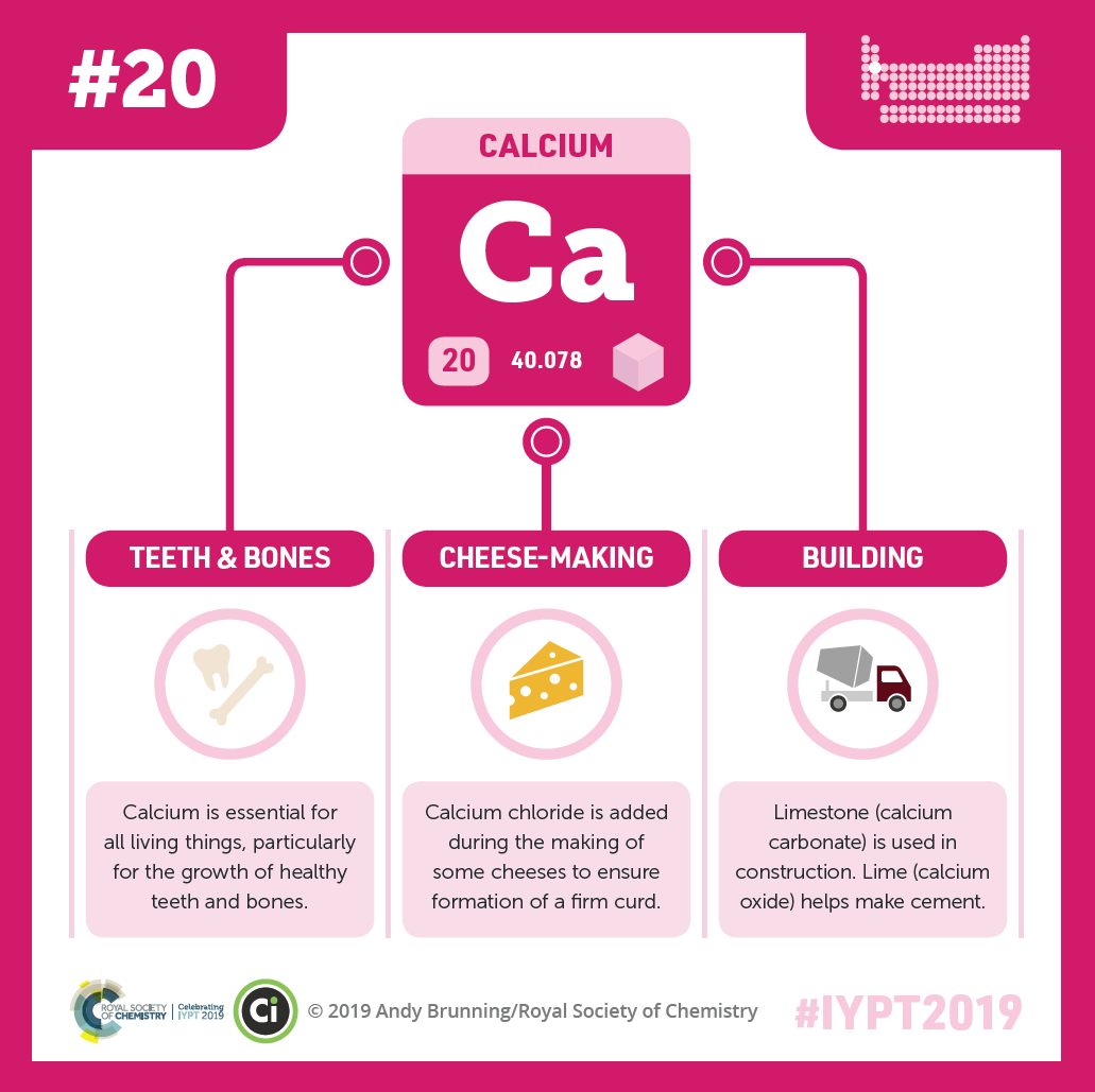
Someone’s hand moving to scroll through this blog post is possible because of a mineral that both gives bones their strength and allows muscles to move: calcium. As the most abundant mineral in our bodies, it’s essential for lots of important functions. It’s found in many foods, medicines, and dietary supplements.
Calcium keeps your bones strong, allows your muscles to move, and is important for many other bodily functions. The element is found in foods, medicines, and the world around us. Credit: Compound Interest CC BY-NC-ND 4.0. Click to enlarge.
You may know that antioxidants can help protect your cells from oxidative damage, but do you know about selenium—an element often found in special proteins called antioxidant enzymes? Selenium is essential to your body, which means you must get it from the food you eat. But it’s a trace element so you only need a small amount to benefit from its effects. In addition to its antioxidant properties, it’s also important for reproduction, DNA synthesis, and hormonemetabolism.
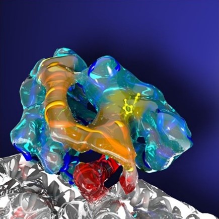
ATP (yellow) powering a protein (blue) that moves material within cells and helps them divide. Credit: Charles Sindelar, Yale University.
Just as electricity powers almost every modern gadget, the tiny moleculeadenosine triphosphate (ATP) is the major source of energy for organisms’ biochemical reactions. ATP stores energy in the chemical bonds that hold its three phosphate groups together—the triphosphate part of its name. In the human body, ATP powers processes such as cell signaling, muscle contraction, nerve firing, and DNA and RNA synthesis. Because our cells are constantly using and producing ATP, each of us turns over roughly our body weight in the molecule every day!
Our bodies can produce ATP in several ways, but the most common is cellular respiration—a multistep process in which glucose molecules from our diet and oxygen react to form water and carbon dioxide. The breakdown of a single molecule of glucose in this way releases energy, which the body captures and stores in around 32 ATP molecules. Along with oxygen, mitochondria are crucial for producing ATP through cellular respiration, which is why they’re sometimes called the powerhouses of cells.
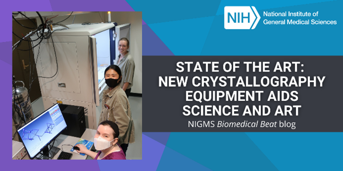
Upgrading X-ray crystallography equipment at the University of Arkansas in Fayetteville has had an unexpected benefit: enabling analyses that could help art museums authenticate, restore, and learn more about their pieces.
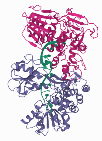
Two copies of a protein (pink and purple) produced by the hepatitis C virus interacting with the same strand of DNA (green). This structure was solved using equipment at the University of Arkansas X-ray crystallography center. Credit: PDB 2F55.
Scientists use X-ray crystallography to determine the detailed 3D structures of molecules. In biomedical contexts, researchers often apply X-ray crystallography to map the structures of proteins and other biomolecules like DNA and RNA. A molecule’s structure can shed light on its function and help answer scientific questions. For example, knowing the structures of proteins involved in antibiotic resistance can help researchers determine how those molecules work and how to combat bacteria that produce them.
Protein Data Bank’s 50 years logo. Credit: PDB website.
The Protein Data Bank (PDB), established in 1971, is the single global repository for 3D structural data of proteins, DNA, RNA, and even complexes these biological molecules form with drugs or other small molecules. More than 1 million people—including researchers, medical professionals, educators, and students—use the PDB each year. NIGMS and other parts of NIH have helped fund this free digital resource since 1978.
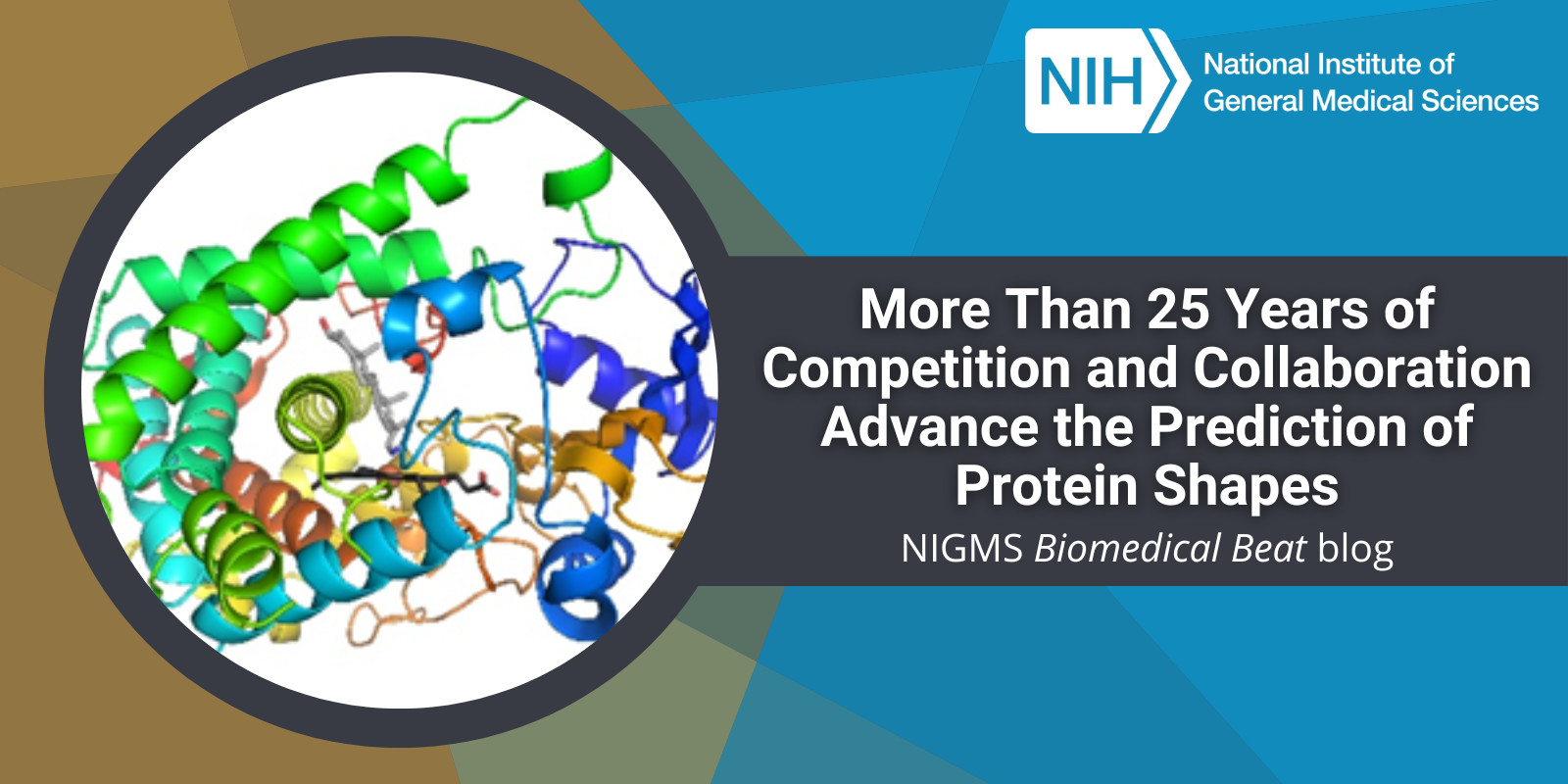
Proteins (such as hemoglobin, actin, and amylase) are workhorse molecules that contribute to virtually every activity in the body. Some of proteins’ many jobs include carrying oxygen from your lungs to the rest of your body (hemoglobin), allowing your muscles to move (actin and myosin), and digesting your food (amylase, pepsin, and lactase). All proteins are made up of chains of amino acids that fold into specific 3D structures, and each protein’s structure allows it to perform its distinct job. Proteins that are misfolded or misshapen can cause diseases such as Parkinson’s or cataracts.
While it’s straightforward to use the genetic code to predict amino acid sequences of proteins from gene sequences, the vast diversity of protein shapes and many factors that influence a protein’s 3D structure make it much more complicated to create simple folding rules that could be used to predict proteins’ structures from these sequences. Scientists have worked on this problem for nearly 50 years, and NIGMS has supported many of their efforts, including the Critical Assessment of Structure Prediction (CASP) program.
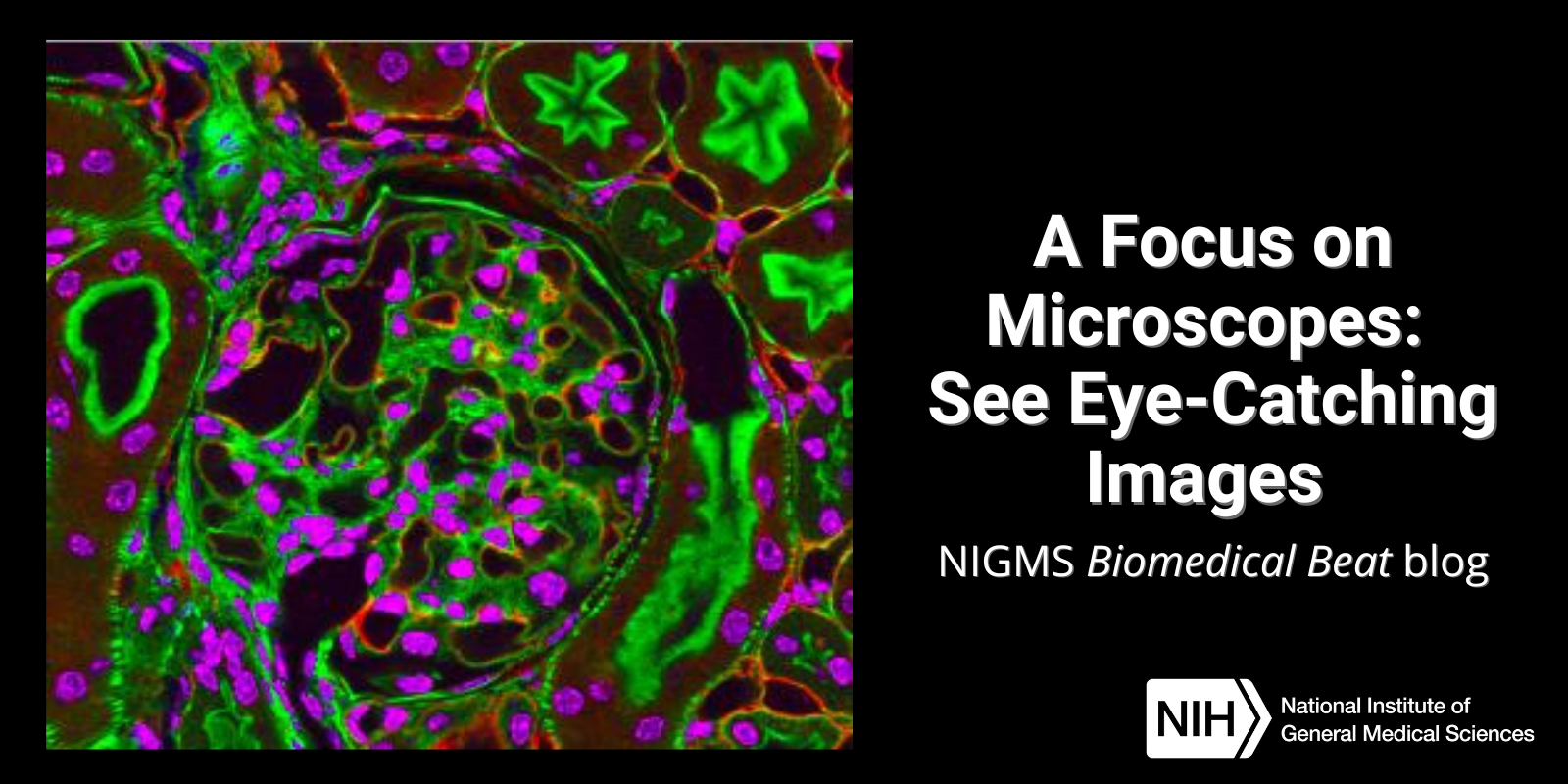
Have you ever wondered what creates striking images of cells and other tiny structures? Most often, the answer is microscopes. Many of us have encountered basic light microscopes in science classes, but those are just one of many types that scientists use. Check out the slideshow to see images researchers have captured using different kinds of microscopes. For even more images of the microscopic world, visit the NIGMS Image and Video Gallery.
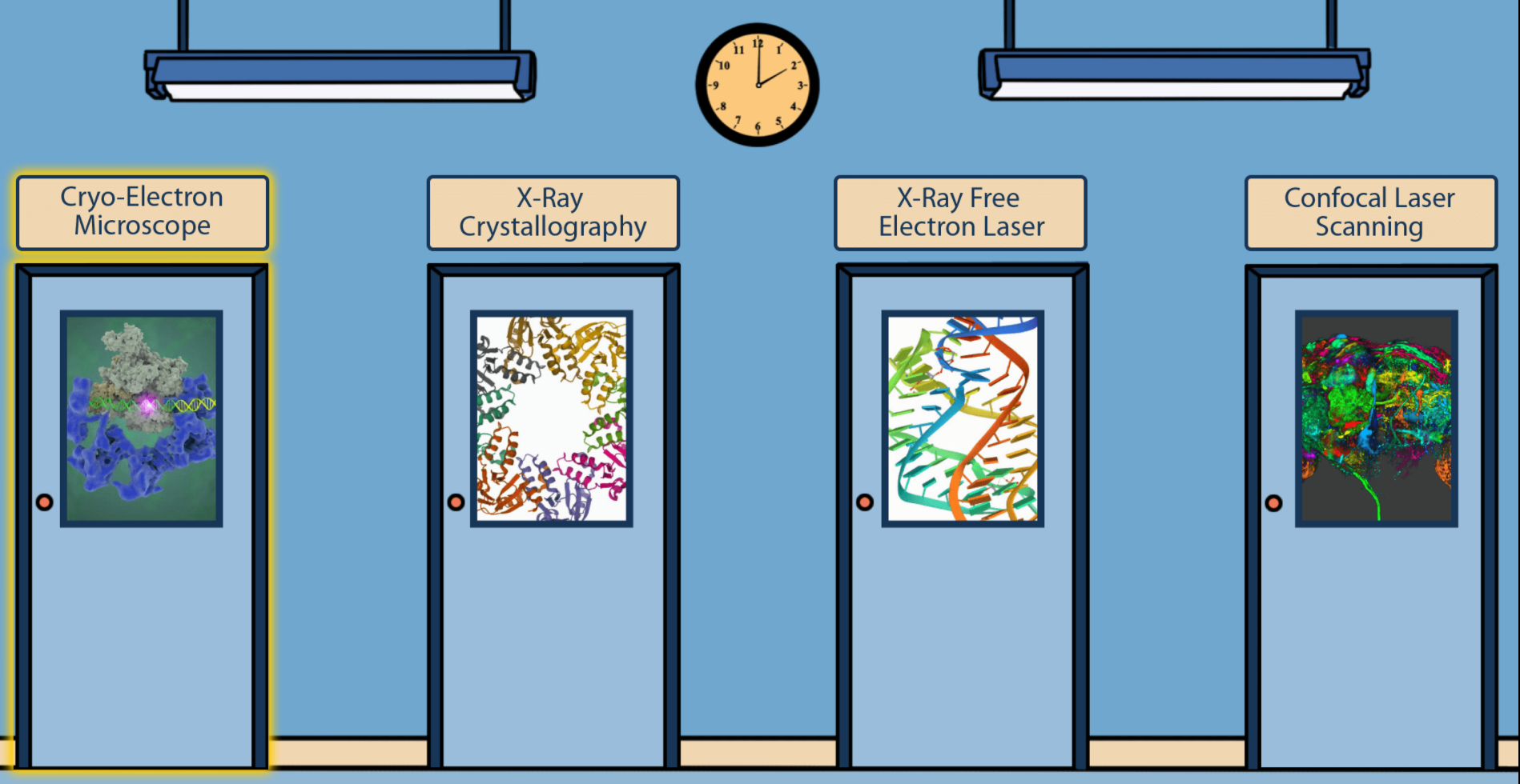
Students, teachers, and other curious minds can step into a scientific imaging lab with a free online interactive developed by NIGMS and Scholastic. Imaging tools help scientists unlock the mysteries of our cells and molecules. A better understanding of this tiny world can help researchers learn about the body’s normal and abnormal processes and lead to more effective, targeted treatments for illnesses.
Proteins play a role in virtually every activity in the body. They make up hair and nails, help muscles move, protect against infection, and more. Many NIGMS-funded researchers study the rich variety of proteins in humans and other organisms to shed light on their roles in health and disease.
NIGMS and Scholastic bring you our latest issue of Pathways, which focuses on imaging tools that help scientists unlock the mysteries of our cells and molecules. A better understanding of this tiny world can help researchers learn about the body’s normal and abnormal processes and lead to more effective, targeted treatments for illnesses.
Pathways is designed for students in grades 6 through 12. This collection of free resources teaches students about basic science and its importance to health, as well as exciting research careers.


 Calcium keeps your bones strong, allows your muscles to move, and is important for many other bodily functions. The element is found in foods, medicines, and the world around us. Credit: Compound Interest CC BY-NC-ND 4.0. Click to enlarge.


 Protein Data Bank’s 50 years logo. Credit: PDB website.
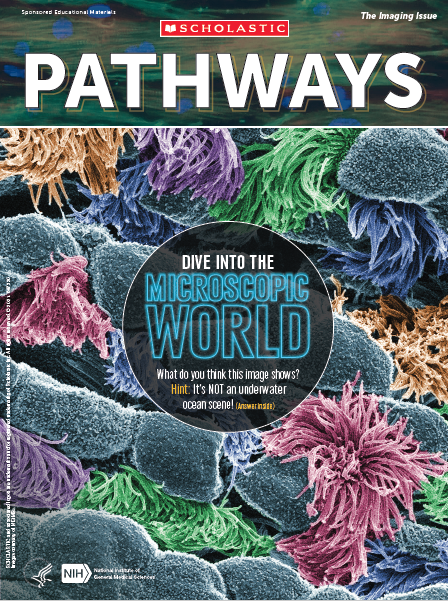 Cover of Pathways student magazine.