Teens Explore Science and Health through Game Design

0 comments

Educators often struggle to teach teens about sexual and reproductive health. Hexacago Health Academy (HHA) Link to external web site, an education program from the University of Chicago, leverages the fun activity of gameplay to impart these lessons to young people from Chicago’s South Side community. Funded by the Student Education Partnership Award (SEPA)Link to external web site, part of the National Institute of General Medical Sciences (NIGMS), in 2015, HHA assists teachers in their goal of helping teen students gain awareness and control over their health and also learn about careers in STEM Link to external web site and health fields.

Woman in a black buisness suit with arms crossed standing against a wall and smiling
Melissa Gilliam, founder of Ci3. Credit: Anna Knott, Chicago Magazine.

Genesis of HHA

HHA was cofounded by Melissa Gilliam Link to external web site, a University of Chicago professor of Obstetrics/Gynecology and Pediatrics and founder of the Center for Interdisciplinary Inquiry & Innovation in Sexual and Reproductive Health (Ci3) Link to external web site. During a 2013 summer program with high school students, Gilliam and Patrick Jagoda Link to external web site, associate professor of English and Cinema & Media Studies, and cofounder of Ci3’s Game Changer Chicago Design Lab, introduced the students to their STEM-based alternate reality game called The Source, in which a young woman crowdsources player help to solve a mystery that her father has created for her.

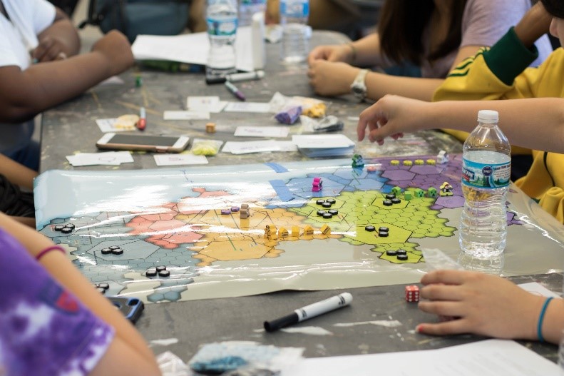
From their experience with The Source, Gilliam and Jagoda quickly learned that students not only wanted to play games but to design them too. What followed was the Game Changer Lab’s creation of the Hexacago game board, as well as the launch of HHA, a SEPA-funded project that the lab oversees.

Hexacago Game Board

At the core of HHA is the Hexacago game board, which displays the city of Chicago, along with Lake Michigan, a train line running through the city, and neighborhoods gridded into a hexagonal pattern.

HHA students not only play games designed from the Hexacago board template, but also design their own games from it that are intended to inspire behavior change in health-related situations and improve academic performance.

High school students seated at a table with a glossy, laminate test model of the Hexacago game and game pieces on top of it
Credit: Ci3 at the University of Chicago.

In this way, HHA is much more than just game design and play. “Students have no idea that what they’re doing is learning. In their minds, they’re really focused on designing games,” says Gilliam. “That’s the idea behind Hexacago Health Academy: helping people acquire deep knowledge of science and health issues by putting on the hat of a game designer.” Moreover, through the process of gameplay and design, students practice all the rich skills that result from teamwork, including collaborative learning, leadership, and communication.

Continue reading “Teens Explore Science and Health through Game Design”

Interview with a Scientist: Michael Summers, Using Nuclear Magnetic Resonance to Study HIV

0 comments

For more than 30 years, NIGMS has supported the structural characterization of human immunodeficiency virus (HIV) enzymes and viral proteins. This support has been instrumental in the development of crucial drugs for antiretroviral therapy such as protease inhibitors. NIGMS continues to support further characterization of viral proteins as well as cellular and viral complexes. These complexes represent the fundamental interactions between the virus and its host target cell and, as such, represent potential new targets for therapeutic development.

In this third in a series of three video interviews with NIGMS-funded researchers probing the structure of HIV, Michael Summers,Link to external web site professor of biochemistry at the University of Maryland, Baltimore County, discusses his use of nuclear magnetic resonance (NMR) technology to study HIV. Of recent interest to Summers has been using NMR to investigate how HIV’s RNA enables the virus to reproduce. His goals for this line of research are to develop treatments against HIV as well as learning how to best engineer viruses to deliver helpful therapies to individuals with a variety of diseases.

Continue reading “Interview with a Scientist: Michael Summers, Using Nuclear Magnetic Resonance to Study HIV”

Interview with a Scientist: Wes Sundquist, How the Host Immune System Fights HIV

1 comment

For more than 30 years, NIGMS has supported the structural characterization of human immunodeficiency virus (HIV) enzymes and viral proteins. This support has been instrumental in the development of crucial drugs for antiretroviral therapy such as protease inhibitors. NIGMS continues to support further characterization of viral proteins as well as cellular and viral complexes. These complexes represent the fundamental interactions between the virus and its host target cell and, as such, represent potential new targets for therapeutic development.

In this second in a series of three video interviews with NIGMS-funded researchers probing the structure of HIV, Wes Sundquist, professor of biochemistry at the University of Utah, discusses his lab’s studies of how HIV uses factors in host cells to replicate itself. In particular, Sundquist focuses on the ESCORT pathway that enables HIV to leave infected cells and spread infection elsewhere.

Continue reading “Interview with a Scientist: Wes Sundquist, How the Host Immune System Fights HIV”

Interview With a Scientist: Irwin Chaiken, Rendering HIV Inert

0 comments

For more than 30 years, NIGMS has supported the structural characterization of human immunodeficiency virus (HIV) enzymes and viral proteins. This support has been instrumental in the development of crucial drugs for antiretroviral therapy such as protease inhibitors. NIGMS continues to support further characterization of viral proteins as well as cellular and viral complexes. These complexes represent the fundamental interactions between the virus and its host target cell and, as such, represent potential new targets for therapeutic development.

Continue reading “Interview With a Scientist: Irwin Chaiken, Rendering HIV Inert”

CLAMP Helps Lung Cells Pull Together

0 comments
ALT TEXT
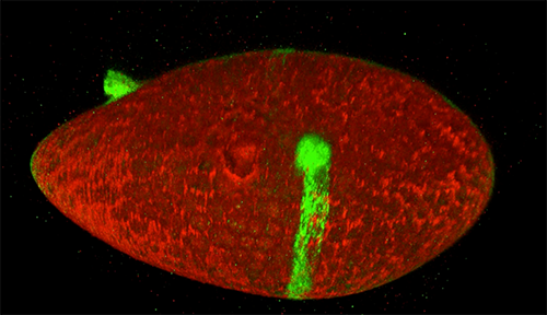
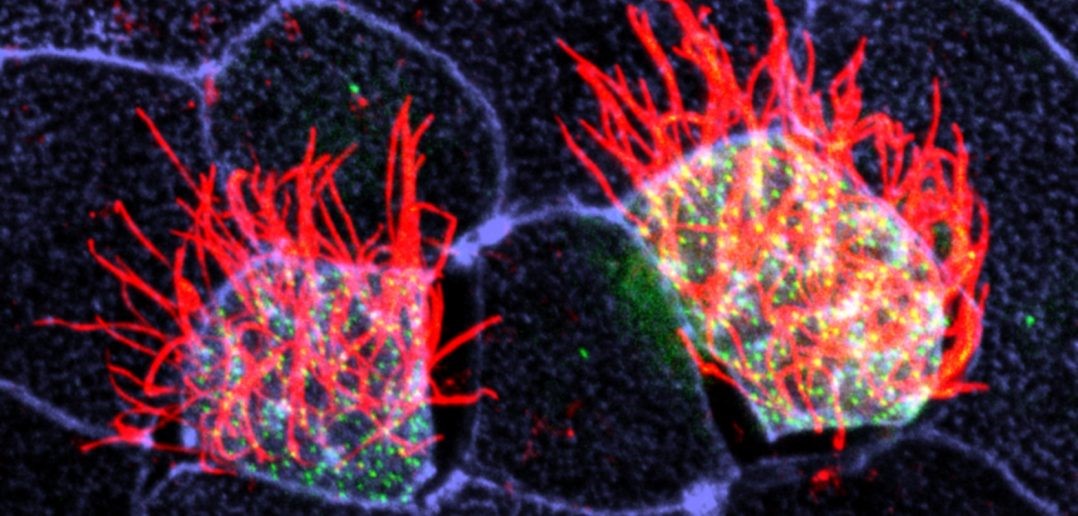
Cells covered with cilia (red strands) on the surface of frog embryos. Credit: The Mitchell Lab.

The outermost cells that line blood vessels, lungs, and other organs also act like guards, alert and ready to thwart pathogens, toxins, and other invaders that can do us harm. Called epithelial cells, they don’t just lie passively in place. Instead, they communicate with each other and organize their internal structures in a single direction, like a precisely drilled platoon of soldiers lining up together and facing the same way.

Lining up this way is crucial during early development, when tissues and organs are forming and settling into their final positions in the developing body. In fact, failure to line up in the correct way is linked to numerous birth defects. In the lungs, for instance, epithelial cells’ ability to synchronize with one another is important since these cells have special responsibilities such as carrying mucus up and out of lung tissue. When these cells can’t coordinate their functions, disease results.

Some lung epithelial cells are covered in many tiny, hair-like structures called cilia. All the cilia on lung epithelial cells must move uniformly in a tightly choreographed way to be effective in their mucus-clearing job. This is a unique example of a process called planar cell polarity (PCP) that occurs in cells throughout the body. Researchers are seeking to identify the signals cells use to implement PCP. Continue reading “CLAMP Helps Lung Cells Pull Together”

Interview with a Scientist: Jeramiah Smith on the Genomic Antics of an Ancient Vertebrate

0 comments

The first known descriptions of cancer come from ancient Egypt more than 3,500 years ago. Early physicians attributed the disease to several factors, including an imbalance in the body’s humoral fluids, trauma, and parasites. Only in the past 50 years or so have we figured out that mutations in critical genes are often the trigger. The sea lamprey, a slimy, snake-like blood sucker, is proving to be an ideal tool for understanding these mutations.

The sea lamprey, often called the jawless fish, is an ancient vertebrate whose ancestor diverged from the other vertebrate lineages (fish, reptiles, birds and mammals) more than 500 million years ago. Jeramiah Smith,Link to external web site associate professor of biology at the University of Kentucky, has discovered that lamprey have two separate genomes: a complete genome specific to their reproductive cells, consisting of 99 chromosomes (humans have 23 pairs) and another genome in which about 20 percent of genes have been deleted after development. Using the lamprey model, Smith and his colleagues have learned that many of these deleted genes—such as those that initiate growth pathways—are similar to human oncogenes (i.e., cancer-causing genes).

Continue reading “Interview with a Scientist: Jeramiah Smith on the Genomic Antics of an Ancient Vertebrate”

Pericytes: Capillary Guardians in the Brain

0 comments
ALT TEXT
The long arms of pericytes cells (red) stretch along capillaries (blue) in a mouse brain. Credit: Andy Shih.

Nerve cells, or neurons, in our brains do amazing work, from telling our hearts to beat to storing our memories. But neurons cannot operate alone. Many kinds of cells support and regulate neurons and—like neurons—they can come under attack due to injuries or disorders, such as stroke or Alzheimer’s disease. Learning what jobs these cells do and how they respond to disease may show researchers new ways to treat central nervous system disorders. One type of support cell, the pericyte, plays some key roles in brain health. These cells are readily adaptable, even in adult brains, and can support a variety of functions.

Pericytes help with blood flow to nerve cells in the brain. They lie wrapped all along the huge networks of capillaries—the tiniest blood vessels—that both feed neurons and form the blood-brain barrier, which filters out certain substances from blood to protect the brain. Pericytes have a body that appears as a bump protruding from a capillary surface. Pericytes also have long thin arms that stretch along each capillary like a snake on a tree branch. These arms, called processes, reach almost to where the next pericyte process begins, without overlapping. This creates a pericyte chain that covers nearly the entire capillary network.

Pericytes are critical for blood vessel stability and blood-brain barrier function. They’re also known to die off as a result of trauma and disease. Andy ShihLink to external web site, Andree-Ann Berthiaume, and colleagues at the Medical University of South Carolina in Charleston, set up an imaging technique in mouse brains that allowed them to see what pericytes do under normal conditions as well as how these cells respond when some are damaged.

Continue reading “Pericytes: Capillary Guardians in the Brain”

Optogenetics Sparks New Research Tools

1 comment

Imagine if scientists could zap a single cell (or group of cells) with a pulse of light that makes the cell move, or even turns on or off the cell’s vital functions.

Scientists are working toward this goal using a technology called optogenetics. This tool draws on the power of light-sensitive molecules, called opsins and cryptochromes, which are naturally occurring molecules found in the cell membranes of a wide variety of species, from microscopic bacteria and algae to plants and humans. These light-reacting molecules change their shape or activity when they sense light, so they can be used to trigger cellular activity, such as turning on or off ion flow into the cell and other regulatory pathways. The ability to induce changes in cells has a broad range of practical applications, from enabling scientists to see how cells function to providing the basis for potential therapeutic applications for blindness, cancer, and epilepsy.

Opsins first gained a foothold in research about a decade ago when scientists began using them to study specific electrical networks in the brain. This research relied on channelrhodopsins, opsins that could be used to control the flow of charged ions into and out of the cell. Normally, when a neuron reaches a certain ion concentration, it is triggered to fire, but neuron firing can be changed by inserting opsins in the membrane. Neuroscientists figured out how to incorporate light-sensitive opsin proteins by inserting the opsin gene into the host’s DNA. The genetically encoded opsin proteins in the neuronal membranes could be turned on or off by shining light into the brain itself, using optical fibers or micro-LEDs, to switch on or off the flow of ions and neuron firing.

Since those early studies in the brain, the optogenetics field has come a long way. But the leap from brain cells to other cells has been challenging. Scientists first needed to find a way to deliver light into tissues deep in the body. And, unlike stationary brain cells, they needed a way to target cells that are on the move (such as immune cells). They also needed to develop a way to study not only cell networks but also individual cells and cell parts. The NIGMS-funded researchers highlighted below are among the scientists working to overcome these obstacles and are using optogenetics in new and inventive ways.

Illustration showing how bridges can be built within a cell using light-reacting molecules
Illustration shows how “bridges” can be built within a cell through the use of light-reacting molecules. The light triggers proteins to line up within the cell, making it easier to shuttle molecules between the membranes of two subcellular organelles. This optogenetic strategy is helping scientists to control cell function with a simple beam of light. Illustration courtesy of Yubin Zhou.

Building Bridges

Yubin ZhouLink to external web site of Texas A&M is using optogenetics to control the way cells communicate and to study immune cell function. In one line of research, Zhou is using light to make it easier for calcium ions to enter cells. The ions carry instructions for the cell and also help tether small cellular structures (called organelles).  Those inter-membrane tethers allow for the movement of  proteins and lipids back and forth across the cell, and are critical for sending chemical messengers to communicate information (see illustration). When this process is disrupted, it can lead to extreme changes in cell function and even cell death. Using this technology to “switch on” normal pathways enables the scientists to better understand how such processes can be disrupted.

Continue reading “Optogenetics Sparks New Research Tools”

The Skull’s Petrous Bone and the Rise of Ancient Human DNA: Q & A with Genetic Archaeologist David Reich

1 comment
A macro image of the petrous bone. 3 sections are color coded A (green), B (blue), and C (red)
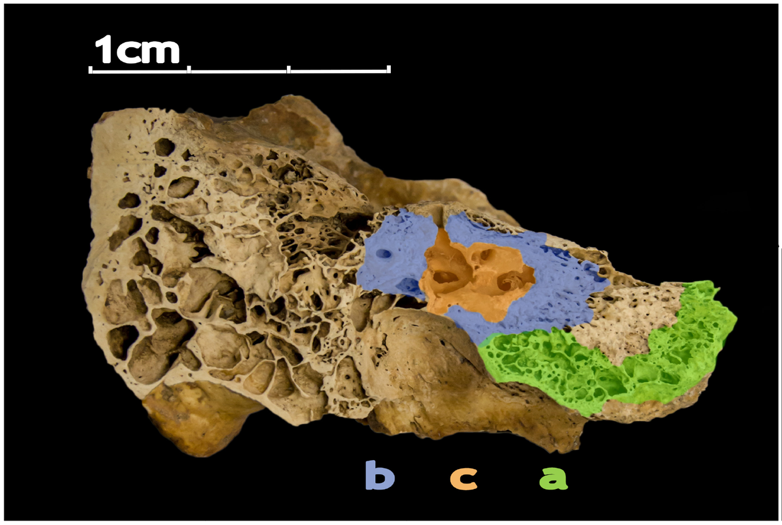
The human petrous bone in the skull protects the inner ear structures. Though it is one of the hardest, densest bones in the body, some portions (such as the area in orange, protecting the cochlea) are denser than others. Possibly because the petrous bone is so dense, DNA within the petrous bone is better preserved than in other bones. In some cases, scientists have extracted more than 100 times more DNA from the petrous bone than other bones, including teeth. Credit: Pinhasi et al., 2015, PLOS ONE.

For the past few decades, new evidence about ancient humans—in the form of skeletal remains, tools, and other artifacts—has trickled in, inching us closer to an understanding of how our species evolved and spread out across the planet. In just the past few years, however, knowledge of our deep past expanded significantly thanks to a series of technological breakthroughs in sequencing of ancient human genomes. This technology can be used to find genetic links among populations of human ancestors dating back hundreds of thousands of years.

In addition to advances in genomic technology, another factor is driving the explosion of new discoveries—an inch-long section of the human skull. Found near our ears, this pyramid-shaped portion of the temporal bone is nicknamed the petrous bone. The bone is very hard, possibly because it needs to protect fragile structures such as the cochlea, which translates sound into brain signals, and the semicircular canals, which help us maintain our balance. Perhaps because the petrous bone is so dense, it also is the bone in the body that best preserves DNA after a person dies. As a result, archaeologists are scrambling to study samples taken from this pyramid-shaped structure to unlock the mysteries of our species’ formative years.

Here’s a sampling of headlines declaring new findings about ancient peoples from around the globe that were based on genetic information obtained from the petrous bone (NIGMS-funded research indicated in black):

“How the introduction of farming changed the human genome” November 2015

“Fourth strand’ of European ancestry originated with hunter-gatherers isolated by Ice Age” November 2015

“Scientists sequence first ancient Irish human genomes” December 2015

“Genetic studies provide insight into ancient Britain’s diversity” January 2016

“The world’s first farmers were surprisingly diverse” June 2016

“Study reveals Asian ancestry of Pacific islanders” October 2016

“Ancient DNA solves mystery of the Canaanites, reveals the biblical people’s fate” July 2017

“Ancient DNA data fills in thousands of years of human prehistory in Africa” September 2017

“European Hunter-Gatherers Interbred With Farmers From the Near East” November 2017

“Surprise as DNA reveals new group of Native Americans: the ancient Beringians” January 2018

“Ancient DNA reveals genetic replacement despite language continuity in the South Pacific” February 2018

“Stone Age Moroccan Genomes Reveal Sub-Saharan African, Near Eastern Ancestry” March 2018

“Some early modern populations in Britain may have had dark skin” March 2018

Continue reading “The Skull’s Petrous Bone and the Rise of Ancient Human DNA: Q & A with Genetic Archaeologist David Reich”

Cellular Footprints: Tracing How Cells Move

0 comments
ALT TEXT
An engineered cell (green) in a fruit fly follicle (red), or egg case, leaves a trail of fluorescent material as it moves across a fruit fly egg chamber, allowing scientists to trace its path and measure how long it took to complete its journey. Credit: David Bilder, University of California, Berkeley.

Cells are the basis of the living world. Our cells make up the tissues and organs of our bodies. Bacteria are also cells, living sometimes alone and sometimes in groups called biofilms. We think of cells mostly as staying in one spot, quietly doing their work. But in many situations, cells move, often very quickly. For example, when you get a cut, infection-fighting cells rally to the site, ready to gobble up bacterial intruders. Then, platelet cells along with proteins from blood gather and form a clot to stop any bleeding. And finally, skin cells surrounding the wound lay down scaffolding before gliding across the cut to close the wound.

This remarkable organization and timing is evident right from the start. Cells migrate within the embryo as it develops so that body tissues and organs end up in the right places. Harmful cells use movement as well, as when cells move and spread (metastasize) from an original cancer tumor to other parts of the body. Learning how and why cells move could give scientists new ways to guide those cells or turn off or slow down the movement when needed.

Glowing Breadcrumbs

Scientists studying how humans and animals form, from a single cell at conception to a complex body at birth, are particularly interested in how and when cells move. They use research organisms like the fruit fly, Drosophila, to watch movements by small populations of cells. Still, watching cells migrate inside a living fly is challenging because the tissue is too dense to see individual cell movement. But moving those cells to a dish in the lab might cause them to behave differently than they do inside the fly. To solve this problem, NIGMS-funded researcher David BilderLink to external web site and colleagues at the University of California, Berkeley, came up with a way to alter fly cells so they could track how the cells behave without removing them from the fly. They engineered the cells to lay down a glowing track of proteins behind them as they moved, leaving a traceable path through the fly’s tissue. The technique, called M-TRAIL (matrix-labeling technique for real-time and inferred location), allows the researchers to see where a cell travels and how long it takes to get there.

Bilder and his team first used M-TRAIL in flies to confirm the results of past studies of Drosophila ovaries in the lab using other imaging techniques. In addition, they found that M-TRAIL could be used to study a variety of cell types. The new technique also could allow a cell’s movement to be tracked over a longer period than other imaging techniques, which become toxic to cells in just a few hours. This is important, because cells often migrate for days to reach their final destinations.

Continue reading “Cellular Footprints: Tracing How Cells Move”