Tag: Cellular Processes

A Heart-Shaped Protein

1 comment
Structure of the serum albumin protein

The structure of the serum albumin protein is shaped like a heart. Credit: Wladek Minor, University of Virginia.

From cookies and candies to balloons and cards, heart-shaped items abound this time of year. They’re even in our blood. It turns out that the most abundant protein molecule in blood plasma—serum albumin (SA)—is shaped very much like a heart. Continue reading “A Heart-Shaped Protein”

Cool Images: A Holiday-Themed Collection

1 comment

Here are some images from our gallery that remind us of the winter holidays—and showcase important findings and innovations in biomedical research.

Ribbons and Wreaths

Wreath

This wreath represents the molecular structure of a protein, Cas4, which is part of a system, known as CRISPR, that bacteria use to protect themselves against viral invaders. The green ribbons show the protein’s structure, and the red balls show the location of iron and sulfur molecules important for the protein’s function. Scientists have harnessed Cas9, a different protein in the bacterial CRISPR system, to create a gene-editing tool known as CRISPR-Cas9. Using this tool, researchers can study a range of cellular processes and human diseases more easily, cheaply and precisely. Last week, Science magazine recognized the CRISPR-Cas9 gene-editing tool as the “breakthrough of the year.”

Continue reading “Cool Images: A Holiday-Themed Collection”

Sugar Rush in Research

0 comments
Cookies
Sugar sprinkled on cookies and other treats is often an attractive—and sweet tasting—finishing touch. But the sugar-rich coating that surrounds most cells is far more—it’s a vital ingredient for many basic cellular processes. Credit: Stock image.

Simple sugars such as sucrose (found in the sugar bowl) and fructose (in fruits and honey) provide the sweet finishing touches on many holiday treats. But did you know that versions of these molecules also serve important functions in our cells?

Cells assemble sugar molecules into chains known as glycans. These glycans, which can be linear or branching, play an astounding number of biological roles. When bound to proteins called lectins, they enable a fertilized egg to attach properly onto a woman’s uterine wall and help immune cells move out of a blood vessel to the site of an infection. When decorated with specific patterns of molecules called sulfates, glycans can help direct the growth of nerves. And it’s the glycans found on our blood cells that define blood type (A, B, AB or O). Continue reading “Sugar Rush in Research”

Bacterial Biofilms: A Charged Environment

1 comment
Bacillus subtilis biofilm
A Bacillus subtilis biofilm grown in a Petri dish. Credit: Süel Lab, UCSD.

Last summer, we shared findings from Gürol Süel Exit icon and colleagues at the University of California, San Diego, that bacterial cells in tight-knit microbial communities called biofilms expand in a stop-and-go pattern. The researchers concluded that this pattern helps make food at the nutrient-rich margin available to the cells in the starved center, but they didn’t know how. They’ve now shown that the cells use electrochemical signaling to communicate and cooperate with each other.

Because nutrients and other signals cells use to sense each other and their environment move rather slowly, the researchers looked for a faster, more active communication system in biofilms of the bacterium B. subtilis. They focused on electrical signaling via potassium, a positively charged ion that, for example, our nerve and muscle cells use to send or receive signals. Continue reading “Bacterial Biofilms: A Charged Environment”

Cracking a Ubiquitous Code

0 comments

We asked the heads of our scientific divisions to tell us about some of the big questions in fundamental biomedical science that researchers are investigating with NIGMS support. This article is the third in an occasional series that explores these questions and explains how pursuing the answers could advance understanding of important biological processes.

Ubiquitin (Ub) molecules
Ubiquitin (Ub) molecules attached to proteins can form possibly hundreds of different shapes. Credit: NIGMS.

Researchers are on a quest to crack a code made by ubiquitin, a small protein that plays a big role in coordinating cellular function. By attaching to other proteins, ubiquitin determines what those proteins should do next.

Just as zip codes direct letters to specific towns, the ubiquitin code might direct one protein to help with DNA repair, another to assist in cell division, and a third to transport molecules into and out of cells. Continue reading “Cracking a Ubiquitous Code”

Seeing Telomerase’s ‘Whiskers’ and ‘Toes’

1 comment
Telomerase and its components.

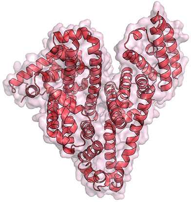
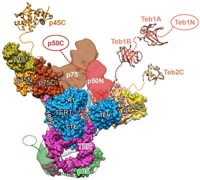
The image here is the “front view” of telomerase, with the enzyme’s components shown in greater detail than ever before. Credit: UCLA Department of Chemistry and Biochemistry.

Like the features of a cat in a dark alley, those of an important enzyme called telomerase have been elusive. Using a combination of imaging techniques, a research team led by Juli Feigon Exit icon of the University of California, Los Angeles, has now captured the clearest view ever of the enzyme.

Telomerase maintains the DNA at the ends of our chromosomes, known as telomeres, which act like the plastic tips on the ends of shoelaces. In the absence of telomerase activity, telomeres get shorter each time our cells divide. Eventually, the telomeres become so short that the cells stop dividing or die. On the other hand, cells with abnormally high levels of telomerase activity can constantly rebuild their protective chromosomal caps. Telomerase is particularly active within cancer cells. Continue reading “Seeing Telomerase’s ‘Whiskers’ and ‘Toes’”

Cool Image: Tracing Proteins in Action

2 comments
Bright amorphous loops
These bright, amorphous loops represent a never-before-seen glimpse at how proteins that play a key role in cell duplication are themselves duplicated. Credit: Sue Jaspersen, Zulin Yu and Jay Unruh, Stowers Institute for Medical Research.

Looking like necklaces stacked on a dresser, these bright, amorphous loops show the outlines of yeast proteins that make up the spindle pole, a cellular component found in organisms as diverse as yeast and humans. Each cell starts with a single spindle pole, which must somehow duplicate to form the pair that works together to pull matching chromosomes apart during cell division. Scientists don’t completely understand how this duplication occurs, but they do know that errors in spindle pole copying can lead to a number of health conditions, including cancer.

Continue reading “Cool Image: Tracing Proteins in Action”

Interview With a Worm: We’re Not So Different

2 comments
Planarian
Credit: Alejandro Sánchez Alvarado, Stowers Institute for Medical Research.
Schmidtea mediterranea
Home: Freshwater habitats along the Mediterranean
Party trick: Regenerating its head
Most charismatic feature: Eyespots
Work site: Science labs worldwide

The planarian has a power few creatures can match. Remove its head, its tail or nearly any of its body parts, and this aquatic flatworm will simply grow it back. Humans can’t do that, of course. And yet many of the genes that help the planarian regenerate are also found in us. To learn more about this tiny marvel, we “interviewed” a representative. Continue reading “Interview With a Worm: We’re Not So Different”

Cool Images: A Halloween-Inspired Cell Collection

3 comments

As Halloween approaches, we turned up some spectral images from our gallery. The collection below highlights some spooky-sounding—but really important—biological topics that researchers are actively investigating to spur advances in medicine.

Cell Skeleton
Fibroblast
The cell skeleton, or cytoskeleton, is the framework that gives a cell its shape, helps it move and keeps its contents organized for proper function. A cell that lacks a cytoskeleton becomes misshapen and immobile. This fibroblast, a cell that normally makes connective tissues and travels to the site of a wound to help it heal, is lacking a cytoskeleton. Researchers have associated faulty cytoskeletons and resulting abnormal cell movement with birth defects and weakened immune system functioning. See fibroblasts with healthy skeletons.

Continue reading “Cool Images: A Halloween-Inspired Cell Collection”

Turning Back Every Clock

0 comments
Clock
Scientists are studying which genes control biological clock gears and which genes are controlled by them. Credit: Stock image.

When daylight savings time ends this Sunday, we’ll need to adjust every clock in our homes, cars and offices. Our internal clocks will need to adjust too.

The body has a master clock in the brain, as well as others in nearly every tissue and organ. These biological clocks drive circadian rhythms, the physical, mental and behavioral changes we experience on a roughly 24-hour cycle. Your hunger in the morning and sleepiness at night, for example, are caused partly by clock gears in motion. These gears can get out of synch with the day-night cycle when the time changes or when we travel through time zones.

Continue reading “Turning Back Every Clock”