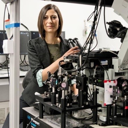
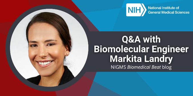
Tag: Cool Tools/Techniques

In Other Words: Not All Cultures Are Human

0 comments

The word culture may make you think of a flag, style of clothing, celebration, or some other tradition associated with a particular group of people. But in biomedical science, a culture is a group of cells grown in a lab. Scientists use cultures to learn about basic biological processes and to develop and test new medicines.

Below the title “Culture: In Other Words,” two images are separated by a jagged line. On the left are outlines of faces surrounding a globe of the Earth. On the right is a hand holding a Petri dish with cells growing in it. Under the images, text reads: “Did you know? In biomedical science, a culture is a group of cells grown in a lab.”
Credit: NIGMS.

The Birth of a Culture

Scientists can grow many types of cells as cultures, from bacteria to human cells. To create a culture, a researcher adds cells to a container such as a Petri dish along with a mix of nutrients the cells need to grow and divide. The exact recipe varies depending on the cell type. (Because many lab containers were historically made of glass, researchers sometimes refer to studies that use cultures as in vitro—Latin for “in glass.”) Once the cells multiply and fill their container, researchers split the culture into new containers to produce more.

Continue reading “In Other Words: Not All Cultures Are Human”

State of the Art: New Crystallography Equipment Aids Science and the Study of Artifacts

0 comments

Upgrading X-ray crystallography equipment at the University of Arkansas in Fayetteville has had an unexpected benefit: enabling analyses that could help art museums authenticate, restore, and learn more about their pieces.

Intertwined curled ribbons.
Two copies of a protein (pink and purple) produced by the hepatitis C virus interacting with the same strand of DNA (green). This structure was solved using equipment at the University of Arkansas X-ray crystallography center. Credit: PDB 2F55.

Scientists use X-ray crystallography to determine the detailed 3D structures of molecules. In biomedical contexts, researchers often apply X-ray crystallography to map the structures of proteins and other biomolecules like DNA and RNA. A molecule’s structure can shed light on its function and help answer scientific questions. For example, knowing the structures of proteins involved in antibiotic resistance can help researchers determine how those molecules work and how to combat bacteria that produce them.

Continue reading “State of the Art: New Crystallography Equipment Aids Science and the Study of Artifacts”

Career Conversations: Q&A With Immunoengineer Caroline Jones

0 comments
A headshot of Dr. Jones.
Dr. Caroline Jones. Credit: Moises Gomez.

“I find it fulfilling to be a scientist because I know that even if at some points it seems like I’m working on an incremental experiment, eventually it’s going to help solve a bigger problem,” says Caroline Jones, Ph.D., an assistant professor of bioengineering at the University of Texas at Dallas. Check out the highlights of our interview with Dr. Jones to learn about her career path, her passion for sharing science with the public, and her research on sepsis—an overwhelming or impaired whole-body immune response to an insult, such as an infection or injury that’s responsible for the deaths of nearly 270,000 Americans every year.

Q: How did you first become interested in science?

A: My mother was a high school math teacher, so I had that role model growing up. I also had a math and engineering teacher in high school who encouraged me and sparked my interest in the quantitative side of science. I decided to study biomedical engineering in college because I wanted to apply quantitative tools in a way that helped people.

Continue reading “Career Conversations: Q&A With Immunoengineer Caroline Jones”

Cloudy With a Chance of Scientific Discoveries

0 comments

The cloud. To many, it’s a mysterious black hole that somehow transports photos and files from their old or lost phone to their new one. To some researchers, though, it’s an invaluable resource that allows them access to data analytics tools they wouldn’t otherwise have.

Hands typing on a laptop with a digitized cloud and computer icons floating above them.
Credit: iStock.

Scientists have begun using cloud computing to store, process, and analyze their data through online bioinformatics tools. Biological data sets are often large and hard to interpret, requiring complex calculating instructions—or algorithms—to understand them. Fortunately, these algorithms can run on local computers or remotely through cloud computing.

One advantage of cloud-based programs over local computers is the ability to analyze data without taking up the user’s personal storage space. With cloud-based storage, researchers can store their large data files, including their labeled notes called annotations. Another benefit is that users have easy access to software packages within the cloud for data analysis. The cloud also encourages collaboration among scientists by making it easy to share large amounts of data.

Continue reading “Cloudy With a Chance of Scientific Discoveries”

Photographing the Physics of Cells

0 comments
Dr. Lakadamyali sitting behind a large, complex microscope in a lab.
Dr. Melike Lakadamyali with a microscope. Credit: Courtesy of Dr. Lakadamyali.

“It would be a dream come true if I could look at a cell within a tissue and have a Google Maps view to zoom in until I saw individual molecules,” says Melike Lakadamyali, Ph.D., an associate professor of physiology at the University of Pennsylvania’s Perelman School of Medicine in Philadelphia. Her lab is helping make part of that dream a reality by developing super-resolution microscopy tools that visualize cells at a near-molecular level.

Blending Physics and Biology

Science and math fascinated Dr. Lakadamyali since childhood, and she felt especially drawn to physics because she enjoyed using logic to solve problems. After graduating high school in her native country of Cyprus, she chose to study physics at the University of Texas, Austin. She never gave much thought to applying physics methods to biological
questions—a field known as biophysics—until her third year as an undergraduate, when she gained her first research experience in the lab of Josef Käs, Ph.D.

Continue reading “Photographing the Physics of Cells”

Quiz: What Can Research Organisms Reveal About Health?

0 comments

Scientists often use research organisms to study life. Examples range from simple organisms like bacteria to more complex ones such as mice. NIGMS funds studies of research organisms to understand biological processes that are common to all organisms, including humans. Errors in these fundamental processes can cause disease, and better understanding of these malfunctions can aid in the development of potential treatments.

Research organisms may also reveal novel biological processes that can lead to important scientific or medical technologies. For example, researchers studying interactions between viruses and bacteria made a discovery that led to the CRISPR (clustered regularly interspaced short palindromic repeats) gene-editing system, which was recognized by the 2020 Nobel Prize in chemistry.

Continue reading “Quiz: What Can Research Organisms Reveal About Health?”

Biology Beyond the Lab: Using Computers to Study Life

1 comment
A headshot of Dr. Melissa Wilson.
Learn more about Dr. Melissa Wilson’s computational biology research in another Biomedical Beat blog post. Credit: Jacob Sahertian, ASU.

“You’re not going to be able to do biology without understanding programming in the future,” Melissa Wilson, Ph.D., an associate professor of genomics, evolution, and bioinformatics at Arizona State University, said in her 2019 NIGMS Early Career Investigator Lecture. “You don’t have to be an expert programmer. But without understanding programming, I can assert you won’t be able to do biology in the next 20 years.”

A growing number of researchers, like Dr. Wilson, are studying biology using computers and mathematical methods. Some of them started in traditional biology or other life science labs, while others studied computer science or math first. Here, we’re featuring two researchers who took different paths to computational biology.

Continue reading “Biology Beyond the Lab: Using Computers to Study Life”

More Than 25 Years of Competition and Collaboration Advance the Prediction of Protein Shapes

0 comments

Proteins (such as hemoglobin, actin, and amylase) are workhorse molecules that contribute to virtually every activity in the body. Some of proteins’ many jobs include carrying oxygen from your lungs to the rest of your body (hemoglobin), allowing your muscles to move (actin and myosin), and digesting your food (amylase, pepsin, and lactase). All proteins are made up of chains of amino acids that fold into specific 3D structures, and each protein’s structure allows it to perform its distinct job. Proteins that are misfolded or misshapen can cause diseases such as Parkinson’s or cataracts.

While it’s straightforward to use the genetic code to predict amino acid sequences of proteins from gene sequences, the vast diversity of protein shapes and many factors that influence a protein’s 3D structure make it much more complicated to create simple folding rules that could be used to predict proteins’ structures from these sequences. Scientists have worked on this problem for nearly 50 years, and NIGMS has supported many of their efforts, including the Critical Assessment of Structure Prediction (CASP) program.

Continue reading “More Than 25 Years of Competition and Collaboration Advance the Prediction of Protein Shapes”

A Focus on Microscopes: See Eye-Catching Images

3 comments

Have you ever wondered what creates striking images of cells and other tiny structures? Most often, the answer is microscopes. Many of us have encountered basic light microscopes in science classes, but those are just one of many types that scientists use. Check out the slideshow to see images researchers have captured using different kinds of microscopes. For even more images of the microscopic world, visit the NIGMS Image and Video Gallery.

Continue reading “A Focus on Microscopes: See Eye-Catching Images”

Explore Scientific Imaging Through a Virtual “Internship”

1 comment

Students, teachers, and other curious minds can step into a scientific imaging lab with a free online interactive developed by NIGMS and Scholastic. Imaging tools help scientists unlock the mysteries of our cells and molecules. A better understanding of this tiny world can help researchers learn about the body’s normal and abnormal processes and lead to more effective, targeted treatments for illnesses.

Entrances to the virtual imaging labs.
Continue reading “Explore Scientific Imaging Through a Virtual “Internship””