Year: 2015

Cool Images: A Holiday-Themed Collection

1 comment

Here are some images from our gallery that remind us of the winter holidays—and showcase important findings and innovations in biomedical research.

Ribbons and Wreaths

Wreath

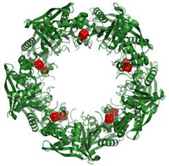
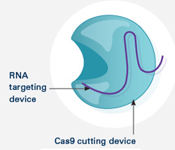
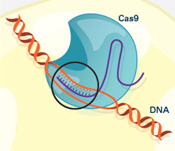
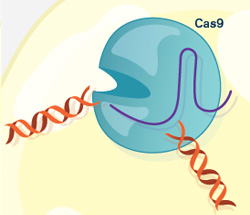
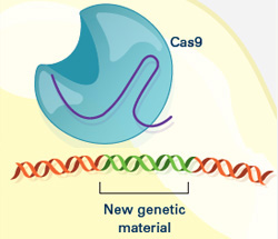
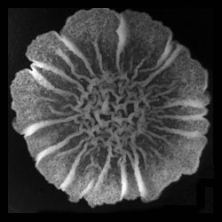
This wreath represents the molecular structure of a protein, Cas4, which is part of a system, known as CRISPR, that bacteria use to protect themselves against viral invaders. The green ribbons show the protein’s structure, and the red balls show the location of iron and sulfur molecules important for the protein’s function. Scientists have harnessed Cas9, a different protein in the bacterial CRISPR system, to create a gene-editing tool known as CRISPR-Cas9. Using this tool, researchers can study a range of cellular processes and human diseases more easily, cheaply and precisely. Last week, Science magazine recognized the CRISPR-Cas9 gene-editing tool as the “breakthrough of the year.”

Continue reading “Cool Images: A Holiday-Themed Collection”

Recognition for CRISPR Gene-Editing Tool

3 comments

The CRISPR gene-editing tool was recognized today by Science magazine as its “breakthrough of the year.” We support a number of researchers working in this exciting area and have featured it on this blog. To learn more about this exceptionally promising new method, see below for our illustrated explanation of the CRISPR system and its possible applications.

How the CRISPR System Works

Illustration of CRISPR system

The CRISPR system has two components joined together: a finely tuned targeting device (a small strand of RNA programmed to look for a specific DNA sequence) and a strong cutting device (an enzyme called Cas9 that can cut through a double strand of DNA).

CRISPR system in a cell

Once inside a cell, the CRISPR system locates the DNA it is programmed to find. The CRISPR seeking device recognizes and binds to the target DNA (circled, black).

The Cas9 enzyme cuts both strands of the DNA.

New genetic material incorporated into the broken DNA

Researchers can insert into the cell new sections of DNA. The cell automatically incorporates the new DNA into the gap when it repairs the broken DNA.

CRISPR has many possible uses, including:

  • Insert a new gene so the organism produces useful medicines.
  • Help treat genetic diseases.
  • Create tailor-made organisms to study human diseases.
  • Help produce replacements for damaged or diseased tissues and organs.

Sugar Rush in Research

0 comments
Cookies
Sugar sprinkled on cookies and other treats is often an attractive—and sweet tasting—finishing touch. But the sugar-rich coating that surrounds most cells is far more—it’s a vital ingredient for many basic cellular processes. Credit: Stock image.

Simple sugars such as sucrose (found in the sugar bowl) and fructose (in fruits and honey) provide the sweet finishing touches on many holiday treats. But did you know that versions of these molecules also serve important functions in our cells?

Cells assemble sugar molecules into chains known as glycans. These glycans, which can be linear or branching, play an astounding number of biological roles. When bound to proteins called lectins, they enable a fertilized egg to attach properly onto a woman’s uterine wall and help immune cells move out of a blood vessel to the site of an infection. When decorated with specific patterns of molecules called sulfates, glycans can help direct the growth of nerves. And it’s the glycans found on our blood cells that define blood type (A, B, AB or O). Continue reading “Sugar Rush in Research”

Bacterial Biofilms: A Charged Environment

1 comment
Bacillus subtilis biofilm
A Bacillus subtilis biofilm grown in a Petri dish. Credit: Süel Lab, UCSD.

Last summer, we shared findings from Gürol Süel Exit icon and colleagues at the University of California, San Diego, that bacterial cells in tight-knit microbial communities called biofilms expand in a stop-and-go pattern. The researchers concluded that this pattern helps make food at the nutrient-rich margin available to the cells in the starved center, but they didn’t know how. They’ve now shown that the cells use electrochemical signaling to communicate and cooperate with each other.

Because nutrients and other signals cells use to sense each other and their environment move rather slowly, the researchers looked for a faster, more active communication system in biofilms of the bacterium B. subtilis. They focused on electrical signaling via potassium, a positively charged ion that, for example, our nerve and muscle cells use to send or receive signals. Continue reading “Bacterial Biofilms: A Charged Environment”

Cracking a Ubiquitous Code

0 comments

We asked the heads of our scientific divisions to tell us about some of the big questions in fundamental biomedical science that researchers are investigating with NIGMS support. This article is the third in an occasional series that explores these questions and explains how pursuing the answers could advance understanding of important biological processes.

Ubiquitin (Ub) molecules
Ubiquitin (Ub) molecules attached to proteins can form possibly hundreds of different shapes. Credit: NIGMS.

Researchers are on a quest to crack a code made by ubiquitin, a small protein that plays a big role in coordinating cellular function. By attaching to other proteins, ubiquitin determines what those proteins should do next.

Just as zip codes direct letters to specific towns, the ubiquitin code might direct one protein to help with DNA repair, another to assist in cell division, and a third to transport molecules into and out of cells. Continue reading “Cracking a Ubiquitous Code”

Seeing Telomerase’s ‘Whiskers’ and ‘Toes’

1 comment
Telomerase and its components.

The image here is the “front view” of telomerase, with the enzyme’s components shown in greater detail than ever before. Credit: UCLA Department of Chemistry and Biochemistry.

Like the features of a cat in a dark alley, those of an important enzyme called telomerase have been elusive. Using a combination of imaging techniques, a research team led by Juli Feigon Exit icon of the University of California, Los Angeles, has now captured the clearest view ever of the enzyme.

Telomerase maintains the DNA at the ends of our chromosomes, known as telomeres, which act like the plastic tips on the ends of shoelaces. In the absence of telomerase activity, telomeres get shorter each time our cells divide. Eventually, the telomeres become so short that the cells stop dividing or die. On the other hand, cells with abnormally high levels of telomerase activity can constantly rebuild their protective chromosomal caps. Telomerase is particularly active within cancer cells. Continue reading “Seeing Telomerase’s ‘Whiskers’ and ‘Toes’”

Cool Image: Tracing Proteins in Action

2 comments
Bright amorphous loops
These bright, amorphous loops represent a never-before-seen glimpse at how proteins that play a key role in cell duplication are themselves duplicated. Credit: Sue Jaspersen, Zulin Yu and Jay Unruh, Stowers Institute for Medical Research.

Looking like necklaces stacked on a dresser, these bright, amorphous loops show the outlines of yeast proteins that make up the spindle pole, a cellular component found in organisms as diverse as yeast and humans. Each cell starts with a single spindle pole, which must somehow duplicate to form the pair that works together to pull matching chromosomes apart during cell division. Scientists don’t completely understand how this duplication occurs, but they do know that errors in spindle pole copying can lead to a number of health conditions, including cancer.

Continue reading “Cool Image: Tracing Proteins in Action”

Sharing ‘Behind the Scene’ Stories About Scientific Discoveries

0 comments

If a picture is worth a thousand words, what’s a video worth? For cell biologist Ron Vale, it’s priceless.

Screen shot from the video
In this iBiology Exit icon “discovery talk,” Ron Vale describes the twists and turns that led him to unexpected findings, including a motor protein involved in important cellular processes.

In 2006, Vale started a video-based science outreach project called iBiology Exit icon to give people around the world broader access to research seminars. The free online videos, which cover a range of biomedical fields and career-related topics, take viewers behind the scenes of scientific findings and convey the excitement of the discovery process.

While geared mostly for undergraduate students, graduate students and postdoctoral researchers, the videos are also a rich resource for anyone who wants a better understanding of many biomedical areas, including those we cover on this blog. Continue reading “Sharing ‘Behind the Scene’ Stories About Scientific Discoveries”

Interview With a Worm: We’re Not So Different

2 comments
Planarian
Credit: Alejandro Sánchez Alvarado, Stowers Institute for Medical Research.
Schmidtea mediterranea
Home: Freshwater habitats along the Mediterranean
Party trick: Regenerating its head
Most charismatic feature: Eyespots
Work site: Science labs worldwide

The planarian has a power few creatures can match. Remove its head, its tail or nearly any of its body parts, and this aquatic flatworm will simply grow it back. Humans can’t do that, of course. And yet many of the genes that help the planarian regenerate are also found in us. To learn more about this tiny marvel, we “interviewed” a representative. Continue reading “Interview With a Worm: We’re Not So Different”

El Niño Season Temperatures Linked to Dengue Epidemics

1 comment
Screen shot from a video showing dengue incidence in Southeast Asia.
Incidence of dengue fever across Southeast Asia, 1993-2010. Note increasing incidence (red) starting about June 1997, which corresponds to a period of higher temperatures driven by a strong El Niño season. At the end of the El Niño event, in January 1999, dengue incidence is much lower (green). Credit: Wilbert van Panhuis, University of Pittsburgh.

Weather forecasters are already warning about an intense El Niño season that’s expected to alter precipitation levels and temperatures worldwide. El Niño seasons, characterized by warmer Pacific Ocean water along the equator, may impact the spread of some infectious diseases transmitted by mosquitoes.

In a study published last month in the Proceedings of the National Academy of Sciences, researchers reported a link between intense dengue fever epidemics in Southeast Asia and the high temperatures that a previous El Niño weather event brought to that region.

Dengue fever, a viral infection transmitted by the Aedes mosquito, can cause life-threatening high fever, severe joint pain and bleeding. Infection rates soar every two to five years. Interested in understanding why, an international team of researchers collected and analyzed incidence reports including 3.5 million dengue fever cases across eight Southeast Asian countries spanning an 18-year period. The study is part of Project Tycho, an effort to study disease transmission dynamics by mining historical data and making that data freely available to others. Continue reading “El Niño Season Temperatures Linked to Dengue Epidemics”