Category: Cells

The Changing Needs of a Cell: No Membrane? No Problem!

0 comments

Russian nesting dolls. Credit: iStock.

How “membrane-less” organelles help with key cellular functions

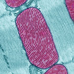
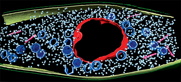
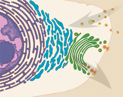
Scientists have long known that animal and plant cells have specialized subdivisions called organelles. These organelles are surrounded by a semi-permeable barrier, called a membrane, that both organizes the organelles and insulates them from the rest of the cell’s mix of proteins, salt, and water. This set-up helps to make cells efficient and productive, aiding in energy production and other specialized functions. But, because of their semi-permeable membranes, organelles can’t regroup and reform in response to stress or other outside changes. Cells need a rapid response team working alongside the membrane-bound organelles to meet these fluctuating needs. Until recently, who those rapid responders were and how they worked has been a mystery.

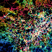
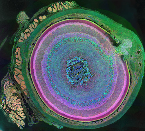
Recent research has led biologists to learn that the inside of a cell or an organelle is not just a lot of different molecules dissolved in water. Instead, we now know that cells contain many pockets of liquid droplets (one type of liquid surrounded by a liquid of different density) with specialized composition and function that are not surrounded by membranes. Because these “membrane-less organelles” are not confined, they can rapidly come together in response to chemical signals, such as those that indicate stress, and equally rapidly fall apart when they are no longer needed, or when the cell is about to divide. This enables membrane-less organelles to be “rapid responders.” They can have complex, multilayered structures that help them to perform many critical cell functions with multiple steps, just like membrane-bound organelles. Scientists even suspect that the way these organelles form as droplets may shed light on how life on Earth first took shape (see sidebar “Could This Be How Life First Took Shape?” at bottom of page).

The Many Membrane-less Organelles

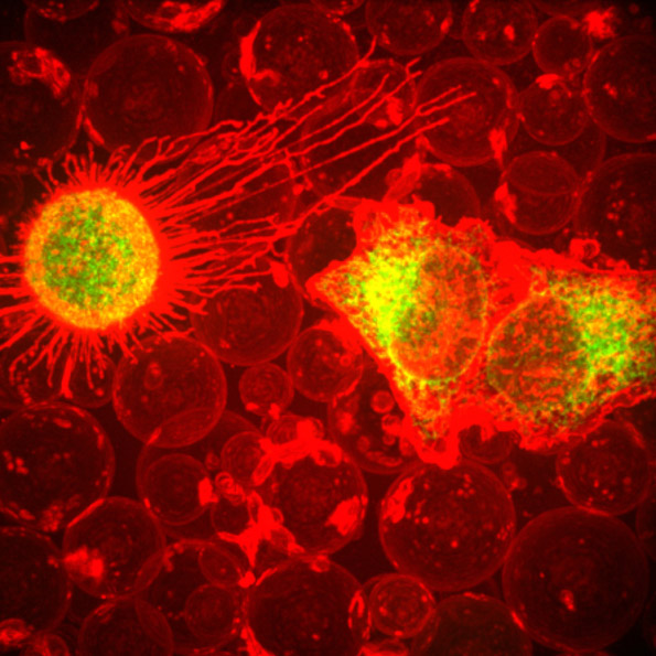
Scientists have identified more than a dozen membrane-less organelles at work in mammalian cells. Several kinds found inside the nucleus—including nuclear speckles, paraspeckles, and Cajal bodies—help with cell growth, stress response, the metabolizing (breaking down) of RNA, and the control of gene expression—the process by which information in a gene is used in the synthesis of a protein. Out in the cytoplasm, P-bodies, germ granules, and stress granules are membrane-less organelles that are involved in metabolizing or protecting messenger RNA (mRNA), controlling which mRNAs are made into proteins, and in maintaining balance, or homeostasis, of the cell’s overall health.

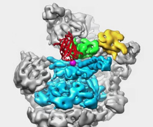
The nucleolus, located inside the nucleus, is probably the largest of the membrane-less organelles. It acts as a factory to assemble ribosomes, the giant molecular machines that “translate” messenger RNAs to make all cellular proteins.

Continue reading “The Changing Needs of a Cell: No Membrane? No Problem!”

Fall 2017 Issue of Findings Magazine

0 comments

It’s back! Check out the new issue of Findings magazine.

Findings Magazine Cover for Fall 2017.Findings presents cutting-edge research from scientists in diverse biomedical fields. The articles are aimed at high school students with the goal of making science—and the people who do it—interesting and exciting, and to inspire young readers to pursue careers in biomedical research. In addition to putting a face on science, Findings offers activities such as quizzes and crossword puzzles and, in its online version, video interviews with scientists.

The Fall 2017 issue profiles Yale University biologist Enrique De La Cruz, who studies how actin—a protein chain that supports cell structure—breaks so easily. Also profiled is University of California, Berkeley, biologist Rebecca Heald and her study of developmental factors that control an animal’s size.

This issue also features:

  • A virtual reality program designed to help burn patients manage pain
  • The promise of gene therapy for glaucoma
  • The many ways scientists categorize the biological world using “omics”
  • What researchers know—and don’t know—about how general anesthetics work
  • How animation helps researchers visualize interactions between biological molecules
  • How cells use sugary outer coatings to distinguish friend from foe
  • What makes our tissues stiff, squishy, solid, or see-through (hint: its initials are ECM)
  • How super-powerful microscopes are revealing views of biology never possible before

View Findings online, or order a print copy (classroom sets of up to 30 copies are available for educators).

Happy Birthday, BioBeat

0 comments

This month, our blog that highlights NIGMS-funded research turns four years old! For each candle, we thought we’d illuminate an aspect of the blog to offer you, our reader, an insider’s view.

Who are we?

Over the years, the editorial team has included onsite science writers, office interns, staff scientists and guest authors from universities. Kathryn, who’s a regular contributor, writes entirely from her home office. Chris, who has a Ph.D. in neuroscience and now manages the blog, used to do research in a lab. Alisa has worked in NIGMS’ Bethesda-based office the longest: 22 years! She and I remember when we first launched Biomedical Beat as an e-newsletter in 2005. You can read more about each of the writers on the contributors page and if you know someone who’s considering a career in science communications, tell them to drop us a line.

How do we come up with the stories?

We get our story ideas from a range of sources. For instance, newspaper articles about an experimental pest control strategy in Florida and California prompted us to write about NIGMS-funded studies exploring the basic science of the technique. A beautiful visual from a grantee’s institution inspired a short post on tissue regeneration research. And an ongoing conversation with NIGMS scientific staff about the important role of research organisms in biological studies sparked the idea for a playful profile of one such science superstar.

A big change in our storytelling has been shifting the focus from a single finding to broader progress in a lab or field. So instead of reporting on a study just published in a scientific journal, we may write about the scientist’s career path or showcase a collection of recent findings in that particular field. These approaches help us demonstrate that scientific understanding usually progresses through the slow and steady work undertaken by many labs.

What are our favorite posts?

I polled the writers on posts they liked, and the list is really long! Here are the top picks.


Four Ways Inheritance Is More Complex Than Mendel Knew


The Endoplasmic Reticulum: Networking in the Cell


Interview With a Scientist: Janet Iwasa, Molecular Animator


From Basic Research to Bioelectric Medicine


An Insider’s Look at Life: Magnified, an Airport Exhibit of Stunning Microscopy Images

What are your favorite posts?

We regularly review data about the number of times a blog post has been viewed to identify the ones that interest readers the most. That information also helps guide our decisions about other topics to feature on the blog. The Cool Image posts are among the most popular! Below are some other chart-topping posts.


Our Complicated Relationship With Viruses


The Proteasome: The Cells Trash Processor in Action


Demystifying General Anesthetics


Meet Sarkis Mazmanian and the Bacteria That Keep Us Healthy


5 Reasons Biologists Love Math

We always like hearing from readers! If there’s a basic biomedical research topic you’d like us to write about, or if you have feedback on a story or the blog in general, please leave your suggestions in the comment field below.

Cool Image: Biological Bubbles

1 comment
Cells are in the process of pinching off parts of their membranes to produce bubbles filled with a mix of proteins and RNAs. Researchers are harnessing this process to develop better drug delivery techniques. The image, courtesy of Chi Zhao, David Busch, Connor Vershel and Jeanne Stachowiak of the University of Texas at Austin, was entered in the Biophysical Society’s 2017 Art of Science Image contest and featured on the NIH Director’s Blog.

This fiery-looking image shows animal cells caught in the act of making bubbles, or blebbing.

Certain cells regularly pinch off parts of their membranes to produce bubbles filled with a mix of proteins and RNAs. The green and yellow portions in the image show the cell membranes as they separate from the cell’s skeleton and bleb from the main cell. The bubbles, shown in red, are called plasma-derived membrane vesicles, or PMVs. PMVs can travel to other parts of the body where they may aid in cell-to-cell communication.

The University of Texas at Austin researchers who produced this image are exploring ways to use PMVs to deliver medicines to precise locations in the body.

Blebbing for Drug Delivery

Drug delivery research tries to find ways to carry medicines to only the tissues in the body that need them with the goal of reducing side effects. To achieve this, delivery methods need to recognize just the cells they target, usually by finding a unique protein on the cell’s surface. Scientists can make proteins that recognize and attach to targets such as cancer cells, but they’ve had trouble attaching medicines to the proteins they made. To get around this problem, scientists could employ PMV-making cells in a laboratory, perhaps even cells taken from a patient who is receiving treatment. They could engineer the cells to make the targeting proteins and then attach the targeting proteins to the PMV surface. The cell’s own protein-making machinery does the hardest job.

The Texas scientists have engineered such donor cells with proteins on their surfaces that precisely target certain kinds of breast cancer cells. When the donor cells are induced to bleb, they produce PMVs laden with the target proteins that locate and bind to the cancer cells.

Researchers hope that eventually PMVs with surface targeting proteins could be filled with medicines and infused into the patient to deliver the drugs specifically to cancerous cells while leaving healthy tissues untouched. Research will continue to investigate this possibility.

This research was funded in part by NIH under grant R01GM112065.

Viruses: Manufacturing Tycoons?

0 comments
Pseudomonas chlororaphis

A computer image shows a bacterial cell invaded by a virus. The virus uses the cell to copy itself many times. It has built a protein compartment (red, rough circle surrounding the center) to house its DNA. Viral heads (blue, smaller pentagonal shapes spread through out) and tails (pink, rod shaped near the edges) are essential parts of a finished viral particle. The small, light blue particles are the bacterium’s own protein-making ribosomes. Credit: Vorrapon Chaikeeratisak, Kanika Khanna, Axel Brilot and Katrina Nguyen.

As inventors and factory owners learned during the Industrial Revolution, the best way to manufacture a lot of products is with an assembly line that follows a set of precisely organized steps employing many copies of identical and interchangeable parts. Some viruses are among life’s original mass producers: They use sophisticated organization principles to turn bacterial cells into virus particle factories.

Scientists at the University of California, San Diego, and the University of California, San Francisco, used cutting-edge techniques to watch a bacteria-infecting virus (bacteriophage) set up its particle-making factory inside a host cell.

The image above shows this happening inside a Pseudomonas chlororaphis, a soil-borne bacterium that protects plants against fungal pathogens. The virus builds a compartment (red, rough circle surrounding the center) that helps organize an assembly line for making copies of itself. The compartment looks like a cell nucleus, which bacteria do not have, and it functions like a nucleus by keeping activities that directly involve DNA separate from other cellular functions. Continue reading “Viruses: Manufacturing Tycoons?”

The Drama of Cell Death

1 comment
spermatids

Spermatids—one stage in the formation of sperm—in the fruit fly (Drosophila). Credit: Sigi Benjamin-Hong, Rockefeller University (modified).

Although it looks like a bursting firework from a Fourth of July celebration, this image actually was created from pictures of spermatids—one stage in the formation of sperm—in the fruit fly (Drosophila). Drosophila is an organism that scientists often use as a model for studying how cells accomplish their amazing tasks. Drosophila studies can help reveal where an essential cellular process goes wrong in diseases such as autoimmune conditions or cancer. Cell death, or apoptosis, is one of these processes.

Almost every animal cell has the ability to destroy itself via apoptosis. Apoptosis is important because it allows the body both to develop normally and get rid of dangerous and unwanted cells when it needs to later in life, such as when cells become cancerous. Many different signals both within and outside the cell influence whether apoptosis happens when it should, and abnormal regulation of this process is associated with some diseases. Hermann Steller Exit icon and colleagues at Rockefeller University in New York City study Drosophila and mammalian cells to tease apart the steps of apoptosis and the many molecular signals that regulate it. Continue reading “The Drama of Cell Death”

Beauty is in the Eye

2 comments

Our eyes are the gateway to countless brilliant sights. However, as evidenced by the images on this page, the eye itself can be breathtakingly exquisite as well. This May, as we celebrate Healthy Vision Month with the National Eye Institute, we hope sharing the beauty hidden in your eyes will inspire you to take the necessary steps to protect your vision, prevent vision loss and make the most of the vision you have remaining.

Visit NEI to learn more about caring for your eyes.

Happy Healthy Vision Month!

Mammalian eye
Eyes are beautiful, and they take on a whole new look in this agate-like image, which highlights just how complex mammalian eyes really are. Researchers used staining and imaging techniques to turn each of the 70-plus cell types in this mouse eye a different color. The image won first place in the 2011 International Science and Engineering Visualization Challenge. Credit: Bryan William Jones, University of Utah Moran Eye Center.
Mouse eye
This burst of starry points is actually part of the retina from a mouse eye. The image comes from a research project investigating the promise of gene therapy for glaucoma. Untreated glaucoma is a leading cause of blindness. The disease is characterized by the death of cells called retinal ganglion cells. Scientists are hoping to deliver gene therapy to these cells as a treatment for glaucoma. In this photo, a fluorescent protein (GFP) lights up to show the location of retinal ganglion cells—and to reveal how well the proposed gene therapy technique might work. Credit: Kenyoung Kim, Wonkyu Ju and Mark Ellisman, National Center for Microscopy and Imaging Research, University of California, San Diego.
Mouse eye
What appears as a tree branch painstakingly wrapped in green wire is a microscopic blood vessel from the retina at the back of a mouse eye. These vessels can help diagnose conditions such as glaucoma and diabetic eye disease. The vessels also have a characteristic appearance in people with high blood pressure. This detailed image was created to help scientists understand what happens in a genetic disease called neurofibromatosis, in which tumors begin to form on nerve tissue. Credit: National Center for Microscopy and Imaging Research, University of California, San Diego.
Mouse eye
Like a colorful fiber-optic network, this microscopic layer from a mouse’s eye relays information from the retina to the brain. Retinal ganglion neurons (orange) and their associated optic nerve fibers (red) are overlaid with blood vessels (blue) and spidery glial cells (green). By comparing detailed images of healthy eye tissues with similar images of a diseased eye, researchers can learn about changes in biology that occur as eye diseases develop. Credit: National Center for Microscopy and Imaging Research, University of California, San Diego.

The Endoplasmic Reticulum: Networking Inside the Cell

5 comments

Like a successful business networker, a cell’s endoplasmic reticulum (ER) is the structure that reaches out—quite literally—to form connections with many different parts of a cell. In several important ways, the ER enables those other parts, or organelles, to do their jobs. Exciting new images of this key member of the cellular workforce may clarify how it performs its roles. Such knowledge will also help studies of motor neuron and other disorders, such as amyotrophic lateral sclerosis (ALS), that are associated with abnormalities in ER functioning.

Structure Follows Function

Illustration of some of the jobs that the ER performs in the cell.

An illustration of some of the jobs that the endoplasmic reticulum (ER) performs in the cell. Some ER membranes (purple) host ribosomes on their surface. Other ER membranes (blue) extending into the cytoplasm are the site of lipid synthesis and protein folding. The ER passes on newly created lipids and proteins to the Golgi apparatus (green), which packages them into vesicles for distribution throughout the cell. Credit: Judith Stoffer.

Initiated in 1965, the Postdoctoral Research Associate Program (PRAT) is a competitive postdoctoral fellowship program to pursue research in one of the laboratories of the National Institutes of Health. PRAT is a 3-year program providing outstanding laboratory experiences, access to NIH’s extensive resources, mentorship, career development activities and networking. The program places special emphasis on training fellows in all areas supported by NIGMS, including cell biology, biophysics, genetics, developmental biology, pharmacology, physiology, biological chemistry, computational biology, immunology, neuroscience, technology development and bioinformatics

The ER is a continuous membrane that extends like a net from the envelope of the nucleus outward to the cell membrane. Tiny RNA- and protein-laden particles called ribosomes sit on its surface in some places, translating genetic code from the nucleus into amino acid chains. The chains then get folded inside the ER into their three-dimensional protein structures and delivered to the ER membrane or to other organelles to start their work. The ER is also the site where lipids—essential elements of the membranes within and surrounding a cell—are made. The ER interacts with the cytoskeleton—a network of protein fibers that gives the cell its shape—when a cell divides, moves or changes shape. Further, the ER stores calcium ions in cells, which are vital for signaling and other work.

To do so many jobs, the ER needs a flexible structure that can adapt quickly in response to changing situations. It also needs a lot of surface area where lipids and proteins can be made and stored. Scientists have thought that ER structure combined nets of tubules, or small tubes, with areas of membrane sheets. However, recent NIGMS PRAT (Postdoctoral Research Associate; see side bar) fellow Aubrey Weigel, working with her mentor and former PRAT fellow Jennifer Lippincott-Schwartz of the Eunice Kennedy Shriver National Institute of Child Health and Human Development (currently at the Howard Hughes Medical Institute in Virginia) and colleagues, including Nobel laureate Eric Betzig, wondered whether limitations in existing imaging technologies were hiding a better answer to how the ER meets its surface-area structural needs in the periphery, the portion of the cell not immediately surrounding the nucleus. Continue reading “The Endoplasmic Reticulum: Networking Inside the Cell”

Birthdays, Nobel Prizes and Basic Research

1 comment
James D. Watson
James D. Watson. Credit: Wikimedia Commons, Cold Spring Harbor Laboratory.

April 6 is the birthday of two Nobel Prize winners in physiology or medicine—James Watson and Edmond H. Fischer. They have also both been NIGMS-supported researchers.

Double helix model
In 1953, Watson and Crick created their historic model of the shape of DNA: the double helix. Credit: Cold Spring Harbor Laboratory archives.

James D. Watson, born on this day in 1928, was honored with the Nobel Prize in 1962. He shared it with Francis H. Compton Crick and Maurice Wilkins “for their discoveries concerning the molecular structure of nucleic acids and its significance for information transfer in living material.” This laid the groundwork for future discoveries. In the early 1950s, Wilkins and another scientist, Rosalind Franklin, worked to determine DNA’s structure. In 1953, Watson and Crick discovered its shape as a double helix. This twisted ladder structure enabled other researchers to unlock the secret of how genetic information is stored, transferred and copied. Franklin is widely recognized as having played a significant role in revealing the physical structure of DNA; due to her death at age 37 in 1958, Franklin did not earn a share of the prize. Read more about DNA.

Continue reading “Birthdays, Nobel Prizes and Basic Research”

Actin’s Many Roles

2 comments
Skin cancer cells

Skin cancer cells from a mouse. Credit: Catherine and James Galbraith, Oregon Health and Science University, Center for Spatial Systems Biomedicine, Knight Cancer Institute.

This heart-shaped image shows two mouse skin cancer cells connected to each other with actin, a protein that is part of the cellular skeleton. Researchers use mouse cells like these to tease out the molecular methods that cancer uses to invade new tissues in the body. It turns out that actin plays an essential role.

Cells can move as a collective, or independently. Movement of an individual cell requires a series of carefully controlled steps. Among them, a cell must break contacts with its neighbor cells and change its connections to the proteins and fibers around it. In addition, it must sense and follow a chemical path through the tissue it lies in. To do this, a cell changes shape, molding its membrane into flaps or feet called protrusions reaching in the direction it is traveling. Actin, among a variety of other molecules, is involved in all of these steps, but especially the shape change, when it gathers inside the cell membrane to help form the protrusions. Continue reading “Actin’s Many Roles”