Tag: Cells

CLAMP Helps Lung Cells Pull Together

0 comments
ALT TEXTCells covered with cilia (red strands) on the surface of frog embryos. Credit: The Mitchell Lab.

The outermost cells that line blood vessels, lungs, and other organs also act like guards, alert and ready to thwart pathogens, toxins, and other invaders that can do us harm. Called epithelial cells, they don’t just lie passively in place. Instead, they communicate with each other and organize their internal structures in a single direction, like a precisely drilled platoon of soldiers lining up together and facing the same way.

Lining up this way is crucial during early development, when tissues and organs are forming and settling into their final positions in the developing body. In fact, failure to line up in the correct way is linked to numerous birth defects. In the lungs, for instance, epithelial cells’ ability to synchronize with one another is important since these cells have special responsibilities such as carrying mucus up and out of lung tissue. When these cells can’t coordinate their functions, disease results.

Some lung epithelial cells are covered in many tiny, hair-like structures called cilia. All the cilia on lung epithelial cells must move uniformly in a tightly choreographed way to be effective in their mucus-clearing job. This is a unique example of a process called planar cell polarity (PCP) that occurs in cells throughout the body. Researchers are seeking to identify the signals cells use to implement PCP. Continue reading “CLAMP Helps Lung Cells Pull Together”

Pericytes: Capillary Guardians in the Brain

0 comments
ALT TEXT
The long arms of pericytes cells (red) stretch along capillaries (blue) in a mouse brain. Credit: Andy Shih.

Nerve cells, or neurons, in our brains do amazing work, from telling our hearts to beat to storing our memories. But neurons cannot operate alone. Many kinds of cells support and regulate neurons and—like neurons—they can come under attack due to injuries or disorders, such as stroke or Alzheimer’s disease. Learning what jobs these cells do and how they respond to disease may show researchers new ways to treat central nervous system disorders. One type of support cell, the pericyte, plays some key roles in brain health. These cells are readily adaptable, even in adult brains, and can support a variety of functions.

Pericytes help with blood flow to nerve cells in the brain. They lie wrapped all along the huge networks of capillaries—the tiniest blood vessels—that both feed neurons and form the blood-brain barrier, which filters out certain substances from blood to protect the brain. Pericytes have a body that appears as a bump protruding from a capillary surface. Pericytes also have long thin arms that stretch along each capillary like a snake on a tree branch. These arms, called processes, reach almost to where the next pericyte process begins, without overlapping. This creates a pericyte chain that covers nearly the entire capillary network.

Pericytes are critical for blood vessel stability and blood-brain barrier function. They’re also known to die off as a result of trauma and disease. Andy ShihLink to external web site, Andree-Ann Berthiaume, and colleagues at the Medical University of South Carolina in Charleston, set up an imaging technique in mouse brains that allowed them to see what pericytes do under normal conditions as well as how these cells respond when some are damaged.

Continue reading “Pericytes: Capillary Guardians in the Brain”

Optogenetics Sparks New Research Tools

1 comment

Imagine if scientists could zap a single cell (or group of cells) with a pulse of light that makes the cell move, or even turns on or off the cell’s vital functions.

Scientists are working toward this goal using a technology called optogenetics. This tool draws on the power of light-sensitive molecules, called opsins and cryptochromes, which are naturally occurring molecules found in the cell membranes of a wide variety of species, from microscopic bacteria and algae to plants and humans. These light-reacting molecules change their shape or activity when they sense light, so they can be used to trigger cellular activity, such as turning on or off ion flow into the cell and other regulatory pathways. The ability to induce changes in cells has a broad range of practical applications, from enabling scientists to see how cells function to providing the basis for potential therapeutic applications for blindness, cancer, and epilepsy.

Opsins first gained a foothold in research about a decade ago when scientists began using them to study specific electrical networks in the brain. This research relied on channelrhodopsins, opsins that could be used to control the flow of charged ions into and out of the cell. Normally, when a neuron reaches a certain ion concentration, it is triggered to fire, but neuron firing can be changed by inserting opsins in the membrane. Neuroscientists figured out how to incorporate light-sensitive opsin proteins by inserting the opsin gene into the host’s DNA. The genetically encoded opsin proteins in the neuronal membranes could be turned on or off by shining light into the brain itself, using optical fibers or micro-LEDs, to switch on or off the flow of ions and neuron firing.

Since those early studies in the brain, the optogenetics field has come a long way. But the leap from brain cells to other cells has been challenging. Scientists first needed to find a way to deliver light into tissues deep in the body. And, unlike stationary brain cells, they needed a way to target cells that are on the move (such as immune cells). They also needed to develop a way to study not only cell networks but also individual cells and cell parts. The NIGMS-funded researchers highlighted below are among the scientists working to overcome these obstacles and are using optogenetics in new and inventive ways.

Illustration showing how bridges can be built within a cell using light-reacting molecules
Illustration shows how “bridges” can be built within a cell through the use of light-reacting molecules. The light triggers proteins to line up within the cell, making it easier to shuttle molecules between the membranes of two subcellular organelles. This optogenetic strategy is helping scientists to control cell function with a simple beam of light. Illustration courtesy of Yubin Zhou.

Building Bridges

Yubin ZhouLink to external web site of Texas A&M is using optogenetics to control the way cells communicate and to study immune cell function. In one line of research, Zhou is using light to make it easier for calcium ions to enter cells. The ions carry instructions for the cell and also help tether small cellular structures (called organelles).  Those inter-membrane tethers allow for the movement of  proteins and lipids back and forth across the cell, and are critical for sending chemical messengers to communicate information (see illustration). When this process is disrupted, it can lead to extreme changes in cell function and even cell death. Using this technology to “switch on” normal pathways enables the scientists to better understand how such processes can be disrupted.

Continue reading “Optogenetics Sparks New Research Tools”

Cellular Footprints: Tracing How Cells Move

0 comments
ALT TEXT
An engineered cell (green) in a fruit fly follicle (red), or egg case, leaves a trail of fluorescent material as it moves across a fruit fly egg chamber, allowing scientists to trace its path and measure how long it took to complete its journey. Credit: David Bilder, University of California, Berkeley.

Cells are the basis of the living world. Our cells make up the tissues and organs of our bodies. Bacteria are also cells, living sometimes alone and sometimes in groups called biofilms. We think of cells mostly as staying in one spot, quietly doing their work. But in many situations, cells move, often very quickly. For example, when you get a cut, infection-fighting cells rally to the site, ready to gobble up bacterial intruders. Then, platelet cells along with proteins from blood gather and form a clot to stop any bleeding. And finally, skin cells surrounding the wound lay down scaffolding before gliding across the cut to close the wound.

This remarkable organization and timing is evident right from the start. Cells migrate within the embryo as it develops so that body tissues and organs end up in the right places. Harmful cells use movement as well, as when cells move and spread (metastasize) from an original cancer tumor to other parts of the body. Learning how and why cells move could give scientists new ways to guide those cells or turn off or slow down the movement when needed.

Glowing Breadcrumbs

Scientists studying how humans and animals form, from a single cell at conception to a complex body at birth, are particularly interested in how and when cells move. They use research organisms like the fruit fly, Drosophila, to watch movements by small populations of cells. Still, watching cells migrate inside a living fly is challenging because the tissue is too dense to see individual cell movement. But moving those cells to a dish in the lab might cause them to behave differently than they do inside the fly. To solve this problem, NIGMS-funded researcher David BilderLink to external web site and colleagues at the University of California, Berkeley, came up with a way to alter fly cells so they could track how the cells behave without removing them from the fly. They engineered the cells to lay down a glowing track of proteins behind them as they moved, leaving a traceable path through the fly’s tissue. The technique, called M-TRAIL (matrix-labeling technique for real-time and inferred location), allows the researchers to see where a cell travels and how long it takes to get there.

Bilder and his team first used M-TRAIL in flies to confirm the results of past studies of Drosophila ovaries in the lab using other imaging techniques. In addition, they found that M-TRAIL could be used to study a variety of cell types. The new technique also could allow a cell’s movement to be tracked over a longer period than other imaging techniques, which become toxic to cells in just a few hours. This is important, because cells often migrate for days to reach their final destinations.

Continue reading “Cellular Footprints: Tracing How Cells Move”

Genomic Gymnastics of a Single-Celled Ciliate and How It Relates to Humans

0 comments
Laura Landweber
Credit: Denise Applewhite.
Laura Landweber
Grew up in: Princeton, New Jersey
Job site: Columbia University, New York City
Favorite food: Dark chocolate and dark leafy greens
Favorite music: 1940’s style big band jazz
Favorite hobby: Swing dancing
If I weren’t a scientist I would be a: Chocolatier (see “Experiments in Chocolate” sidebar at bottom of story)

One day last fall, molecular biologist Laura Landweber surveyed the Princeton University lab where she’d worked for 22 years. She and her team members had spent many hours that day laboriously affixing yellow Post-it notes to the laboratory equipment—microscopes, centrifuges, computers—they would bring with them to Columbia University, where Landweber had just been appointed full professor. Each Post-it specified the machinery’s location in the new lab. Items that would be left behind—glassware, chemical solutions, furniture, office supplies—were left unlabeled.

As Landweber viewed the lab, decorated with a field of sunny squares, her thoughts turned to another sorting process—the one used by her primary research subject, a microscopic organism, to sift through excess DNA following mating. Rather than using Post-it notes, the creature, a type of single-celled organism called a ciliate, uses small pieces of RNA to tag which bits of genetic material to keep and which to toss.

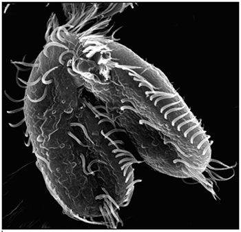
Landweber is particularly fond of Oxytricha trifallax, a ciliate with relatives that live in soil, ponds and oceans all over the world. The kidney-shaped cell is covered with hair-like projections called cilia that help it move around and devour bacteria and algae. Oxytricha is not only bizarre in appearance, it’s also genetically creative.

Unlike humans, whose cells are programmed to die rather than pass on genomic errors, Oxytricha cells appear to delight in genomic chaos. During sexual reproduction, the ciliate shatters the DNA in one of its two nuclei into hundreds of thousands of pieces, descrambles the DNA letters, throws most away, then recombines the rest to create a new genome.

Landweber has set out to understand how—and possibly why—Oxytricha performs these unusual genomic acrobatics. Ultimately, she hopes that learning how Oxytricha rearranges its genome can illuminate some of the events that go awry during cancer, a disease in which the genome often suffers significant reorganization and damage.

Oxytricha’s Unique Features

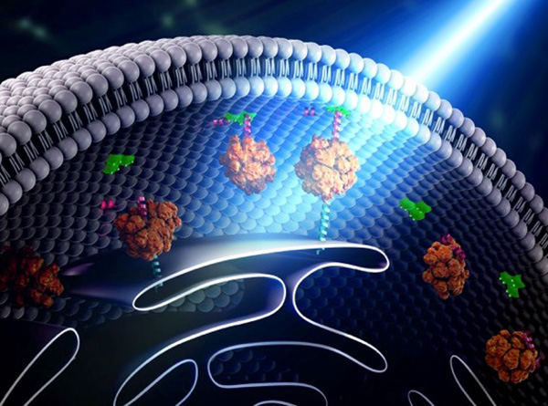
Oxytricha carries two separate nuclei—a macronucleus and a micronucleus. The macronucleus, by far the larger of the two, functions like a typical genome, the source of gene transcription for proteins. The tiny micronucleus only sees action occasionally, when Oxytricha reproduces sexually.

Oxytricha trifallax cells in the process of mating
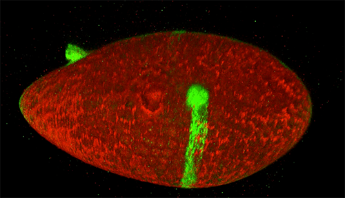
Two Oxytricha trifallax cells in the process of mating. Credit, Robert Hammersmith.

What really makes Oxytricha stand out is what it does with its DNA during the rare occasions that it has sex. When food is readily available, Oxytricha procreates without a partner, like a plant grown from a cutting. But when food is scarce, or the cell is stressed, it seeks a mate. When two Oxytricha cells mate, the micronuclear genomes in each cell swap DNA, then replicate. One copy of the new hybrid micronucleus remains intact, while the other breaks its DNA into hundreds of thousands of pieces, some of which are tagged, recombined, then copied another thousand-fold to form a new macronucleus. Continue reading “Genomic Gymnastics of a Single-Celled Ciliate and How It Relates to Humans”
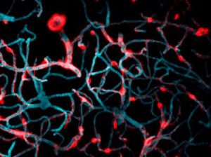
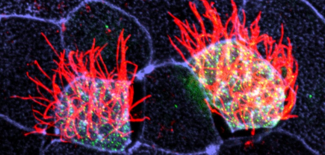
Have Nucleus, Will Travel (in Three Dimensions)

0 comments
A closeup of two human cells with the cells dyed green and the necleaus dyed red.These two human cells are nearly identical, except that the cell on the left had its nucleus (dyed red) removed. The structures dyed green are protein strands that give cells their shape and coherence. Credit: David Graham, UNC-Chapel Hill.

Both of the cells above can scoot across a microscope slide equally well. But when it comes to moving in 3D, the one without the red blob in the center (the nucleus) stalls out. That’s sort of like an Olympic speed skater who wouldn’t be able to perform even a single leap in a figure skating competition.

Scientists have known for some time that the nucleus is involved in moving cells across a flat surface—it slides to one side of the cell and “pushes” from behind. However, scientists have also shown that cells with their nuclei removed can migrate along a flat surface just as well as their brethren with intact nuclei. But they had no idea that, without a nucleus, a cell could no longer move in three dimensions.

This discovery was made by UNC-Chapel Hill biologists Keith Burridge and James Bear and their colleagues. These NIGMS-funded researchers also observed that cells whose nuclei had been disconnected from the cytoskeleton could not move through 3D matrices. The cytoskeleton is the microscopic network of actin protein filaments and tubules in the cytoplasm of many cells that provides the cell’s shape and coherence. It has also has been thought to play a major role in cell movement.

Two views of cells one on top of the other. The top animation shows a cell moving across the frame while the cells in the bottom box are static.The gray, stringy background of these videos is a 3D jello-like matrix. The cell in the top half of this video has a nucleus and can migrate through the matrix. Both cells in the bottom half have been enucleated (a fancy term for having its nucleus removed) and cannot travel through the matrix. Credit: Graham et al., Journal of Cell Biology, 2018.

The researchers speculate that the reason cells without nuclei (or those whose nuclei have been disconnected from the cytoskeleton) don’t navigate in 3D has to do with complex mechanical interactions between the cytoskeleton and the nucleoskeleton. The nucleoskeleton is a molecular scaffold within the nucleus supporting many functions such as DNA replication and transcription, chromatin remodeling, and mRNA synthesis. The interface between the cytoskeleton and nucleoskeleton consists of interlocking proteins that together provide the physical traction that cells need to push their way through 3D environments. Disrupting this interface is the equivalent of breaking the clutch in a car: the motor revs, but the wheels don’t spin, and the car goes nowhere.

A better understanding of the physical connections between the nucleus and the cytoskeleton and how they influence cell migration may provide additional insight into the role of the nucleus in diseases, such as cancer, in which the DNA-containing organelle is damaged or corrupted.

This research was funded in part by NIGMS grants 5R01GM029860-35, 5P01GM103723-05, and 5R01GM111557-04.

What Zombie Ants Are Teaching Us About Fungal Infections: Q & A with Entomologists David Hughes and Maridel Fredericksen

0 comments

I can still remember that giddy feeling I had seven years ago, when I first read about the “zombie ant.” The story was gruesome and fascinating, and it was everywhere. Even friends and family who aren’t so interested in science knew the basics: in a tropical forest somewhere there’s a fungus that infects an ant and somehow takes control of the ant’s brain, forcing it to leave its colony, crawl up a big leaf, bite down and wait for the sweet relief of death. A grotesque stalk then sprouts from the poor creature’s head, from which fungal spores rain down to infect a new batch of ants.

A fungal fruiting body erupts through the head of a carpenter ant infected by a parasitic fungus in Thailand. Credit: David Hughes, Penn State University.

The problem is, it doesn’t happen quite like that. David Hughes, the Penn State University entomologist who reported his extensive field observations of the fungus/ant interactions in BMC Ecology Exit icon, which caused much excitement back in 2011, has continued to study the fungus, Ophiocordyceps unliateralis, and its carpenter ant host, Camponotus leonardi.

In late 2017, Hughes and his colleagues published an article in PNAS Exit icon in which they used sophisticated microscopy and image-processing techniques to describe in great detail how the fungus invades various parts of the ant’s body including muscles in its legs and head.

Although Hughes’s earlier BMC Ecology paper showed fungus in the head of an ant, the new study reveals that the fungus never actually enters the brain.

To me, the new finding somehow made the fungus’ control over the ant even more baffling. What exactly was going on?

To find out, I spoke with Hughes and his graduate student Maridel Fredericksen.

Continue reading “What Zombie Ants Are Teaching Us About Fungal Infections: Q & A with Entomologists David Hughes and Maridel Fredericksen”

Interview With a Scientist: Joel Kralj, Electromicist

0 comments

Every one of our thoughts, emotions, sensations, and movements arise from changes in the flow of electricity in the brain. Disruptions to the normal flow of electricity within and between cells is a hallmark of many diseases, especially neurological and cardiac diseases.

The source of electricity within nerve cells (i.e., neurons) is the separation of charge, referred to as voltage, across neuronal membranes. In the past, scientists weren’t able to identify all the molecules that control neuronal voltage. They simply lacked the tools. Now, University of Colorado biologist Joel Kralj Exit icon has developed a way to overcome this hurdle. His new technique—combining automated imaging tools and genetic manipulation of cells—is designed to measure the electrical contribution of every protein coded by every gene in the human genome. Kralj believes this technology will usher in a new field of “electromics” that will be of enormous benefit to both scientists studying biological processes and clinicians attempting to treat disease.

In 2017, Kralj won a New Innovator Award from the National Institutes of Health for his work on studying voltage in neurons. He is using the grant money to develop a new type of microscope that will be capable of measuring neuronal voltage from hundreds of cells simultaneously. He and his research team will then attempt to identify the genes that encode any of the 20,000 proteins in the human body that are involved in electrical signaling. This laborious process will involve collecting hundreds of nerve cells, genetically removing a single protein from each cell, and using the new microscope to see what happens. If the voltage within a cell is changed in any way when a specific protein is removed, the researchers can conclude that the protein is essential to electrical signaling.

In this video, Kralj discusses how he plans to use his electromics platform to study electricity-generating cells throughout the body, as well as in bacterial cells (see our companion blog post “Feeling Out Bacteria’s Sense of Touch” featuring Kralj’s research for more details).

Dr. Kralj’s work is funded in part by the NIH under grant 1DP2GM123458-01.

The Changing Needs of a Cell: No Membrane? No Problem!

0 comments

Russian nesting dolls. Credit: iStock.

How “membrane-less” organelles help with key cellular functions

Scientists have long known that animal and plant cells have specialized subdivisions called organelles. These organelles are surrounded by a semi-permeable barrier, called a membrane, that both organizes the organelles and insulates them from the rest of the cell’s mix of proteins, salt, and water. This set-up helps to make cells efficient and productive, aiding in energy production and other specialized functions. But, because of their semi-permeable membranes, organelles can’t regroup and reform in response to stress or other outside changes. Cells need a rapid response team working alongside the membrane-bound organelles to meet these fluctuating needs. Until recently, who those rapid responders were and how they worked has been a mystery.

Recent research has led biologists to learn that the inside of a cell or an organelle is not just a lot of different molecules dissolved in water. Instead, we now know that cells contain many pockets of liquid droplets (one type of liquid surrounded by a liquid of different density) with specialized composition and function that are not surrounded by membranes. Because these “membrane-less organelles” are not confined, they can rapidly come together in response to chemical signals, such as those that indicate stress, and equally rapidly fall apart when they are no longer needed, or when the cell is about to divide. This enables membrane-less organelles to be “rapid responders.” They can have complex, multilayered structures that help them to perform many critical cell functions with multiple steps, just like membrane-bound organelles. Scientists even suspect that the way these organelles form as droplets may shed light on how life on Earth first took shape (see sidebar “Could This Be How Life First Took Shape?” at bottom of page).

The Many Membrane-less Organelles

Scientists have identified more than a dozen membrane-less organelles at work in mammalian cells. Several kinds found inside the nucleus—including nuclear speckles, paraspeckles, and Cajal bodies—help with cell growth, stress response, the metabolizing (breaking down) of RNA, and the control of gene expression—the process by which information in a gene is used in the synthesis of a protein. Out in the cytoplasm, P-bodies, germ granules, and stress granules are membrane-less organelles that are involved in metabolizing or protecting messenger RNA (mRNA), controlling which mRNAs are made into proteins, and in maintaining balance, or homeostasis, of the cell’s overall health.

The nucleolus, located inside the nucleus, is probably the largest of the membrane-less organelles. It acts as a factory to assemble ribosomes, the giant molecular machines that “translate” messenger RNAs to make all cellular proteins.

Continue reading “The Changing Needs of a Cell: No Membrane? No Problem!”

Beauty is in the Eye

2 comments

Our eyes are the gateway to countless brilliant sights. However, as evidenced by the images on this page, the eye itself can be breathtakingly exquisite as well. This May, as we celebrate Healthy Vision Month with the National Eye Institute, we hope sharing the beauty hidden in your eyes will inspire you to take the necessary steps to protect your vision, prevent vision loss and make the most of the vision you have remaining.

Visit NEI to learn more about caring for your eyes.

Happy Healthy Vision Month!

Mammalian eye

Eyes are beautiful, and they take on a whole new look in this agate-like image, which highlights just how complex mammalian eyes really are. Researchers used staining and imaging techniques to turn each of the 70-plus cell types in this mouse eye a different color. The image won first place in the 2011 International Science and Engineering Visualization Challenge. Credit: Bryan William Jones, University of Utah Moran Eye Center.
Mouse eye

This burst of starry points is actually part of the retina from a mouse eye. The image comes from a research project investigating the promise of gene therapy for glaucoma. Untreated glaucoma is a leading cause of blindness. The disease is characterized by the death of cells called retinal ganglion cells. Scientists are hoping to deliver gene therapy to these cells as a treatment for glaucoma. In this photo, a fluorescent protein (GFP) lights up to show the location of retinal ganglion cells—and to reveal how well the proposed gene therapy technique might work. Credit: Kenyoung Kim, Wonkyu Ju and Mark Ellisman, National Center for Microscopy and Imaging Research, University of California, San Diego.
Mouse eye

What appears as a tree branch painstakingly wrapped in green wire is a microscopic blood vessel from the retina at the back of a mouse eye. These vessels can help diagnose conditions such as glaucoma and diabetic eye disease. The vessels also have a characteristic appearance in people with high blood pressure. This detailed image was created to help scientists understand what happens in a genetic disease called neurofibromatosis, in which tumors begin to form on nerve tissue. Credit: National Center for Microscopy and Imaging Research, University of California, San Diego.
Mouse eye

Like a colorful fiber-optic network, this microscopic layer from a mouse’s eye relays information from the retina to the brain. Retinal ganglion neurons (orange) and their associated optic nerve fibers (red) are overlaid with blood vessels (blue) and spidery glial cells (green). By comparing detailed images of healthy eye tissues with similar images of a diseased eye, researchers can learn about changes in biology that occur as eye diseases develop. Credit: National Center for Microscopy and Imaging Research, University of California, San Diego.