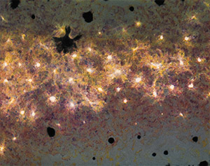
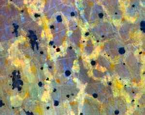
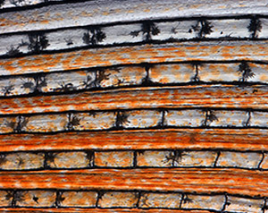
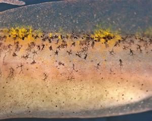
If you’ve ever visited an aquarium or snorkeled along a coral reef, you’ve witnessed the dazzling colors and patterns on tropical fish. The iridescent stripes and dots come from pigment cells, which also tint skin, hair and eyes in all kinds of animals, including humans. Typically, bright colors help attract mates, while dull ones provide camouflage. In humans, pigment helps protect skin from DNA-damaging UV light.
Researchers study cellular hues not only to decipher how they color our world, but also to understand skin cancers that originate from pigment cells. Some of these researchers work their way back, developmentally speaking, to focus on the type of cell, known as a neural crest cell, that is the precursor of pigment cells.
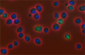
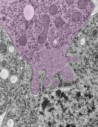
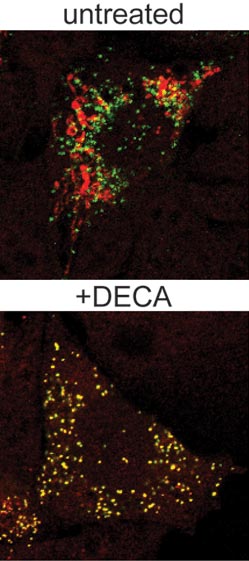
Present at the earliest stages of development, neural crest cells migrate throughout an embryo and transform into many different types of cells and tissues, including nerve cells, cartilage, bone and skin. The images here, from research on neural crest cells in fish and salamanders, showcase the beauty and versatility of pigment cells in nature’s palette.