The tiny roundworm Caenorhabditis elegans is one of the most common research organisms—creatures scientists use to study life. While C. elegans may seem drastically different from humans, it shares many genes and molecular pathways with us. Viewed with a microscope, the worm can also be surprisingly beautiful. Aside from the stunning imagery, these examples from our Image and Video Gallery show how C. elegans helps scientists advance our understanding of living systems and find new ways to improve our health.
 Credit: Keir Balla and Emily Troemel, University of California San Diego.
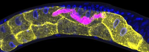
This C. elegans has been infected with microsporidia (purple), parasites closely related to fungi. The yellow shapes are the worm’s gut cells, and the blue dots are nuclei. Some microsporidia can infect people, so studying the parasites in worms could help researchers devise strategies to prevent or treat infections.
Continue reading “Cool Images: Wondrous Worms”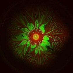 Credit: Liyang Xiong and Lev Tsimring, BioCircuits Institute, UCSD.

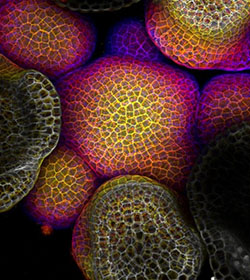
 Credit: James E. Hayden, The Wistar Institute, Philadelphia, PA.
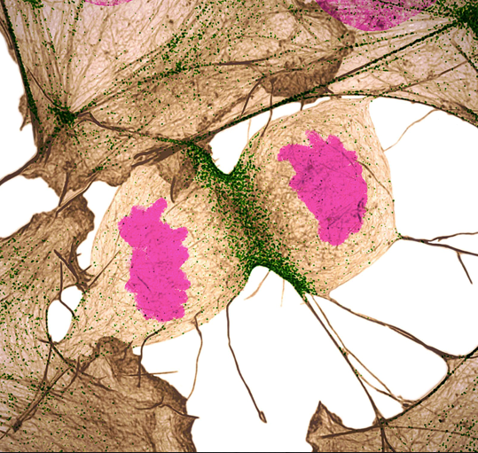
 Credit: Arun Sampathkumar and Elliot Meyerowitz, California Institute of Technology.
 Credit: Nilay Taneja, Vanderbilt University, and Dylan T. Burnette, Ph.D., Vanderbilt University School of Medicine.
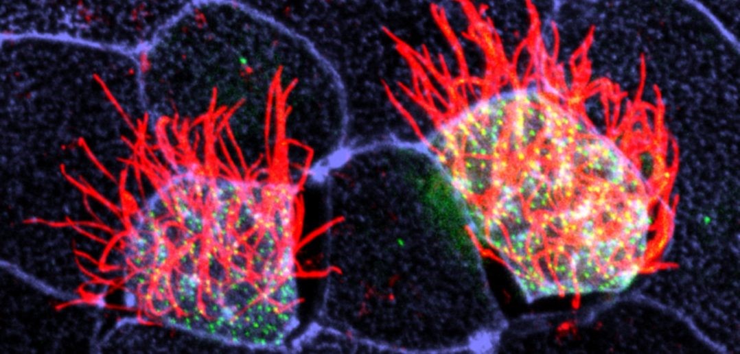 Cells covered with cilia (red strands) on the surface of frog embryos. Credit: The Mitchell Lab.